

Bioregenerative Engineering Principles and Applications - Shu Q. Liu
..pdf286 FUNDAMENTAL CELLULAR FUNCTIONS
Hatada I, Mukai T: Genomic imprinting of p57(KIP2), a cyclin-dependent kinase inhibitor, in mouse, Nature Genet 11:204–6, 1995.
Hatada I, Ohashi H, Fukushima Y, Kaneko Y, Inoue M et al: An imprinted gene p57(KIP2) is mutated in Beckwith-Wiedemann syndrome, Nature Genet 14:171–3, 1996.
Matsuoka S, Thompson JS, Edwards MC, Barletta JM, Grundy P et al: Imprinting of the gene encoding a human cyclin-dependent kinase inhibitor, p57(KIP2), on chromosome 11p15, Proc Natl Acad Sci USA 93:3026–30, 1996.
Scandura JM, Boccuni P, Massague J, Nimer SD: Transforming growth factor beta-induced cell cycle arrest of human hematopoietic cells requires p57KIP2 upregulation, Proc Natl Acad Sci USA 101:15231–6, 2004.
Human protein reference data base, Johns Hopkins University and the Institute of Bioinformatics, at http://www.hprd.org/protein.
6.4. Meiosis
Gilbert Scott F: Developmental Biology, 7th ed, Sinauer Associates, Sunderland, UK, 2003, pp 183–219.
Alberts B, Bray D, Lewis J, Raff M, Roberts K, Watson JD: Molecular Biology of the Cell, 3rd ed, Garland Publishing, New York, 1994.
6.5. Experimental Assessment of Cell Division
Gratzner HG: Monoclonal antibody to 5-bromo- and 5-iododeoxyuridine: A new reagent for determination of DNA replication, Science 218:474–5, 1982.
Krauss G: Biochemistry of Signal Transduction and Regulation, 3rd ed, Wiley-VCH GmbH, 2003. Liu SQ: Focal activation of angiotensin II type 1 receptor and smooth muscle cell proliferation in the neointima of experimental vein grafts: Relation to eddy blood flow, Arterioscl Thromb Vasc
Biol 19:2630–9, 1999.
Liu SQ, Tieche C, Alkema PK: Neointima formation on elastic lamina and collagen matrix scaffolds implanted in the rat aorta, Biomaterials 25:1869–82, 2004.
6.6. Cell Migration
Jaffe AB, Hall A: Rho GTPases: Biochemistry and biology, Annu Rev Cell Dev Biol 21:247–69, 2005.
Raftopoulou M, Hall A: Cell migration: Rho GTPases lead the way, Dev Biol 265:23–32, 2004. Etienne-Manneville S, Hall A: Rho GTPases in cell biology, Nature 420:629–35, 2002.
Hall A: Rho GTPases and the actin cytoskeleton, Science 279:509–14, 1998.
Even-Ram S, Yamada KM: Cell migration in 3D matrix, Curr Opin Cell Biol 17:524–32, 2005. Keller R: Cell migration during gastrulation, Curr Opin Cell Biol 17:533–41, 2005.
Small JV, Resch GP: The comings and goings of actin: coupling protrusion and retraction in cell motility, Curr Opin Cell Biol 17:517–23, 2005.
Yamaguchi H, Wyckoff J, Condeelis J: Cell migration in tumors, Curr Opin Cell Biol 17:559–64, 2005.
Li S, Guan JL, Chien S: Biochemistry and biomechanics of cell motility, Annu Rev Biomed Eng 7:105–50, 2005.
Craig SW, Johnson RP: Assembly of focal adhesions: Progress, paradigms, and proteins, Curr Opin Cell Biol 8:74–85, 1996.
Lauffenburger DA, Horwitz AF: Cell migration: A physically integrated molecular process, Cell 84:359–69, 1996.
BIBLIOGRAPHY 287
Mitchison TJ, Cramer LP: Actin-based cell motility and cell locomotion, Cell 84:371–9, 1996. Kiosses WB, Shattil SJ, Pampori N, Schwartz MA: Rac recruits high-affinity integrin alphavbeta3
to lamellipodia in endothelial cell migration, Nature Cell Biol 3(3):316–20, 2001.
Ridley AJ, Schwartz MA, Burridge K, Firtel RA, Ginsberg MH et al: Cell migration: Integrating signals from front to back, Science 302:1704–9, 2003.
Raftopoulou M, Hall A: Cell migration: Rho GTPases lead the way, Dev Biol 265:23–32, 2004. Schwartz MA: Integrin signaling revisited, Trends Cell Biol 11:466–70, 2001.
Sieg DJ, Hauck CR, Ilic D, Klingbeil CK, Schlaefer E et al: FAK integrates growth-factor and integrin signals to promote cell migration, Nature Cell Biol 2:249–56, 2000.
Klemke RL, Cai S, Giannini AL, Gallagher PJ, de Lanerolle P et al: Regulation of cell motility by mitogen-activated protein kinase, J Cell Biol 137:481–92, 1997.
Cramer LP, Mitchison TJ: Myosin is involved in postmitotic cell spreading, J Cell Biol 131:179–89, 1995.
RhoA
Cannizzaro LA, Madaule P, Hecht F, Axel R, Croce CM et al: Chromosome localization of human ARH genes, a ras-related gene family, Genomics 6:197–203, 1990.
Kiss C, Li J, Szeles A, Gizatullin RZ, Kashuba VI et al: Assignment of the ARHA and GPX1 genes to human chromosome bands 3p21.3 by in situ hybridization and with somatic cell hybrids,
Cytogenet Cell Genet 79:228–30, 1997.
Maekawa M, Ishizaki T, Boku S, Watanabe N, Fujita A et al: Signaling from Rho to the actin cytoskeleton through protein kinases ROCK and LIM-kinase, Science 285:895–8, 1999.
Wang HR, Zhang Y, Ozdamar B, Ogunjimi AA, Alexandrova E et al: Regulation of cell polarity and protrusion formation by targeting RhoA for degradation, Science 302:1775–9, 2003.
Wu KY, Hengst U, Cox LJ, Macosko EZ, Jeromin A et al: Local translation of RhoA regulates growth cone collapse, Nature 436:1020–4, 2005.
Zhou Y, Su Y, Li B, Liu F, Ryder JW et al: Nonsteroidal anti-inflammatory drugs can lower amyloidogenic A-beta(42) by inhibiting Rho, Science 302:1215–7, 2003.
Ridley AJ, Allen WE, Peppelenbosch M, Jones GE: Rho family proteins and cell migration, Biochem Soc Symp 65:111–24, 1999.
Michiels F, Collard JG: Rho-like GTPases: their role in cell adhesion and invasion, Biochem Soc Symp 65:125–146, 1999.
Sheetz MP, Felsenfeld D, Galbraith CG, Choquet D: Cell migration as a five-step cycle, Biochem Soc Symp 65:233–44, 1999.
Human protein reference data base, Johns Hopkins University and the Institute of Bioinformatics, at http://www.hprd.org/protein.
6.7. Immunoglobulin-Like Domain-Containing Cell Adhesion Molecules
NCAM
Bello MJ, Salagnon N, Rey JA, Guichaoua MR, Berge-Lefranc JL et al: Precise in situ localization of NCAM, ETS1, and D11S29 on human meiotic chromosomes, Cytogenet Cell Genet 52:7–10, 1989.
Cunningham BA, Hemperly JJ, Murray BA, Prediger EA, Brackenbury R et al: Neural cell adhesion molecule: Structure, immunoglobulin-like domains, cell surface modulation, and alternative RNA splicing, Science 236:799–806, 1987.
D’Eustachio P, Owens GC, Edelman GM, Cunningham BA: Chromosomal location of the gene encoding the neural cell adhesion molecule (N-CAM) in the mouse, Proc Natl Acad Sci USA 82:7631–5, 1985.
288 FUNDAMENTAL CELLULAR FUNCTIONS
Nguyen C, Mattei MG, Mattei JF, Santoni MJ, Goridis C et al: Localization of the human NCAM gene to band q23 of chromosome 11: The third gene coding for a cell interaction molecule mapped to the distal portion of the long arm of chromosome 11, J Cell Biol 102:711–5, 1986.
Rabinowitz JE, Rutishauser U, Magnuson T: Targeted mutation of Ncam to produce a secreted molecule results in a dominant embryonic lethality, Proc Natl Acad Sci USA 93:6421–4, 1996.
Rutishauser U, Acheson A, Hall AK, Mann DM, Sunshine J: The neural cell adhesion molecule (NCAM) as a regulator of cell-cell interactions, Science 240:53–7, 1988.
Rutishauser U, Goridis C: NCAM: The molecule and its genetics, Trends Genet 2:72–6, 1986.
Telatar M, Lange E, Uhrhammer N, Gatti RA: New localization of NCAM, proximal to DRD2 at chromosome 11q23, Mam Genome 6:59–60, 1995.
VCAM
Besemer J, Harant H, Wang S, Oberhauser B, Marquardt K et al: Selective inhibition of cotranslational translocation of vascular cell adhesion molecule 1, Nature 436:290–3, 2005.
Cybulsky MI, Fries JWU, Williams AJ, Sultan P, Eddy R et al: Gene structure, chromosomal location, and basis for alternative mRNA splicing of the human VCAM1 gene, Proc Natl Acad Sci USA 88:7859–63, 1991.
Garmy-Susini B, Jin H, Zhu Y, Sung RJ, Hwang R et al: Integrin alpha-4-beta-1–VCAM-1–medi- ated adhesion between endothelial and mural cells is required for blood vessel maturation, J Clin Invest 115:1542–51, 2005.
Kumar AG, Dai XY, Kozak CA, Mims MP, Gotto AM et al: Murine VCAM-1: Molecular cloning, mapping, and analysis of a truncated form, J Immun 153:4088–98, 1994.
Lu TT, Cyster JG: Integrin-mediated long-term B cell retention in the splenic marginal zone, Science 297:409–12, 2002.
Minn AJ, Gupta GP, Siegel PM, Bos PD, Shu W et al: Genes that mediate breast cancer metastasis to lung, Nature 436:518–24, 2005.
Olson E, Srivastava D: Molecular pathways controlling heart development, Science 272:671–6, 1996.
ICAM
Bella J, Kolatkar PR, Marlor CW, Greve JM, Rossmann MG: The structure of the two amino-ter- minal domains of human ICAM-1 suggests how it functions as a rhinovirus receptor and as an LFA-1 integrin ligand, Proc Natl Acad Sci USA 95:4140–5, 1998.
Greve JM, Davis G, Meyer AM, Forte CP, Yost SC et al: The major human rhinovirus receptor is ICAM-1, Cell 56:839–47, 1989.
Le Beau MM, Ryan D Jr, Pericak-Vance MA: Report of the committee on the genetic constitution of chromosomes 18 and 19, Cytogenet Cell Genet 51:338–57, 1989.
Lu TT, Cyster JG: Integrin-mediated long-term B cell retention in the splenic marginal zone, Science 297:409–12, 2002.
Simmons D, Makgoba MW, Seed B: ICAM, an adhesion ligand of LFA-1, is homologous to the neural cell adhesion molecule NCAM, Nature 331:624–7, 1988.
Sligh JE Jr, Ballantyne CM, Rich SS, Hawkins HK, Smith CW et al: Inflammatory and immune responses are impaired in mice deficient in intercellular adhesion molecule 1, Proc Natl Acad Sci USA 90:8529–33, 1993.
PTPRd
Mizuno K, Hasegawa K, Katagiri T, Ogimoto M, Ichikawa T et al: MPTP-delta, a putative murine homolog of HPTP-delta, is expressed in specialized regions of the brain and in the B-cell lineage, Mol Cell Biol 13:5513–23, 1993.
SELECTINS 289
Schaapveld RQJ, van den Maagdenberg AMJM, Schepens JTG, Olde Weghuis D, Geurts van Kessel A et al: The mouse gene Ptprf encoding the leukocyte common antigen-related molecule LAR: Cloning, characterization, and chromosomal localization, Genomics 27:124–30, 1995.
Uetani N, Kato K, Ogura H, Mizuno K, Kawano K et al: Impaired learning with enhanced hippocampal long-term potentiation in PTP-delta-deficient mice, EMBO J 19:2775–85, 2000.
PTPRk
Fuchs M, Muller T, Lerch MM, Ullrich A: Association of human protein-tyrosine phosphatase kappa with members of the armadillo family, J Biol Chem 271:16712–9, 1996.
Yang Y, Gil MC, Choi EY, Park SH, Pyun KH et al: Molecular cloning and chromosomal localization of a human gene homologous to the murine R-PTP-kappa, a receptor-type protein tyrosine phosphatase, Gene 186:77–82, 1997.
Zhang Y, Siebert R, Matthiesen P, Yang Y, Ha H et al: Cytogenetical assignment and physical mapping of the human R-PTP-kappa gene (PTPRK) to the putative tumor suppressor gene region 6q22.2-q22.3, Genomics 51:309–11, 1998.
PTPRm
Gebbink MFBG, van Etten I, Hateboer G, Suijkerbuijk R, Beijersbergen RL et al: Cloning, expression and chromosomal localization of a new putative receptor-like protein tyrosine phosphatase, FEBS Lett 290:123–30, 1991.
Suijkerbuijk RF, Gebbink MFGB, Moolenaar WH, Geurts van Kessel A: Fine mapping of the human receptor-like protein tyrosine phosphatase gene (PTPRM) to 18p11.2 by fluorescence in situ hybridization, Cytogenet Cell Genet 64:245–6, 1993.
Human protein reference data base, Johns Hopkins University and the Institute of Bioinformatics, at http://www.hprd.org/protein.
Selectins
Classification and Structure [6.8]. Selectins (Table 6.6) are lectin-type adhesion molecules expressed in the membrane of several cell types, including vascular endothelial cells, leukocytes, and platelets. Selectins are classified into several groups: E-selectin, L- selectin, and P-selectin. E-selectin is found in endothelial cells, and its function is to mediate the interaction of endothelial cells with leukocytes via binding to corresponding ligands. L-selectin is expressed in leukocytes and is responsible for binding to ligands on endothelial cells and other leukocytes. P-selectin is expressed in platelets and endothelial cells, and is responsible for binding to ligands on leukocytes and endothelial cells. Selectins are involved in the regulation of several basic leukocyte activities, including leukocyte adhesion to, rolling on, and migration through the endothelium.
A typical selectin is composed of several domains: an N-terminal lectin-like domain, an epidermal growth factor (EGF)-like domain, several consensus repeats, a transmembrane domain, and a cytoplasmic domain (Fig. 6.8). The lectin-like and EGF-like domains are similar in amino acid sequence among different selectins, while other domains differ between different selectins. The N-terminal lectin domain is responsible for the adhesion properties of selectins in a Ca2+ -dependent manner.
Function [6.9]. The primary function of selectins is to mediate interaction between leukocytes, platelets, and endothelial cells. Selectins can selectively bind to the oligosaccharides of glycoproteins in the membrane of a target cell. Endothelial cells express various selectin ligands, including glycosylation cell adhesion molecule-1, CD34, mucosal addressin cell adhesion molecule-1, and podocalyxin. Leukocytes express primarily E-selectin glycoprotein ligand-1 and P-selectin glycoprotein ligand-1. E-selectin and

290
TABLE 6.6. Characteristics of Selected Selectins*
|
|
Amino |
Molecular |
|
|
Proteins |
Alternative Names |
Acids |
Weight (kDa) |
Expression |
Functions |
|
|
|
|
|
|
E-Selectin |
Selectin-E, endothelial leukocyte adhesion |
610 |
67 |
Vascular endothelial cells |
Regulating leukocyte adhesion to |
|
molecule 1, ELAM1, ELAM, leukocyte |
|
|
|
endothelial cells and mediating |
|
endothelial cell adhesion molecule 2, |
|
|
|
inflammatory reactions |
|
LECAM2, CD62E |
|
|
|
|
L-Selectin |
Lymphocyte adhesion molecule 1, LYAM1, |
385 |
44 |
Leukocytes, bone marrow |
Regulating leukocyte adhesion to |
|
LAM1, CD62 antigen ligand, CD62L, |
|
|
cells |
endothelial cells |
|
leukocyte adhesion molecule 1, leukocyte |
|
|
|
|
|
endothelial cell adhesion molecule 1, |
|
|
|
|
|
LECAM1, SELL |
|
|
|
|
P-selectin |
Platelet α granule membrane protein, SELP, |
830 |
91 |
Platelets, endothelial cells |
Regulating platelet adhesion to |
|
CD62, granulocyte membrane protein, |
|
|
|
endothelial cells |
|
GRMP, PSGL1 |
|
|
|
|
|
|
|
|
|
|
*Based on bibliography 6.8.

SELECTINS 291
|
|
Lectin |
|
|
EGF |
Lectin |
|
|
EGF |
|
|
|
|
Consensus |
|
Lectin |
repeats |
Consensus |
EGF |
|
repeats |
|
|
|
|
|
|
Consensus |
|
|
repeats |
|
E-selectin |
L-selectin |
P-selectin |
Figure 6.8. Schematic representation of the structure of selectins. Based on bibliography 6.8.
P-selectin of endothelial cells can bind to E-selectin glycoprotein ligand-1 and P-selectin glycoprotein ligand-1 of leukocytes, respectively. L-selectin of leukocytes can bind to glycosylation cell adhesion molecule-1, CD34, mucosal addressin cell adhesion molecule- 1, and podocalyxin of endothelial cells. When leukocyte-leukocyte interaction takes place, the L-selectin of one cell can bind to the P-selectin glycoprotein ligand-1 of another cell. Similarly, the P-selectin of platelets can bind to the P-selectin glycoprotein ligand-1 of leukocytes and to the glycosylation cell adhesion molecule-1, CD34, and mucosal addressin cell adhesion molecule-1 of endothelial cells. The binding of selectins with corresponding ligands is the basis for leukocyte adhesion to and rolling on the endothelium.
Under physiological conditions, the constitutive level of selectins in the cell membrane is considerably low, and thus leukocytes and platelets rarely adhere to endothelial cells. Endothelial cells and platelets can synthesize and maintain a constitutive pool of selectins (primarily P-selectin). The synthesized selectin molecules are not deployed to the cell membrane, but stored in the α-granules of platelets and the Weibel–Palade bodies of endothelial cells. These selectin molecules can be redistributed to the cell membrane rapidly in response to inflammatory stimulation. The expression of selectins and ligands are also upregulated in inflammatory reactions. Increased selectin level enhances selectin–ligand interaction and thus facilitates leukocyte and platelet adhesion to the endothelium.
Since leukocytes and endothelial cells are subject to bloodflow, the formation of selectin and ligand bonds must be rapid and the bonds must be sufficiently strong to resist shearing forces imposed by the bloodflow. Under certain shearing conditions, adhered leukocytes may roll on the endothelium. Such a process requires coordination between shear stress and the adhesion bond dynamics, so that the formation of adhesion bonds at the cell leading edge due to selectin–ligand interaction is associated with an equal level
292 FUNDAMENTAL CELLULAR FUNCTIONS
of disruption of adhesion bonds at the cell trailing edge due to shear stress. Leukocyte adhesion to and rolling on the endothelium are critical processes in inflammatory responses. These processes prepare leukocytes for transmigration into the interstitial space, where inflammatory reactions take place.
Selectin–ligand interaction may contribute to signal transduction in leukocytes and endothelial cells. Although a complete mechanism is not yet demonstrated, preliminary studies have shown that leukocyte adhesion to L-selectin ligands induces calcium redistribution and activation of mitogen-activated protein kinases. The level of activation is related to the density of the selectin ligands. There is also evidence that selectin–ligand interaction induces activation of integrins. Further investigations are needed to clarify selectin-related signaling pathways.
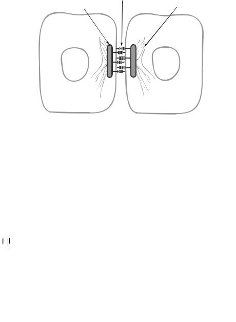
Cadherins
Classification and Structure [6.10]. Cadherins (Table 6.7) are a family of calciumdependent cell adhesion molecules, which are characterized by the presence of cadherinspecific repeats in the extracellular region of the molecule. Cadherins are traditionally classified into several subfamilies: classical cadherins, protocadherins, and desmosomal cadherins. Cadherins are usually associated with a class of molecules known as catenins. These adhesion molecules are involved in the regulation cell–cell interaction, tissue morphogenesis, as well as mitogenic activities such as cell proliferation and migration.
The classical cadherin subfamily includes E-, P-, and N-cadherins (Fig. 6.9). These molecules are localized to the zonula adherens or adherens junctions, which are intercellular contacts required for cell adhesion, cell–cell communication, and tissue formation and organization. These cadherins share similar amino acid sequences and mediate Ca2+ - dependent cell–cell interaction and connection. A typical classical cadherin is composed of an N-terminal precursor sequence, which contains a proteolytic processing signal sequence K/RRXKR, four characteristic cadherin repeats immediately following the N- terminal precursor sequence, a transmembrane domain, and a well-conserved cytoplasmic domain. Cleavage of the N-terminal precursor sequence is required for the activation of cadherin. Each cadherin repeat in the extracellular region contains consensus Ca2+ - binding sites. The binding of Ca2+ induces dimerization of cadherins and protection of the molecule from degradation. The cytoplasmic domain of cadherins interacts with the actin cytoskeleton via cadherin-associated proteins known as catenins.
Protocadherins constitute another cadherin subfamily. Compared with the classical cadherins, these adhesion molecules are characterized by the lack of the proteolytic precursor sequence and the presence of more than four cadherin repeats in the extracellular region (Fig. 6.10). In addition, unlike the classical cadherins, the cytoplasmic domain of protocadherins is considerably heterogeneous in structure. The structural difference suggests different mechanisms in regulating cell adhesion between protocadherins and classical cadherins. It appears that cell adhesion mediated by protocadherins is not as strong as that mediated by classical cadherins.
The third subfamily of cadherins is found in desmosomes and is defined as desmosomal cadherins. Desmosomes are intercellular structures identified in epithelial and cardiac muscular cells (Fig. 6.11) and are responsible for cell–cell interaction and connection, which play a critical role in regulating the formation and integrity of tissues and organs. A typical desmosome appears under an electron microscope as a complex with two parallel plaques (one from each cell) and a narrow gap ( 30 nm in width) between two cell

293
TABLE 6.7. Characteristics of Selected Cadherins*
|
|
Amino |
Molecular |
|
|
Proteins |
Alternative Names |
Acids |
Weight (kDa) |
Expression |
Functions |
|
|
|
|
|
|
E-Cadherin |
Calcium-dependent |
901 |
100 |
Ubiquitous |
A glycoprotein that regulates cell adhesion, which is a |
|
adhesion protein, |
|
|
|
calcium-dependent process |
|
cadherin 1, epithelial |
|
|
|
|
|
E-cadherin, ECAD, |
|
|
|
|
|
calcium-dependent |
|
|
|
|
|
adhesion protein, |
|
|
|
|
|
epithelial liver cell |
|
|
|
|
|
adhesion molecule, |
|
|
|
|
|
LCAM |
|
|
|
|
P-Cadherin |
Placental P cadherin, |
829 |
91 |
Ubiquitous |
A glycoprotein that regulates cell–cell adhesion (note that |
|
cadherin 3, PCAD, |
|
|
|
its action is dependent on calcium) |
|
placental calcium- |
|
|
|
|
|
dependent adhesion |
|
|
|
|
|
protein, CDHP |
|
|
|
|
N-Cadherin |
Cadherin 2, neuronal |
906 |
100 |
Nervous system, |
A glycoprotein that regulates calcium-dependent cell–cell |
|
cadherin, N cadherin, |
|
|
ovary, testis |
adhesion |
|
NCAD, CDHN. |
|
|
|
|
Protocadherin 1 |
Protocadherin 42, |
1237 |
134 |
Brain |
A membrane protein found at cell–cell junctions, |
|
PCDH42, PC42, |
|
|
|
regulating neural cell adhesion and development |
|
cadherin-like protein 1 |
|
|
|
|
Desmoglein 1 |
Desmosomal glycoprotein |
1049 |
114 |
Skin, esophagus |
Serving as a calcium-binding glycoprotein component of |
|
1, DG1, pemphigus |
|
|
|
desmosomes in epithelial cells, and regulating cell–cell |
|
foliaceus antigen, |
|
|
|
adhesion and interaction |
|
desmoglein 1 |
|
|
|
|
|
preproprotein |
|
|
|
|
Desmocollin |
Desmocollin 1A/1B, type |
894 |
100 |
Skin, thymus |
Serving as a calcium-binding glycoprotein component of |
|
I, desmocollins |
|
|
|
desmosomes in epithelial cells and regulating cell–cell |
|
desmosomal |
|
|
|
adhesion and interaction |
|
glycoprotein 2/3, |
|
|
|
|
|
DG2/DG3 |
|
|
|
|
|
|
|
|
|
|
*Based on bibliography 6.10.

294 FUNDAMENTAL CELLULAR FUNCTIONS
|
|
|
|
|
|
|
|
|
|
|
|
|
|
|
|
|
|
|
|
|
|
Cadherin |
|
|
Cadherin |
|
|
|
|
|
|
|
||
|
|
|
repeats |
|
Cadherin |
|
|
|
|
|
|||
|
repeats |
|
|
|
||
|
|
|
|
|
||
repeats
E-cadherin |
N-cadherin |
|
|
|
P-cadherin |
||||
Figure 6.9. Schematic representation of the structure of cadherins. Based on bibliography 6.10.
|
|
|
|
|
|
Cadherin |
|
|
|
|
|
|
|
|
|
|
|
|
|
|
|
|
|
|
|
|
|
|
|
|
|
|
|
|
|
|
|
Cadherin |
|
|
|
|
|
|
|
|||
|
Cadherin |
|
|
|
repeats |
|
|
|
repeats |
|
|
repeats
|
|
|
|
|
|
|
|
|
|
|
|
|
|
|
|
|
|
|
|
|
|
|
|
|
|
|
Protocadherin 1 |
Protocadherin 2 |
Protocadherin μ |
||||||
Figure 6.10. Schematic representation of the structure of Protocadherins. Based on bibliography 6.10.
membranes. Such a structure contains several components, including desmosomal cadherins, plakoglobin, plakins, and plakophilins.
There are two types of desmosomal cadherin in desmosomes: desmogleins and desmocollins. These are glycoproteins containing amino acid sequences that are similar to
CADHERINS |
295 |
|
Desmogleins |
Attachment plaque |
Keratin filaments |
|
Epithelial cell |
Epithelial cell |
Figure 6.11. Schematic representation of the structure of desmosomes. Based on bibliography 6.10.
Cadherin
Cadherin  repeats repeats
Cadherin repeats
Desmoglein 1 Desmoglein 2 Desmocollin 1
Figure 6.12. Schematic representation of the structure of desmogleins. Based on bibliography 6.10.
the classical cadherins described above. However, desmogleins contain three additional domains in their cytoplasmic tail, including a proline-rich linker, a repeating unit domain, and a terminal domain (Fig. 6.12). Each type of desmosomal cadherin exists in three isoforms. The isoforms for desmogleins are desmoglein-1,2,3, and those for desmocollins are