

Bioregenerative Engineering Principles and Applications - Shu Q. Liu
..pdf266
TABLE 6.2. Characteristics of Selected Cell Cycle Regulatory Molecules*
|
|
Amino |
Molecular |
|
|
Proteins |
Alternative Names |
Acids |
Weight (kDa) |
Expression |
Functions |
|
|
|
|
|
|
Cyclin A |
CCNA1 |
465 |
52 |
Brain, testis, leukocytes |
Binding to CDK2 and CDC2 kinases, mediating |
|
|
|
|
|
the activity of these kinases, regulating the |
|
|
|
|
|
transition of cell division cycle from the S to |
|
|
|
|
|
G2 phases |
Cyclin B G2/mitotic specific cyclin B1, |
433 |
48 |
Liver, lung, bone marrow |
Binding to CDC2 to form the M-phase-promoting |
|
|
CCNB |
|
|
|
factor and regulating the transition of cell cycle |
|
|
|
|
|
from G2 to M phases |
CDC2 |
Cell division cycle 2, cell cycle |
297 |
34 |
Skin |
A Ser/Thr protein kinase that serves as the |
|
controller CDC2, p34(CDC2), |
|
|
|
catalytic subunit of the M-phase-promoting |
|
cyclin-dependent kinase 1 |
|
|
|
factor (MPF); phosphorylates proteins such as |
|
(CDK1), p34 protein kinase, |
|
|
|
lamin, vimentin, and caldelsom; and regulates |
|
Cdc2 kinase |
|
|
|
the transition of cell cycle from G1 to S phases |
|
|
|
|
|
and from G2 to M phases |
|
|
|
|
|
|
*Based on bibliography 6.3.

267
TABLE 6.3. Characteristics of Selected Cell Cycle Inhibitory Molecules*
|
|
Amino |
Molecular |
|
|
Proteins |
Alternative Names |
Acids |
Weight (kDa) |
Expression |
Functions |
|
|
|
|
|
|
p15(INK4B) |
p15 inhibitsCDK4, cyclin-dependent |
138 |
15 |
Skin, placenta |
Acting as a cyclin-dependent kinase inhibitor, |
|
kinase inhibitor2B, CDKN2B, p15 |
|
|
|
forming a complex with CDK4 or CDK6, |
|
INK4b, CDK4B inhibitor, cyclin- |
|
|
|
suppressing the activation of these kinases, |
|
dependent kinase 4 inhibitor B, |
|
|
|
and inducing the arrest of cell division |
|
multiple-tumor suppressor 2 (MTS2) |
|
|
|
cycle in G1 phase |
p16(INK4A) |
p16 inhibits CDK4, CDKN2A, cyclin- |
173 |
18 |
Ubiquitous |
Serving as an inhibitor for the CDK4 kinase |
|
dependent kinase inhibitor 2A, CDKN2, |
|
|
|
and inducing cell arrest in G1 phase |
|
CDK4 inhibitor, multiple-tumor |
|
|
|
|
|
suppressor 1 (MTS1) |
|
|
|
|
p18(INK4C) |
p18, inhibits CDK4, cyclin-dependent |
168 |
18 |
Brain, liver, |
Serving as a cyclin-dependent kinase inhibitor, |
|
kinase inhibitor 2C, CDKN2C, cyclin- |
|
|
testis, thymus |
suppressing the activity of CDK4 and |
|
dependent kinase 6 inhibitor, cyclin- |
|
|
|
CDK6, and inducing cell arrest in G1 phase |
|
dependent kinase 4 inhibitor C, |
|
|
|
|
|
cyclin-dependent inhibitor, CDK6 |
|
|
|
|
|
inhibitor p18, p18-INK6, p18-INK4c, |
|
|
|
|
|
CDKN6 |
|
|
|
|
p19(INK4D) |
Cyclin-dependent kinase inhibitor 2D, |
166 |
18 |
Ubiquitous |
Serving as a cyclin-dependent kinase inhibitor, |
|
p19 inhibits CDK4, INK4D, p19 INK4D, |
|
|
|
and inducing cell arrest in the G1 phase. |
|
cyclin-dependent kinase 4 inhibitor D, |
|
|
|
|
|
CDK inhibitor p19 INK4D |
|
|
|
|
p21 |
Cyclin-dependent kinase inhibitor 1A, |
164 |
18 |
Heart, bone |
Acting as a cyclin-dependent kinase inhibitor, |
|
cyclin-dependent kinase inhibitor 1, |
|
|
|
inhibiting the activity of cyclin-CDK2 and |
|
DNA synthesis inhibitor, CDK- |
|
|
|
CDK4, and mediating p53-dependent cell |
|
interaction protein 1, wildtype p53- |
|
|
|
arrest in G1 and G2 phases |
|
activated fragment 1, melanoma- |
|
|
|
|
|
differentiation-associated protein 6 |
|
|
|
|
p27 |
Cyclin-dependent kinase inhibitor 1B, |
198 |
22 |
Ubiquitous |
Serving as a cyclin-dependent kinase inhibitor, |
|
cyclin-dependent kinase inhibitor p27, |
|
|
|
binding to and suppressing the cyclin |
|
p27Kip1, KIP1, CDKN4 |
|
|
|
E-CDK2 and cyclin D-CDK4 complexes, |
|
|
|
|
|
and inducing cell arrest in G1 phase |
p57 |
Cyclin-dependent kinase inhibitor 1C, |
316 |
32 |
Heart, skeletal |
Acting as an inhibitor for G1 cyclin/CDK |
|
p57KIP2, KIP2 |
|
|
muscle, B |
complexes and inducing cell arrest in G1 |
|
|
|
|
cells, placenta |
phase |
|
|
|
|
|
|
*Based on bibliography 6.3.
268 |
FUNDAMENTAL CELLULAR FUNCTIONS |
|
|
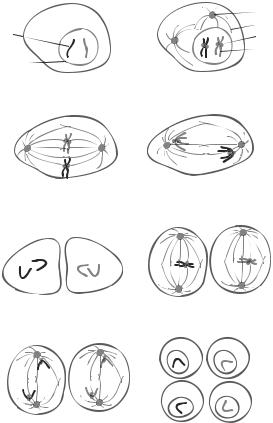
G1 phase |
Prophase I |
|
|
Centrosome |
|
Chromosome |
Microtubule |
|
Kinetochore |
|
|
|
Chromosome |
|
Nuclear envelope |
|
|
Metaphase I |
Anaphase I |
Telophase I |
Metaphase II |
Anaphase II |
Telophase II |
Figure 6.3. Schematic representation of cell meiosis. Meiosis is a process of germ cell division and is composed of two phases, including meiosis I and meiosis II. The phase meiosis I is consists of several stages, including prophase I, metaphase I, anaphase I, and telophase I. The phase meiosis II consists of metaphase II, anaphase II, and telophase II. During prophase I, the nucleus envelope is degraded, centrosomes and microtubule spindles start to form, and scattered chromosomes are organized into apparently double-chromatid structures. During metaphase I, a complete spindle network forms, the two centrosomes are deployed to the mitotic poles, and the chromosomes are aligned in the equator region. During anaphase I, the chromosomes remain paired and are pulled to the mitotic pole of the cell. During telophase I, the chromosomes are completely separated and rearranged near the two poles. The germ progenitor cell is divided into two cells. However, no nucleus envelope is developed. During metaphase II, the chromosomes in each daughter cell are aligned along the equator. The centromeres are connected to the microtubule spindles. During anaphase II, the two identical chromatids for each chromosome are separated and pulled to the mitotic poles. During telophase II, each daughter cell is further divided into two granddaughter cells with a haploid set of chromosomes (a single copy of each chromosome from either the mother or the father). The chromosomes are rearranged and enveloped within the nucleus. Based on bibliography 6.4.
DNA content and measuring the density of cells that undergo DNA synthesis. For DNA content measurement, DNA can be extracted from a given volume of tissue or given area of cultured cells and the DNA content can be measured by DNA extraction and spectrophotometry. Although this method is easy to use, it does not directly give the density of dividing cells.
CELL MIGRATION |
269 |
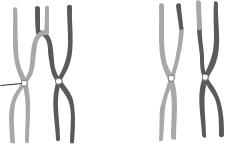
Cross over 
Centromere
Figure 6.4. Schematic representation of chromosomal cross over. During chromosome segregation, chromosomal segments may exchange location between two chromatids of different chromosome complexes. Based on bibliography 6.4.
To directly measure the density of cells that synthesize DNA, a selected type of deoxynucleotides can be tagged with a marker and delivered to cells. Since only dividing cells take up deoxynucleotides, any cells exhibiting the tagged deoxynucleotides can be considered dividing cells. There are two types of tagging markers—radioactive isotopes and molecules—that can be detected by immunohistochemistry. A common radioactive material used for detecting cell division is [3H]-thymidine. This isotope can be delivered to animal models or cultured cells. Tissue specimens or cultured cells can be collected after 24 h, fixed with 4% formaldehyde in phosphate-buffered saline (PBS), and processed for detecting [3H]-thymidine incorporation. Specimens are exposed to X-ray films and cells with positive [3H]-thymidine signals are considered dividing cells. Such a method is referred to as autoradiography. The specimen can be counterstained with hematoxylin and eosin for measuring the total number of cells. The ratio of the number of [3H]-thymidine- labeled cells to that of the total cells can be used as an index for assessing cell division.
Alternatively, 5′-bromodeoxyuridine (BrdU) can be used to detect cell division instead of [3H]-thymidine. BrdU can be directly injected into an animal or delivered to cultured cells. As [3H]-thymidine, BrdU can be taken up only by dividing cells. BrdU can be detected by immunohistochemistry with a BrdU-specific antibody (Fig. 6.5). It is important to address several technical points for the BrdU assay. First, cultured cells or intact tissue specimens without histological sectioning should be treated with a detergent (e.g., 0.5% Triton X-100) to permeabilize cell membrane so that antibody can diffuse through the cell membrane and reach the cell nucleus. For histological tissue sections, since cells are cut open and cell nuclei are exposed, it is not necessary to treat specimens with a detergent. Second, incorporated BrdU is embedded within the cell chromatin, which prevents the anti-BrdU antibody from accessing BrdU. Thus, cultured cells or tissue specimens should be treated with pepsin to digest nucleus proteins and expose DNA, so that the anti-BrdU antibody can access the incorporated BrdU. A DNA-binding fluorochrome, e.g., Hoechst 33258, can be used to counter-staining DNA nonspecifically, allowing the measurement of the total number of cells within a selected specimen. Comparing to the [3H]-thymidine incorporation method, the BrdU method is more advantageous for its simplicity and nonradioactivity.
CELL MIGRATION [6.6]
Cell migration is a fundamental cellular activity observed during development and pathogenic remodeling. During development, cell migration plays a critical role in the initiation
270 FUNDAMENTAL CELLULAR FUNCTIONS
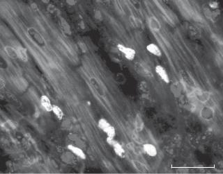
Cardiomyocytes
*
Infarct
Figure 6.5. 5′-Bromodeoxyuridine (BrdU)-positive cells in injured cardiac tissue at day 5 after ischemic injury. In this preparation, cardiac injury was induced by ligating the left anterior descending coronary artery in a mouse model. BrdU was injected into the skeletal muscle of a mouse 24 hrs before observation. Cardiac specimens were fixed in 4% formaldehyde in phosphate-buffered saline (PBS), cut into cryosections, treated subsequently with 0.5% pepsin and 1.5 N HCl, incubated subsequently with an anti-BrdU antibody and a fluorescein-conjugated secondary antibody, and observed by using a fluorescence microscope. Scale bar: 10 μm.
and formation of tissues and organs, such as the nerve and cardiovascular systems. During pathogenic remodeling, cell migration contributes to the initiation and progression of pathogenic disorders, such as atherogenesis (e.g., smooth muscle cell migration from the arterial media to the arterial intima or arterial substitutes), tumorigenesis (e.g., cancer cell migration and metastasis), and inflammation (leukocyte migration to inflammatory sites). Cell migration is a mechanical event that involves a variety of molecular processes and is controlled by a number of known signaling pathways. In this chapter, the mechanics and regulatory mechanisms of cell migration are briefly reviewed.
Mechanics of Cell Migration
Cell migration is accomplished by a number of mechanical processes at the molecular and subcellular levels. Theses processes include protrusion or extension of cell membrane at the cell leading edge, attachment of protruded cell membrane to a substrate via adhesion receptors, contraction and movement of the cell body, retraction at the cell trailing edge, and recycle of adhesion receptors (Fig. 6.6). A variety of regulatory and contractile proteins are involved in the initiation and progression of cell migration. It is important to note that the five processes outlined above are arbitrarily defined. All these processes take place simultaneously and continuously in a cyclic manner. It is difficult to identify the beginning and end of a migration cycle.
Protrusion of Cell Membrane. When a cell is stimulated by a migration-activating factor, such as a chemoattractant, the cell initiates directed membrane protrusion. The direction of membrane protrusion is often determined by the stimulus. For instance, in the presence
CELL MIGRATION |
271 |
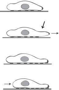
Unstimulated cell
Stimulation
Protrusion
Attachment
Movement and retraction
Figure 6.6. Schematic representation of cell migration. Based on bibliography 6.6.
of a chemoattractant, the cell membrane extends toward the chemoattractant. The forces that drive the membrane protrusion are generated by the actin assembly. Although the mechanisms of actin assembly is under debate, it is thought that controlled sequential extension of actin filaments toward the cell leading edge may provide a propelling force for membrane protrusion. In this model, actin subunits are added to the barbed end of the actin filaments, which point at the leading edge of cell migration. Such a process is controlled by regulatory proteins, including the Arp2/3 complex (see Chapter 3).
Attachment of Cell Membrane to Substrate Matrix at the Leading Edge. Following the protrusion of cell membrane due to directed actin assembly, the next step is the attachment of the extended membrane lamellipodia to substrate. Such a process stabilizes the leading edge of the migrating cell and allows the cell to exert forces on the substrate, a necessary condition for cell migration. The attachment of cell leading edge is mediated by integrins. Unstimulated integrins are freely suspended in the cell membrane. In response to the stimulation of extracellular matrix, integrins are activated and bind to the actin cytoskeleton. Cell membrane protrusion enhances the binding of integrins to extracellular matrix in newly formed lamellipodia. Integrin–matrix interaction further stimulates the binding of integrins to the actin cytoskeleton, facilitating the formation of focal adhesion contacts between the actin cytoskeleton and extracellular matrix. Thus, integrins play a critical role for the attachment of cell lamellipodia to substrate. Since focal adhesion contacts link the actin cytoskeleton to extracellular matrix, forces generated by the actin cytoskeleton can be transmitted to the extracellular matrix, which is critical to cell migration.
Cell Traction and Movement. Cell movement is propelled by traction forces generated by the actin cytoskeleton and exerted on the extracellular matrix substrate. In a fibroblast,
272 FUNDAMENTAL CELLULAR FUNCTIONS
for example, the actin cytoskeleton can generate traction forces about 1 nN/μm2 on a substrate. The generation of traction forces is dependent on the interaction of actin filaments with myosin II in mammalian cells. Actin filaments are distributed around the cell periphery. The barbed ends of the actin filaments are oriented toward the cell periphery in regions near the leading and trailing edges, while those in the middle region are oriented more randomly. The myosin II molecules are distributed more heavily in the middle region than in regions near the leading and trailing edges. With such molecular distributions, the interaction of actin filaments with myosin II results in predominantly peripheral movements. The direction of cell migration may be dependent on the asymmetric distribution of actin filaments and the relationship between actin filaments and the extracellular matrix, which determine the balance of the traction forces between the cell leading and trailing edges. Directed migration can occur only if the traction force at the leading edge exceeds that at the trailing edge.
Retraction of Cell Membrane at the Trailing Edge. To initiate cell migration, the forward movement at the cell leading edge must be accompanied with a retraction at the cell trailing edge. The dynamic interaction of integrins with extracellular matrix may mediate the coordinated leading-edge movement and trailing-edge retraction. The distribution of integrin-containing focal adhesion contacts changes dynamically from the cell leading edge to the trailing edge. The density of focal adhesion contacts is relatively lower at the trailing edge than that at the leading edge. Such a distribution of focal adhesion contacts results in reduced cell membrane adhesion to the extracellular matrix at the cell trailing edge and is in favor of the dissociation of cell membrane from the extracellular matrix. Furthermore, the traction forces generated at the cell leading edge are counterbalanced by those at the trailing edge. A reduction in the density of focal adhesion contacts at the trailing edge likely results in an increase in the traction force per focal adhesion contact, which enhances the disruption of integrin–matrix bonds and thus facilitates the retraction of the cell trailing edge.
In addition to the influence of the physical factors described above, the disruption of the integrin–matrix bonds at the cell trailing edge may be regulated by biochemical processes. For instance, the disruption of αvβ3–vitronectin interaction in migrating neutrophils requires the presence of calcium. The suppression of the calcium-dependent phosphatase calcineurin prevents the disruption of the αvβ3–vitronectin interaction. This observation suggests that calcineurin plays a role in regulating the detachment of cell membrane from matrix substrate at the trailing edage. However, the mechanisms of chemically mediated retraction remain to be investigated.
Replenishment of Integrins. Integrins play a critical role in the mediation of membrane attachment, cell traction, and cell retraction during cell migration. The distribution and activity of integrins vary from the cell leading to trailing edges. This suggests that the cell must replenish active integrins at the cell leading edge. There are two possible ways for the replenishment of integrins: integrin synthesis and recycling. New integrins are continuously synthesized and deployed to the cell membrane. Cells are also able to endocytose and reuse the integrin molecules left behind on the substrate during cell trailing-edge retraction. In addition, cells may actively transport membrane integrins from the cell trailing to leading edges. With these approaches, the cell is able to maintain an appropriate distribution of integrins, which is necessary for the conduction of cell migration.

CELL MIGRATION |
273 |
Regulation of Cell Migration
Role of the Rho family of GTPases. As discussed above, cell migration is accomplished by a number of complex molecular processes. These processes are regulated by a variety of signaling molecules. Among the signaling molecules, the Rho family of small GTPases, including Rho, Rac, and Cdc42, plays a critical role in the regulation of cell migration.
The GTPases of the Rho family are GTP-binding proteins with molecular weight of21 kDa. These proteins belong to the Ras protein superfamily, which include, in addition to the Rho family GTPases, the Rab, the ADP-ribosylation factor (ARF), and the Ran families. The Rab proteins participate in the regulation of vesicle transport, the ARF proteins mediate signal transduction and vesicle transport, whereas the Ran proteins mediate protein transport to the cell nucleus. All proteins of the Ras superfamily are able to bind GTP.
For the Rho family of small GTPases, 11 different isoforms have been identified in mammalian cells, including RhoA, RhoB, RhoC, RhoD, RhoE, RhoG, Rac1, Rac2, Cdc42, TC10, and TTF. Among these proteins, the role of RhoA (see Table 6.4.), Rac1, and Cdc42 has been extensively studies (see Chapter 3 for characteristics of these molecules). RhoA has been shown to regulate the formation and organization of actin filaments, Rac1 mediates the formation of cell lamellipodia and membrane ruffles, whereas Cdc42 is responsible for the formation of filopodia. All these processes are related to cell migration.
All GTPases of the Rho family can bind GTP or GDP. A GTPase is active when GTP is bound, whereas it is inactive when GDP is bound. Nucleotide exchange factors can stimulate the binding of GTP to GTPases, activating the GTPases. A variety of extracellular signals can activate the nucleotide exchange factors and thus the GTPases. While the exact mechanisms of GTPase activation remains poorly understood, GTPase translocation to the cell membrane or cytoskeleton may play a role. For example, the nucleotide exchange factor for Cdc42 is associated with the cell membrane. Activated nucleotide
TABLE 6.4. Characteristics of RhoA*
|
|
Amino |
Molecular |
|
|
Proteins |
Alternative Names |
Acids |
Weight (kDa) |
Expression |
Functions |
|
|
|
|
|
|
RhoA |
Ras homolog gene |
193 |
22 |
Ubiquitous |
Regulating the |
|
family member |
|
|
|
organization and |
|
A, ARHA, aplysia |
|
|
|
remodeling of |
|
ras related |
|
|
|
actin cytoskeleton |
|
homolog1 2 |
|
|
|
during cell |
|
(ARH12), |
|
|
|
morphogenesis |
|
oncogene rho H12, |
|
|
|
and migration |
|
RHOH12, RHO12, |
|
|
|
and mediating |
|
RHOA, |
|
|
|
cell proliferation |
|
transforming |
|
|
|
and |
|
protein RhoA, Ras |
|
|
|
differentiation |
|
homolog gene |
|
|
|
|
|
family member A |
|
|
|
|
|
|
|
|
|
|
*Based on bibliography 6.6.
274 FUNDAMENTAL CELLULAR FUNCTIONS
exchange factor can induce translocation of Cdc42 from the cytoplasm to the cell membrane, which facilitates the activation of Cdc42.
Activated RhoA, Rac1, and Cdc42 can interact with and stimulate downstream signaling molecules, including protein kinases, adapter proteins, and phosphoinositide kinases. For instance, Rho can activate Rho-associated kinase, which in turn phosphorylates myosin II light-chain kinase in smooth muscle cells. Myosin II light-chain kinase can activate myosin light chain and facilitate myosin–actin interaction. The Rho family of small GTPases can also interact with molecules that link to the actin cytoskeleton. An example is the interaction of Rho with p140mDia, which induces the activation of p140mDia. Activated p140mDia can interact with profilin, an actin-binding protein.
Rho, Rac1, and Cdc42 are involved in the regulation of actin polymerization and the formation of stress fibers, which are myosin II-associated contractile actin filamentous bundles. In cultured Swiss 3T3 fibroblasts, cell transfection with active Rho and Rac mutants stimulates the formation of stress fibers and lamellipodia, or wide cell membrane protrusions. Cells transfected with a Rho inhibitor C3 transferase or a dominant-negative mutant for Rac exhibit reduced formation of stress fibers. In addition, Rho, Rac, and Cdc42 promote the formation of focal adhesion contacts. Since actin polymerization, cell membrane protrusion (formation of lamellipodia and filopodia), and formation of focal adhesion contacts are essential processes of cell migration, the small GTPases Rho, Rac, and Cdc42 contribute to the regulation of cell migration.
Role of MAPKs. As discussed in Chapter 5, mitogen-activated protein kinases (MAPKs) are key elements for signaling pathways that respond to the stimulation of growth factors, including platelet-derived growth factor, epidermal growth factor, fibroblast growth factor, and vascular endothelial growth factor. The binding of these growth factors to cognate growth factor receptors induces autophosphorylation of the receptor tyrosine kinase located in the cytoplasmic domain of the receptor. Such a process leads to the activation of a cascade of signaling molecules, including the Ras protein, ERK kinase, and ERK1/2, which belongs to the MAPK family. Activated ERK1/2 can directly phosphorylate myosin light-chain kinase, which activates myosin light chain and promotes myosin–actin interaction. These MAPK-involved processes influence cell migration via mediating the contractility of actin filaments.
CELL ADHESION
Cell adhesion is a molecular process that mediates cell–cell (intercell) and cell–matrix interactions and is involved in the regulation of developmental morphogenesis, physiological adaptation, and pathogenic remodeling. Cells can selectively bind to other cells and extracellular matrix by activating cell adhesion mechanisms. Cell adhesion is related to a variety of molecular and cellular processes, such as cytoskeletal reorganization, alterations in cell geometry, signaling activation, gene expression, and cell mitogenic responses. It is now understood that cell–cell adhesion and cell–matrix adhesion are regulated by several classes of cell adhesion molecules. These include immunoglobulin-like cell adhesion molecules, selectins, cadherins, cell surface heparan sulfate proteoglycans, protein tyrosine phosphatases, and integrins. Cell adhesion is a process that requires coordinated interactions between various cell adhesion molecules as well as between adhesion molecules and the actin cytoskeleton. In this section, the structural and functional characteristics of major classes of adhesion molecules are discussed.

CELL ADHESION |
275 |
Immunoglobulin-Like Domain-Containing Cell Adhesion Molecules [6.7]
ClassiÞcation and Structure. Immunoglobulin (Ig)-like domain-containing cell adhesion molecules (IgCAMs) (see Table 6.5) are cell surface adhesion molecules that belong to the immunoglobulin superfamily, which contains about 100 members. IgCAMs mediate cell–cell and cell–matrix adhesion and play a role in regulating cell signaling. These molecules contribute to the regulation of embryonic development and pathogenic remodeling. In particular, IgCAMs play a critical role in mediating polarized migration of neurons and the development of the nerve system.
A typical IgCAM is composed of one or more Ig-like domains, which are located in the extracellular region of the molecule (Fig. 6.7). Each Ig-like domain is composed of about 100 amino acids, which constitute two opposed β sheets linked by disulfide bonds between cysteine residues. In addition, a typical IgCAM is composed of fibronectin (FN)- like repeats in the extracellular region. Each repeat consists of two β sheets of about 90 amino acids. Some IgCAMs are widely distributed in almost all tissues and organs, while others are expressed in limited tissues and organs. The expression pattern of IgCAMs is regulated to suit the function of various types of tissues and organs during development and remodeling.
Several types of IgCAMs have been identified and characterized. These include neural cell adhesion molecules (NCAMs), vascular cell adhesion molecules (VCAMs), intercellular adhesion molecules (ICAMs), L1-like IgCAMs, and receptor protein tyrosine phosphatases. A typical NCAM is composed of five Ig-like domains and two FN-like repeats in the extracellular region, a transmembrane domain, and a cytoplasmic domain. NCAMs are primarily expressed in neural cells and are involved in the regulation of neural cell adhesion and development. VCAM is composed of seven extracellular Ig-like domains, a transmem-
Ig-like
IgC2
 Ig-like
 FN3
FN3
NCAM |
VCAM |
ICAM |
Tyrosine |
phosphatase |
Figure 6.7. Schematic representation of the structure of immunoglobulin (Ig)-like domain-con- taining cell adhesion molecules. IgC2: Immunoglobulin C2-type domain. FN3: fibronectin type 3 domain. Based on bibliography 6.7.