

Bioregenerative Engineering Principles and Applications - Shu Q. Liu
..pdf356 EMBRYONIC ORGAN DEVELOPMENT
The Notochord [8.4]
The notochord is an embryonic structure established during gastrulation and located underneath the neural tube. The notochord is formed from an early embryonic structure known as the primitive streak, which is initiated at the posterior end of the embryo in the blastocyst stage with two blastoderm layers (epiblast and hypoblast) and plays a critical role in the formation of the mesoderm and the endoderm. The primitive streak progresses toward the anterior end along the anterior-posterior axis. At the same time, the centerline of the primitive streak depresses, forming the primitive groove. When the primitive streak reaches the anterior end, there forms a node structure called Hensen’s node at the anterior end (superior or cranial end in humans) of the primitive streak. The mesoderm and endoderm are formed by the primitive streak cells, which pass through the primitive streak layer, move from the dorsal to the ventral direction, and spread on the inner surface of the epiblast, forming the mesoderm and the endoderm. These cells form two layers ventral to the epiblast: the middle layer and the internal layer. The former develops into the mesoderm and the later endoderm. A group of primitive streak cells migrates through the Hensen’s node and forms the notochord along the anterior–posterior axis. During such a process, the epithelial type of primitive streak cells is transformed into mesenchymal cells. The notochord is a transient structure, which mediates the formation of the neural tube and defines the anterior–posterior axis of the body. In mammals, the notochord cells eventually form part of the endoderm and contribute to the formation of the primitive gut.
The Paraxial Mesoderm
Formation of the Somites [8.5]. The paraxial mesoderm, also known as the somatic dorsal mesoderm, is formed by cells from the primitive streak as described above and organized into two parallel columns that are aligned along the neural tube one at each side. The paraxial mesoderm forms segmented blocks called somites, also known as mesodermal segments. These somites develop into the vertebrae, ribs, skeletal muscles, and dermis. These structures serve as pathways for the extension of the spinal nerve axons and the migration of neural crest cells, which develop into the sympathetic and parasympathetic neurons.
The formation of the somites begins at the anterior portion of the primitive streak. As a first step, paraxial mesodermal cells are organized into two unsegmented presomitic cell clusters called somitomeres, which are aligned along the neural tube one at each side. The somitomere at the anterior end is first separated into individual somites. The tip portion of the somitomere adjacent to an established somite is subsequently separated from the main body of the somitomere. The segmentation process continues from the anterior to the posterior directions (superior to inferior direction in humans) until the entire somitomere is segmented. The segmentation of the somitomere is regulated by several molecules, including hairy 1, ephrin, and ephrin tyrosine kinase receptor. Hairy1 encodes a transcription factor, which can activate the expression of the ephrin and ephrin tyrosine kinase receptor genes. In the somitomere, hairy 1 is expressed in the posterior region of a premature somite, leading to local activation of ephrin and ephrin tyrosine kinase receptor. Ephrin can interact with the ephrin tyrosine kinase receptor, triggering the activation of inhibitory signaling pathways. Such an activity results in local suppression of cell interaction with substrate and enhancement of somite separation
DEVELOPMENT OF MESODERM-DERIVED ORGANS |
357 |
from the somitomere. This pattern of molecular activities repeats along the somitomere from the anterior to the posterior direction until the completion of the whole set of somites. The primitive streak is gradually diminished during the segmentation of the somitomere.
Each pair of somites alongside the neural tube is the origin of the muscular and connective tissue specified for a designated location. Each somite, once established and matured, is committed to the specification of a tissue and its developmental course cannot be altered. This phenomenon has been demonstrated by transplantation of a somite from one location to another. Tissues developed from the transplanted somite will appear in the peripheral location of the tissues that are supposed to form from the original somite. For example, when a somite from the thoracic region is transplanted to the cervical region of a chick, ribs will form in the cervical region on the transplantation side. Overall, somites develop into several types of tissues and structures, including the vertebra, rib, limb, skeletal muscle, connective tissue, and back dermis.
At each level, the somite is organized into distinct regions in the transverse direction, including the ventral region, two lateral regions, and dorsal region. The ventral region develops into a group of mesenchymal stem cells collectively called the sclerotome. Cells in this structure give rise to chondrocytes and osteoblasts of the vertebrae, ribs, and other bones. The two lateral regions, one adjacent to and the other farthest from the neural tube, develop into myotomes, which form myoblasts, precursors of skeletal muscle cells. The lateral myotome adjacent to the neural tube develops into the back skeletal muscles, whereas the lateral myotome farthest from the neural tube form skeletal muscles in the limbs and other body parts. The dorsal region of a somite develops into the dermatome, which is the origin of the dermal tissue in the back area. The formation of these distinct somite regions is mediated by the activation of paracrine molecules, including sonic hedgehog, Wnt1, Wnt3a, neurotrophin 3, and bone morphogenetic protein 4 and 5. These factors are expressed and released from the structures adjacent to the somites, such as the neural tube and the lateral plate mesoderm. The locationand time-dependent activation of these factors may determine the specification and commitment of the cells in the sclerotome, myotomes, and dermatome.
Formation of the Skeletal Muscle System [8.6]. As discussed above, the cell lineages in the lateral myotomes develop into skeletal muscles. The myotome contains muscular progenitor cells called myoblasts. These cells are prompted to enter a process known as myogenesis, the generation of muscular cells. Myogenesis is initiated and enhanced by transcriptional factors MyoD and Myf5, which are myogenic regulatory factors. These factors activate the expression of necessary genes that encode proteins for initiating myogenesis.
During myogenesis, one of the genes that are upregulated is the fibroblast growth factor (FGF) gene. In the presence of increased FGF, myoblasts expand extensively via proliferation. These cells also produce and release extracellular matrix components such as collagen and fibronectin. At a certain time, the FGF level is suppressed, leading to a reduction in the rate of myoblast proliferation. The myoblasts attach to the extracellular matrix through the interaction of the cell membrane integrins, such as α5β1, with selected extracellular matrix components, such as fibronectin. All myoblasts within a muscular bundle are aligned in the same direction by the mediation of cell membrane proteins such as cadherins and cell adhesion molecules (CAMs). The interaction between different myoblasts and between myoblasts and extracellular matrix induce an increase in the level of
358 EMBRYONIC ORGAN DEVELOPMENT
calcium, which initiates and regulates myoblast fusion, the formation of multinucleated muscular cells from individual myoblasts. This process is also mediated by a metalloproteinase, known as meltrin α, which degrades extracellular matrix components and enhances myoblast fusion.
Formation of the Skeleton [8.7]. The skeleton is composed of bones and cartilages. These structures are developed from two mesodermal systems, including the somites and lateral plate mesoderm, and an ectodermal system, the cranial neural crest. The somites give rise to the trunk skeleton (vertebrae and ribs), the lateral plate mesoderm gives rise to the limb skeleton, and the cranial neural crest gives rise to the craniofacial skeleton. For the ectodermal source of the skeleton, the neural crest cells are transformed from an ectodermal cell type to a mesenchymal type, which forms skeletal tissues.
There are two mechanisms for the formation of the bone: direct osteogenesis from soft mesenchymal tissue and ossification of cartilage. The craniofacial bones are formed through direct osteogenesis. In such a process, the cranial neural crest cells migrate from the neural crest to the skull, transform from ectodermal cells to mesenchymal cells, undergo extensive proliferation, and form a mesenchymal tissue. A fraction of the neural crest-derived mesenchymal cells differentiates into osteoblasts or bone progenitor cells. The osteoblasts can produce and release collagen and specific proteoglycans, which serve as a matrix for the formation of the bone. This matrix can bind and retain calcium, which results in the calcification or ossification of the matrix. All osteoblasts within the calcified matrix are considered mature bone cells and are defined as osteocytes. Cells adjacent to the periosteum or the surface membrane of the calcified matrix structure retain the features of osteoblasts and can differentiate into osteocytes and produce bone matrix. Several bone morphogenetic proteins, including BMP2, BMP4, and BMP7, play a critical role in osteogenesis by stimulating the differentiation of osteoblasts and the production of bone matrix components.
Most bones, including the vertebrae, ribs, and limb bones, originate from the somites and lateral plate mesoderm, are developed via cartilage formation and ossification of cartilage. There are several steps for bone morphogenesis: (1) formation of chondrocytes, (2) formation of cartilages, (3) initiation of osteogenesis, and (4) mineralization of bone matrix and formation of bone. Chondrocytes are formed from mesenchymal progenitor cells derived from the somites and lateral plate mesoderm under the stimulation of local osteogenic factors. The newly formed chondrocytes expand extensively via proliferation and generate extracellular matrix components that are necessary for the formation of cartilage. Extracellular matrix and chondrocytes are integrated and organized to form cartilage. Within the cartilage, established chondrocytes are transformed into hypertrophic chondrocytes characterized with an increase in cell volume. These chondrocytes can produce collagen type X and fibronectin. These matrix components absorb calcium and phosphate into the matrix, initiating matrix mineralization or osteogenesis. The hypertrophic chondrocytes also produce and release vascular endothelial growth factor (VEGF), which stimulates angiogenesis or blood vessel formation within the cartilage. The vascular system supplies oxygen and nutrients to the newly generated bone. The hypertrophic chondrocytes undergo apoptosis shortly after these initiating processes for bone formation. As a last step, mesenchymal progenitor cells are transformed into osteoblasts, which generate and release bone-forming extracellular matrix components and stimulate matrix mineralization. A bone forms when a sufficient amount of calcium and phosphate is deposited to the bone matrix.
DEVELOPMENT OF MESODERM-DERIVED ORGANS |
359 |
The Intermediate Mesoderm: Formation of the Kidney [8.8]
The intermediate mesoderm gives rise to the urogenital system, including the kidney and associated duct systems as well as gonads. For kidney development, there are two critical stages: the formation of transient kidneys and the formation of permanent kidneys. For the formation of the transient kidneys, the intermediate mesoderm initiates the formation of two pronephric ducts at the anterior portion of the mesoderm during the early embryonic stage (about 20 days in humans). This process is symmetric with respect to the neural tube from both parts of the intermediate mesoderm. The formation of the pronephric ducts progress continuously toward the posterior end. Established pronephric ducts interact with adjacent mesodermal tissue to form pronephroi, which are side branches attached to the anterior portion of the pronephric ducts. When the formation of the pronephric ducts progresses to the middle portion of the mesoderm, the pronephric ducts stimulate the development of mesonephroi, which constitute the major excretory system of the embryo. The pronephric ducts further progress to the posterior end, where the two pronephric ducts meet to form the cloaca, a presumptive structure for the formation of the bladder, the rectum, and the genital system. During the development of the mesonephroi, the anterior end of the pronephric system undergoes progressive degeneration. This degenerative process continues when the posterior end of the pronephric ducts form new mesonephroi. Shortly after the cessation of mesonephros generation, the mesonephroi are degenerated via cell apoptosis. Thus, the mesonephroi serve only as a temporary excretory system.
While the anterior and middle portions of the pronephric system are degenerated, the posterior portion of the system remains. A side branch, known as the ureteric bud, forms from each of the two pronephric ducts in the posterior portion. The ureteric bud serves as the presumptive structure for the formation of the two permanent kidneys. The ureteric bud interacts with and stimulates surrounding mesenchymal tissue, inducing the formation of the metanephrogenic mesenchyme (Fig. 8.6). At the same time, the metanephrogenic mesenchyme stimulates the ureteric bud to extend and branch into the mesenchyme. The ureteric bud-derived branching system serves as a kidney rudiment, which eventually develops into the kidney. At the end of each established ureteric branch, epithelial cells from the metanephrogenic mesenchyme form a cell cluster and undergo proliferation and differentiation, resulting in the formation of specified kidney cell types, including the glomerular capsule cells, endothelial cells, and tubular cells. These cells are assembled into functional kidney units known as the nephrons. The cells in each nephron are integrated into the structure at the tip of each ureteric branch, establishing a connection between the nephron and the ureteric bud branch, which eventually forms the renal collecting duct. Urine forms in the nephrons and is conveyed to the ureters via the collecting ducts.
The Lateral Plate Mesoderm
The lateral plate mesoderm is located farthest from the central neural tube and is composed of two layers: the somatic mesoderm and the splanchnic mesoderm. The somatic mesoderm is the layer adjacent to the ectoderm, and the splanchnic mesoderm is the layer next to the endoderm. These mesodermal structures develop into the heart, blood vessels, and blood cells. There is a gap between the two mesodermal layers, which is known as the coelom. The coelom forms the three body cavities, including the pleural (thoracic), pericardial (cardiac), and peritoneal (abdominal) cavities.
360 EMBRYONIC ORGAN DEVELOPMENT
A B
Wolffian
duct
Uteric
bud
Metanephric mesenchyme
D |
E |
Comma- |
and S- |
||
|
|
shaped |
|
|
bodies |
Renal vesicle |
|
|
C
F
Bowman's Collecting capsule
duct
Glomerulus
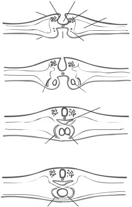
Glomerulor podocytes
Figure 8.6. Processes of nephron formation. (A) The metanephric mesenchyme interacts with the Wolffian duct of the intermediate mesoderm. (B) At about embryonic day 10.5 (E10.5), the ureteric bud forms from the Wolffian duct and invades the metanephric mesenchyme and induces differentiation. (C) At about E11.5, the mesenchymal cells condense to form aggregates near the ureteric bud. Branches form from the ureteric bud. (D) At about E12.5, the condensed mesenchyme undergoes a mesenchymal-to-epithelial transformation and forms epithelial structures (renal vesicles).
(E) At about E13.5, complex epithelial structures, commaand S-shaped bodies, are formed. (F) From E14.5 to E16.5, the S-shaped bodies fuse into the ureteric bud derivatives, inducing the formation of mature nephron. The ureteric bud develops into collecting ducts. Endothelial cells migrate into the nephron and form the glomerulus, which is surrounded by glomerular podocytes (Bowman’s capsule). Selected transcription factors and signaling molecules expressed in the uretic bud and metanephic mesenchyme are shown. WT1 (Wilms’ tumor 1), PAX2 (paired box 2), and EYA1 (eyes absent 1) are transcription factors expressed in the metanephric mesenchyme and regulate ureteric bud induction. GDNF (glial-cell-line-derived neurotrophic factor) is also expressed in the metanephric mesenchyme and stimulates ureteric bud induction and branching by interacting with its receptor RET. WT1 and bone morphogenetic protein 7 (BMP7) play a role in regulating the survival of the metanephric mesenchyme. WNT4 is essential for the mesenchymal-to-epithelial transformation of the metanephric mesenchyme and might be induced by WNT6 signals from the ureteric bud. BMP4 is involved in ureteric bud growth, and WNT11 regulates branching of the ureteric bud. GPC3 modulates signals between the metanephric mesenchyme and the ureteric bud, including bone morphogenic proteins. (Reprinted by permission from Macmillan Publishers Ltd.: Rivera MN, Haber DA: Nature Rev Cancer 5:699–712, copyright 2005.)
DEVELOPMENT OF MESODERM-DERIVED ORGANS |
361 |
||
Neural plate |
|
Neural groove |
|
Ectoderm |
|
Mesenchyme |
|
|
|
Pericardial cavity |
|
25 hours |
|
|
|
Endoderm |
Gut |
Angiogenetic cell cluster |
|
|
|
||
26 hours
Epicardial |
Endocardial |
primordium |
primordium |
|
Foregut |
28 hours |
|
Epimyocardium |
Endocardial tube |
29 hours
Epimyocardium |
Endocardial tube |
Figure 8.7. Early developmental stages of the chick heart. (After Calson BM: Pattern Formation of Embryology, McGraw-Hill, New York, 1981.)
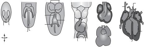
Formation of the Heart [8.9]. The presumptive cardiac cells are specified during the early embryonic stage when the primitive streak forms. These cells migrate through the primitive streak and form two cardiogenic mesodermal clusters, which are symmetrical with respect to the primitive streak, in the splanchnic mesoderm at the level of the Hensen’s node (Fig. 8.7). The two presumptive cardiac cell clusters move toward each other and form two endocardial primordia, the initial form of the heart. At the same time, the adjacent mesodermal cells are stimulated to transform into epithelial cells, forming the pericardium, which encloses the endocardial primordia, and the endocardium, which lines the internal surface of the heart. Within about 3 weeks in the human, the two endocardial primordia meet and fuse into each other to form a single myocardial tube. Cells with cardiomyocyte phenotypes are established during endocardial primordial fusion and initiate the contractile activity, causing pulsations of the endocardial primordia.
Within about 5 weeks, the myocardial tube is developed into a tube with two chambers: the atrium and ventricle (Fig. 8.8). A fraction of cells from the endocardium forms a structure called endocardial cushion, which separates the two-chambered tube into the left and right cardiac canals. At about the same time, the atrial septum develops from the
362 EMBRYONIC ORGAN DEVELOPMENT
A B C
D E F
HFs
|
|
|
HFs |
|
|
|
|
|
PT |
|
|
|
|
|
|
|
LA |
SVG |
|
|
|
|
|
|
|
|
RA OFT |
AA |
PV |
|
|
|
|
Arterial |
|
|
RV LV |
Ao |
LA |
|
|
|
|
pole |
Heart |
|
RA |
||
|
|
|
|
|
|
|
|
||
|
|
|
|
|
|
|
|
|
|
PS |
|
Cardiogenic |
|
Venous |
tube |
OFT |
|
IVC |
PV |
|
|
|
|
|
|||||
|
region |
|
pole |
|
|
|
|||
|
|
ML |
ML |
RV LV |
|
RV |
LV |
||
|
A |
Cardiac |
|
LA IFT RA |
|
Tr |
|||
|
|
|
|
|
|
||||
|
Crescent |
|
|
|
|
|
|
||
R |
|
L |
|
|
|
|
AVC |
|
|
|
|
|
|
|
LV |
|
|
||
|
|
|
|
|
PRA |
PLA |
IVS |
|
|
|
P |
|
|
|
|
|
RA |
|
|
|
|
|
|
|
|
|
|
|
Figure 8.8. Development of the heart. (A) Myocardial progenitor cells originate in the primitive streak (PS) and migrate to the anterior of the embryo at about embryonic day E6.5. (B) The myocardial progenitor cells settle under the head folds (HF) and form the cardiac crescent at about E7.5. (C) The early cardiac tube forms through fusion of the cardiac crescent at the midline (ML) at about E8. (D) The cardiac tube forms a loop at about E8.5. (E) The cardiac tube further develop into chamber structures by about E10.5, but the chambers are still connected. (F) By about E14.5, the cardiac chambers are separated. The left and right ventricles are connected to the pulmonary trunk (PT) and aorta (Ao), respectively. (Abbreviations: A, Anterior; P, posterior; R, right; L, left; AA, aortic arch; AVC, atrioventricular canal; IFT, inflow tract; IVC, inferior vena cava; IVS, interventricular septum; OFT, outflow tract; PLA primitive left atrium; PRA, primitive right atrium; PV, pulmonary vein; SVC, superior vena cava; Tr, trabeculae.) Reprinted by permission from Macmillan Publishers Ltd.: Buckinghamm et al: Nature Rev Genet 6:826–37, copyright 2005.)
atrial portion of the myocardial tube, while the ventricular septum grows from the ventricular portion. Both septa grow toward the endocardial cushion and separate the myocardial tube into a four-chamber structure—the heart. With further development, the heart is connected to the aorta, pulmonary arterial trunk, and vena cava, which are developed simultaneously with the heart.
The embryonic circulation is established when the heart and blood vessels form. The embryonic circulation is different from the postnatal circulation. The difference is caused by distinct functional anatomy of the cardiovascular, pulmonary, and intestinal systems between the prenatal and postnatal stages. During the prenatal stage, the embryo or fetus does not have a functional lung and intestine. Oxygen and nutrients are supplied from the mother’s blood through the placental circulation. During the stage of the two-chamber heart (about 5 weeks in humans), the umbilical venous blood obtains oxygen and nutrients from the placenta, and the veins conduct oxygenated blood to the heart. The heart pumps blood into the arterial system, which supplies blood to the peripheral systems. The arterial system also conveys blood to the placenta, where metabolic wastes are transported to the mother’s circulation.
During the stage of the four-chamber heart when the basic structures of the vascular and other systems are formed, the umbilical veins convey oxygenated blood to the vena cava and subsequently to the right atrium. Unlike the anatomy of the postnatal heart, the right and left atria are connected through the foramen ovale, which is open during the prenatal stage. In addition, the ductus arteriosus between the aorta and pulmonary arterial trunk is also open in the embryo or fetus. Oxygenated blood from the umbilical veins can directly enter the left atrium and left ventricle through the open foramen ovale. The left
DEVELOPMENT OF MESODERM-DERIVED ORGANS |
363 |
ventricle pumps oxygenated blood into the arterial system, which supplies blood to the peripheral systems. Oxygenated blood enters the pulmonary circulation from the right ventricle. A fraction of blood is diverted into the aorta through the ductus arteriosus. Deoxygenated blood with metabolic wastes from the lung returns directly to the left heart, while deoxygenated blood from peripheral organs returns to the right heart. Thus there is a significant degree of mixing between oxygenated and deoxygenated blood in the prenatal circulatory system. Deoxygenated blood with metabolic wastes is conveyed to the placenta for excretion via the umbilical arteries, which bifurcate from the common iliac arteries.
When a baby is born, the umbilical circulation no longer supplies oxygenated blood. A number of anatomical changes take place immediately during the early postnatal period:
(1) the lung starts to expand and function, ensuring a transition of gas exchange from the placenta to the pulmonary system; (2) the foramen ovale closes to stop blood diversion from the right to the left atrium; (3) the ductus arteriosus closes to prevent blood diversion from the pulmonary arterial trunk to the aorta; and (4) the umbilical arteries and veins are committed to degeneration. These drastic anatomical changes ensure the establishment of the postnatal circulation.
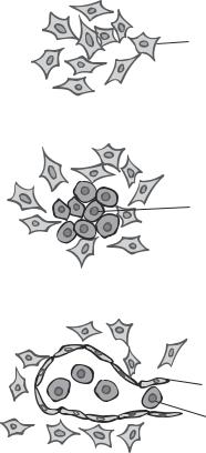
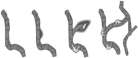
Formation of the Vascular System [8.10]. Blood vessels are formed from the splanchnic mesoderm, occurring simultaneously with heart generation. As a first step, the mesodermal cells differentiate into hemangioblasts, which are progenitor cells for blood vessels as well as blood cells. The hemangioblasts migrate to designated tissues and organs, where they form cell clusters known as blood islands (Fig. 8.9). A blood island is composed of two types of cell: hematopoietic stem cells (inner cells of the blood island) and angioblasts (outer cells). The hematopoietic stem cells develop into blood cell types, including erythrocytes, leukocytes, and platelets, whereas the angioblasts give rise to vascular cells, including endothelial cells, smooth muscle cells, and fibroblasts. There are two basic processes for the formation of blood vessels: vasculogenesis and angiogenesis. Vasculogenesis is the formation of primary endothelial cells and capillaries, which occurs during the embryonic stage. Angiogenesis is the formation of arteries, veins, and capillaries based on the established capillaries (Fig. 8.10). Angiogenesis occurs not only during the embryonic stage but also during the adulthood in response to injury and pathological disorders, such as cancer and hypertrophy. For vasculogenesis, the angioblasts differentiate into endothelial cells, a cell type that lines the internal surface of a blood vessel. The endothelial cells subsequently form tube-shaped capillaries, which are linked together into a capillary network known as the primary capillary plexus. These capillaries are further developed into arteries, veins, and additional capillaries via angiogenesis. During angiogenesis, endothelial cells undergo proliferation and sprout to form new blood vessels. Pericytes are recruited to newly formed capillaries and transformed into smooth muscle cells, eventually leading to the formation arteries and veins.
Several growth factors, including fibroblast growth factor (FGF), vascular endothelial growth factor (VEGF), platelet-derived growth factor (PDGF), and angiopoietin, play important roles in the regulation of vasculogenesis and angiogenesis. Fibroblast growth factor is produced by mesodermal cells and released into the interstitial space. This growth factor can stimulate the differentiation of mesodermal cells into hemangioblasts. Vascular endothelial growth factor is also produced by mesodermal cells. This factor directly stimulates angioblasts to differentiate into endothelial cells and stimulates endothelial cells to form capillaries. The effect of VEGF is dependent on the type of its receptor on
364 EMBRYONIC ORGAN DEVELOPMENT
Mesenchyme cells
Angiogenetic cell cluster or blood island
Endothelial cells
Primitive blood cells
Figure 8.9. Schematic representation of embryonic blood formation and vasculogenesis. (After Langman J: Medical Embryology, 4th ed., Williams & Wilkins, Baltimore, 1981.)
the cell surface. There are two types of VEGF receptor: flk1 (VEGF receptor 2) and flt1 (VEGF receptor 1). The activation of flk1 induces the differentiation of angioblasts into endothelial cells. The activation of flt1 results in the formation of endothelial tubes. Platelet-derived growth factor stimulates the recruitment of pericytes to newly formed blood vessels and the transformation of these cells to smooth muscle cells. Another factor, angiopoietin, interacts with its receptor Tie2 and regulates the recruitment of smooth muscle cells and the formation of blood vessels. Thus, these growth factors are essential for both vasculogenesis and angiogenesis.
Formation of Blood Cells [8.11]. Blood cells are a family of circulating cells, including erythrocytes, leukocytes (monocytes, neutrophils, basophils, eosinophils, and T and B lymphocytes), and platelets. Blood cells are developed from the hematopoietic stem cells derived from the mesodermal hemangioblasts. The process of blood cell formation is defined as hematopoiesis, which is a continuous process through the lifespan because of constant death of blood cells. There are two types of hematopoiesis: embryonic and adult hematopoiesis, which occur during and after the embryonic stage, respectively. During the embryonic stage, hematopoiesis initially occurs in the blood islands derived from the lateral plate mesoderm (Fig. 8.9). The blood islands, however, are not permanent sites for
DEVELOPMENT OF MESODERM-DERIVED ORGANS |
365 |
Tube |
Sprouting |
Sprouting |
Recruitment of |
formation |
MMP activation |
EC proliferation |
pericytes and |
|
|
and migration |
smooth muscle cells |
Figure 8.10. Schematic representation of the blood vessel formation, which involves endothelial cell tube formation, endothelial cell proliferation and migration, sprouting angiogenesis, and pericyte recruitment. (Reprinted from Hallmann R et al: Physiol Rev 85:979–1000, 2005 by permission of the American Physiological Society.)
hematopoiesis. The site of hematopoiesis changes for several times during embryogenesis. The first change occurs when the blood islands are transformed into blood vessels. The site of hematopoiesis is shifted from the blood islands to structures near the aorta known as the aorta–gonad–mesonephroi. With the degeneration and disappearance of the mesonephroi, the site of hematopoiesis is moved to the liver. During the late embryonic stage, hematopoiesis takes place in the bone marrow. During the adulthood, the bone marrow is a permanent site for hematopoiesis. Blood cells die constantly during the lifespan and dead cells are replaced with newly generated cells from the bone marrow.
In the bone marrow, there exist pluripotent hematopoietic stem cells, which can selfrenew, proliferate, and differentiate into all mature blood cell types during the embryonic stage as well as the adulthood. The percentage of hematopoietic stem cells in the bone marrow is about 0.01%. Such a small fraction of cells can replenish the entire family of blood cells. Hematopoietic stem cells can differentiate into lineage progenitor cells, including B- and T-lymphocyte progenitor cells and myeloid progenitor cells. The B- lymphocyte progenitor cells can differentiate into B-lymphocytes and plasma cells through several intermediate cell lineages, including the pro-B cell and pre-B cell lineages. The T-lymphocyte progenitor cells can differentiate into T-lymphocytes through cell lineages, including the pro-T cell, pre-T cell, and immature thymocyte lineages. The myeloid progenitor cells can differentiate into at least five types of lineages: erythroid precursor (CFU-E), platelet precursor (CFU-MK), monocyte precursor (CFU-M), neutrophils precursor (CFU-G), basophil precursor (CFU-BUO), and eosinophil precursor cells (CFUEo). The erythroid precursor cells can differentiate subsequently into proerythroblasts, erythroblasts, reticulocytes (with nuclei removed), and erythrocytes. The platelet precursor cells can develop into immature megakaryocytes and megakaryocytes, which split into platelets. The monocyte precursor cells can give rise to monocytes, which transform