

Bioregenerative Engineering Principles and Applications - Shu Q. Liu
..pdf476 |
BIOMATERIAL ASPECTS OF BIOREGENERATIVE ENGINEERING |
||||
|
|
|
|
|
|
|
|
(A) |
|
(B) |
|
|
|
|
|
|
|
(C) |
|
(D) |
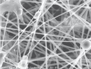
Figure 12.2. Scanning electron microscopic images of poly(3-hydroxybutyrate-co-3- hydroxyhexanoate)-based biomaterials: (A) poly(3-hydroxybutyrate-co-3-hydroxyhexanoate) materials; (B) poly(3-hydroxybutyrate-co-3-hydroxyhexanoate) with 5% gelatin blend; (C) poly(3- hydroxybutyrate-co-3-hydroxyhexanoate) with 10% gelatin blend; (D) poly(3-hydroxybutyrate- co-3-hydroxyhexanoate) with 30% gelatin blend. (Reprinted with permission of the American Chemical Society from Wang YW, Wu Q, Chen GQ: Gelatin blending improves the performance of poly(3-hydroxybutyrate-co-3-hydroxyhexanoate) films for biomedical application, Biomacromolecules 6:566–71, copyright 2005.)
acid is used as an example to demonstrate the principles of the synthesis, material properties, and potential biomedical application of polyamides.
Poly(glutamic acid) can be synthesized from poly(γ-benzyl-L-glutamate) by eliminating the benzyl group by using hydrogen bromide. Poly(glutamic acid) (PGA) can be degraded by enzymatic hydrolysis. In particular, cystein proteases play a critical role in the degradation of PGA. The time course of PGA degradation ranges from several hours to months, depending on the concentration of proteinase, temperature, and the composition of the polymer (with or without additional components). Poly(glutamic acid) exhibits little toxicity when implanted into an animal tissue. Animals can tolerate a single dose of ≤800 mg/kg and an accumulated dose of ≤1.8 g/kg. Polyglutamic acid exhibits little immunogenicity in animal models. This type of materials can be used in various biomedical applications, such as tissue repair and regeneration as well as drug delivery.
Polyphosphazenes [12.9]. Polyphosphazenes are inorganic biodegradable polymers that are constituted with a nitrogen–phosphorus (N==P) backbone. This type of polymer is

SYNTHETIC POLYMERS AS BIOMATERIALS |
477 |
(A) |
|
(B) |
(C) |
|
(D) |
(E) |
|
(F) |
Figure 12.3. Scanning electron microscopic images of MG63 osteoblast-like cells (2-day culture) attached to the polycarbonate membrane surfaces with different micropore sizes: (A) 0.2, (B) 0.4,
(C) 1.0, (D) 3.0, (E) 5.0, and (F) 8.0 μm in diameter of micropore sizes. (Reprinted from Lee SJ et al: Response of MG63 osteoblast-like cells onto polycarbonate membrane surfaces with different micropore sizes, Biomaterials 25:4699–707, copyright 2004 with permission from Elsevier.)
synthesized via reactions of polydichlorophosphazene with amines or alkoxides in tetrahydrofuran or aromatic hydrocarbone solutions. Polymers with various sidegroups can be synthesized by mixing different components. The material properties of polyphosphazenes are dependent on the composition of the polymer. Biodegradable polyphosphazenes can be generated when amino acid derivatives are used as sidegroups. For instance, the ethylglycinato-derived polymers can be degraded into ammonia, phosphate, ethanol, and glycine. Experimental tests in vitro have demonstrated that amino acid-based polyphosphazenes can be degraded within several months. The rate of degradation is dependent on the type of amino acid selected for the sidegroups.
Polyphosphazenes have been used for constructing various structures, such as matrix scaffolds (Fig. 12.4), hydrogels, and microspheres, for drug delivery. The degradation of
478 BIOMATERIAL ASPECTS OF BIOREGENERATIVE ENGINEERING
Figure 12.4. Scanning electron micrograph showing electrospun poly[bis(p-methylphenoxy) phosphazene fiber matrix with arterial endothelial cells after 24 h of culture. (Reprinted with permission of the American Chemical Society from Lakshmi S et al: Fabrication and optimization of methylphenoxy substituted polyphosphazene nanofibers for biomedical applications, Biomacromolecules 5:2212–20, copyright 2004.)
polyphosphazenes is sensitive to changes in temperature. Thus, the degradation rate of these polymers can be regulated by controlling environmental temperature. Such polymers can be used for constructing drug delivery carriers for temperature-related diseases. Other environmental factors, such as pH, may also influence the rate of degradation of polyphosphazenes. For instance, oxybenzoate-containing polyphosphazenes are pH-sensitive. The degradation of this type of polymer can be controlled by altering the content of oxybenzoate within a specified range of pH. Such polymers can be used to deliver drug for diseases that result in a change in pH. In vivo animal tests with subcutaneous implantation have shown that polyphosphazenes materials exhibit low toxicity, induce little inflammatory reactions in host tissues, and are relatively safe for implantation and drug delivery.
Polyanhydrides [12.10]. Polyanhydrides are polymers formed with anhydrides, compounds in which two carbonyl groups are joined with an oxygen atom, RCO—O—COR′, where R and R′ are any organic groups. The polymers are synthesized by reactions of diacids with anhydrides to form acetyl anhydride prepolymers. High-molecular-weight polyanhydrides can be formed from the prepolymers by melt condensation (180ºC for 90 min in vacuo). The addition of coordination catalysts, such as cadmium acetate and metal oxides, can facilitate the polymerization process, increasing the molecular weight of the polymer. Polyanhydrides can be dissolved in organic solvents such as chloroform and dichloromethane. Various components can be copolymerized with anhydrides to alter the solubility. Homopolymers of anhydrides usually exhibit a high level of crystallinity. Copolymerization with different components may reduce the crystallinity, producing more amorphous materials. Copolymerization also influences the mechanical properties of polyanhydrides.
BIOLOGICAL MATERIALS |
479 |
Polyanhydrides are degraded by hydrolytic erosion. A number of factors influence the rate of polyanhydrides degradation. These include pH and copolymerization with different compounds. An increase in pH facilitates polyanhydrides degradation. The incorporation of different aliphatic monomers may facilitate the degradation of the polymer, whereas the addition of methylene groups into the polymer backbone reduces the rate of degradation. Thus, polyanhydrides with various levels of degradation can be synthesized by copolymerization with various compounds. Polyanhydrides can be used to construct various forms of matrix, such as disks and pellets, and can also be injected into target tissues. The injection of mixed polyanhydrides and therapeutic substances is a promising technique for controlled drug delivery. A number of studies have shown that polyanhydrides do not significantly influence the growth of cultured cells and exhibit little toxicity when implanted into target tissues in animal tests.
BIOLOGICAL MATERIALS
Collagen Matrix [12.11]
Collagen matrix is found in mesenchymal and connective tissues, such as the subcutaneous tissue and bone, and the adventitia of tubular organs (blood vessels, airways, esophagus, stomach, and intestines). In mammalian tissues, there exist about 15 types of collagen matrix, namely, collagen types I–XV. Among these types of collagen, types I, II, III, IV, V, IX, XI, and XII are commonly found in connective tissues. Collagen types I, II, III, V, and XI are organized into filamentous structures, known as collagen fibrils, with a diameter of 10–100 nm. These fibrils usually form large collagen bundles as found in subcutaneous tissues and the adventitia of tubular organs. Collagen types I and V are often found in the bone, skin, cornea, tendon, ligament, and internal organs such as the lung, liver, pancreas, and kidney. Collagen types II and XI are found in the cartilage, notochord, and intervertebral disks. Collagen type III is found in blood vessels, skin, and internal organs. Collagen types IX and XII are molecules that link other types of collagen fibril and are known as fibril-associated collagens. These types are found in the cartilage, tendons, and ligaments. In contrast to the filamentous collagen molecules, collagen type IV participates in the construction of a membrane-like structure, known as the basal lamina, which underlies epithelial and endothelial cells. The structure and biochemical features of collagen molecules are discussed on page 103. These aspects will not be repeated here.
Collagen molecules play important roles in the constitution of mammalian tissues or organs. The collagen matrix serves as a structural framework that supports cells, helps organize cells into various forms of tissues and organs, and protects cells from mechanical injury. In addition, collagen matrix participates in the regulation of cellular activities such as cell survival, adhesion, proliferation, and migration. Collagen molecules can directly interact with cells via the cell membrane collagen receptors, or indirectly via the mediation of fibronectin, a matrix component that binds collagen molecules at one side and cell membrane matrix receptors, known as integrins, at the other side. The binding of collagen and fibronectin molecules to the integrin receptors initiate the activation of intracellular signaling pathways that stimulate or activate mitogenic processes, including cell survival, proliferation, and migration.
Given the structural and functional features, collagen matrix has long been used for constructing drug-delivery devices and scaffolds for tissue regeneration. Collagen matrix

480 BIOMATERIAL ASPECTS OF BIOREGENERATIVE ENGINEERING
Figure 12.5. Scanning electron micrograph of electrospun collagen matrix. (Reprinted with permission of the American Chemical Society from Zhang YZ et al: Characterization of the surface biocompatibility of the electrospun PCL-collagen nanofibers using fibroblasts, Biomacromolecules 6:2583–9, copyright 2005.)
has been used in several forms: collagen gels, meshes, composites with different molecules, and decellularized natural matrix. Collagen gels and meshes are suitable for drug delivery, whereas cell-free natural collagen matrix can be used as scaffolds or grafts for the repair or regeneration of various tissues and organs, such as blood vessels, airways, intestines, and bladder. Collagen gel can be spun into collagen fibers, which can be used to construct collagen scaffolds for tissue repair and regeneration (Fig. 12.5).
Native collagen matrix is a suitable material for the construction of tissue scaffolds. Such a material maintains the natural biological and mechanical characteristics and exhibits superior biocompatibility compared to in vitro crosslinked collagen gels or matrix. To prepare a native collagen matrix, mammalian tissue specimens can be collected from the submucosa of intestines, the adventitia of blood vessels, and the subcutaneous tissue. Cells in these specimens can be removed by various enzymatic and hydrolytic methods. Such treatments eliminate the cellular immunogenicity of allogenic tissues (note that extracellular matrix molecules exhibit neglible immunogenicity). The resulting cell-free collagen matrix can be tailored into a scaffold with a desired form and used for tissue repair or regeneration.
Elastic Fibers and Laminae [12.12]
Elastic fibers and laminae are major extracellular matrix components found in mesenchymal and connective tissues. Elastic fibers are present in the lung, connective tissue, the submucosa of intestines, and the wall of veins, whereas elastic laminae are found primarily in the media of large and medium arteries. Elastic fibers and laminae are composed of several proteins, including elastin, microfibrils, and microfibril-associated proteins. Elastin is the most abundant protein in elastic fibers and laminae. Mature elastin is a highly insoluble and hydrophobic protein, and is formed by crosslinking the 72-kDa elastin
BIOLOGICAL MATERIALS |
481 |
precursor, known as tropoelastin. Tropoelastin is produced by several cell types, including the smooth muscle cell, endothelial cell, and fibroblast, and is released into the extracellular space where crosslinking and elastin formation take place. The structure and biochemical features of elastic fibers are discussed on page 109.
Elastic fibers and laminae play an important role in the constitution of tissues and organs as well as in the maintenance of the stability of tissues and organs. For instance, multiple layers of elastic laminae are found in large arteries. These laminae have long been known to contribute to the structural stability and mechanical strength of the arterial wall (44,45). Arteries are subject to extensive mechanical stress induced by arterial blood pressure. Without the support of the elastic laminae, vascular cells may be overstretched under arterial blood pressure. Elastic laminae also contribute to the elasticity of soft tissues, such as connective tissues and arteries. The recoil of the arterial wall is a critical mechanism for the continuation of bloodflow during diastole when cardiac ejection is ceased. Elastic laminae have also been shown to serve as a signaling structure and play a role in regulating arterial morphogenesis and pathogenesis. An important contribution of elastic laminae is to confine smooth muscle cells to the arterial media by inhibiting smooth muscle cell proliferation and migration, thus preventing intimal hyperplasia under physiological conditions. In addition, elastic laminae exhibit antiinflammatory effects and inhibit leukocyte adhesion, activation, and transmigration relative to collagen matrix. These features render elastic laminae a potential material for vascular reconstruction. Furthermore, elastic laminae and elastin-containing structures can be used to prevent inflammatory reactions after surgery.
Polysaccharides [12.13]
Polysaccharides are polymers composed of many monosaccharides bonded together by glycosidic bonds. There are a number of forms of natural polysaccharides, including glycogen, cellulose, alginate, chitosan, starch, and glycosaminoglycan. These polysaccharides are found in animals and plants, and play an important role for the survival and function of animals and plants. Glycogen is a polymer composed of glucose monomers and synthesized in animals for the storage of energy. Alginates are linear polysaccharides composed of β-mannuronic acid and α-guluronic acid, and are found in brown seaweed and in certain bacteria. Starch is a polymer found in plants and synthesized for the storage of energy. Cellulose is found in plants and bacteria. Chitosan is found in the shell of crabs and shrimps. One of the important properties of polysaccharides is their ability to form hydrogel. This property is the basis for polysaccharide-mediated drug delivery. Several types of polysaccharide, such as cellulose, chitosan, and starch, have been used as materials for tissue engineering and drug delivery.
Cellulose. Cellulose is a linear polysaccharide composed of D-glucose units jointed together by 1,4-β-glucosidic bonds. In plants, cellulose participates in the constitution of plant skeleton and cell wall. Cotton is a well-known cellulose-containing material. Cellulose molecules are often arranged in parallel, giving cellulose fibers high mechanical strength. Humans cannot use cellulose as an energy source because of the lack of β- glycosidase, which catalyzes the hydrolysis of β-glycosidic bonds (note that mammals have α-glycosidase that catalyzes the hydrolysis of glycogen and starch). Cellulose can also be produced by bacteria. Bacterial cellulose has been often used for tissue engineering and will be the focus here.
482 BIOMATERIAL ASPECTS OF BIOREGENERATIVE ENGINEERING
Several types of microorganism, including algae (Vallonia), fungi (Saprolegnia, Dictystelium discoideum), and bacteria (Acetobacter, Achromobacter, Aerobacter, Agrobacterium, Pseudomonas, Rhizobium, Sarcina, Alcaligenes, Zoogloea) can synthesize cellulose. Among these microorganisms, the bacterium Acetobacter xylinum, which is usually found in fruits, vegetables, and alcoholic beverages, has been used to generate cellulose for tissue engineering applications. In a culture medium, this bacterium can produce a network of cellulose fibers. The cellulose fibers can be collected, fabricated into desired forms, and used to construct scaffolds for tissue engineering applications.
The bacterial cellulose synthesized by Acetobacter xylinum is similar to the plant cellulose in molecular composition. Both types of cellulose contain D-glucose. However, bacterial cellulose exhibits a higher crystallinity, higher water absorption capacity or lower hydrophobicity, higher mechanical strength, and finer molecular arrangement compared to the plant cellulose. Cellulose and its derivatives, such as cellulose nitrate, cellulose acetate, and cellulose xanthate, can be easily fabricated into desired forms. Unlike other polysaccharides, such as glycogen and starch, cellulose exhibits low water solubility and, therefore, a low rate of degradation when implanted into an animal tissue. A decrease in the crystallinity and hydrophobicity of cellulose usually results in an increase in the biodegradability of cellulose. Given the chemical composition, bacterial cellulose is highly biocompatible and nontoxic to the host. Furthermore, bacterial cellulose is a highly moldable material and can be used to fabricate scaffolds with desired forms. Cellulose-based materials have been used in a number of biomedical applications. These include construction of cellulose membranes for hemodialysis, construction of enzyme carriers for biosensors, drug delivery, construction of scaffolds for the regeneration of various tissue types, such as the bone, cartilage, liver, skin, and blood vessels. These investigations have consistently demonstrated that cellulose-based materials elicit little inflammatory and toxic reactions. Cellulose has been proven a promising material for the construction of tissue regenerating scaffolds.
Alginates [12.14]. Alginates are linear polysaccharides composed of β-mannuronic acid and α-guluronic acid. Alginates are found in brown seaweed and in certain bacteria. The content of β-mannuronic acid and α-guluronic acid may vary depending on the plant or bacterial species from which alginates are obtained. Alginates can be used to form hydrogel and matrix. Divalent cations, such as Ca2+ and Mn2+, can initiate alginate gelation by linking α-guluronic acid units between different polymer chains. The feature of gelation renders alginates a potential material for tissue engineering applications, such as cell seeding and transplantation, tissue repair, and drug delivery.
Alginate gels with various mechanical properties can be generated under different gelling conditions and by using different crosslinkers. Numerous studies have been conducted to test the elastic and shearing mechanical properties. Under compressive forces, alginate matrices exhibit elastic modulus ranging from 1 to 1000 kPa, depending on gelling and experimental conditions. Similarly, the shear modulus of alginate matrices spreads widely from 0.02 to 40 kPa under different experimental conditions. Under tensile forces, the maximal tensile strength or failure stress of alginate gels ranges from 3 to 35 kPa and the maximal or failure strain is from 0.3 to 1.25, depending on the composition of alginates and the strain rate applied.
Alginate gels crosslinked by Ca2+ have been used for a number of biomedical applications. One of the applications is alginate-mediated gene delivery. Alginate microspheres have been fabricated to carry genes of interest. The alginate microspheres can be delivered
BIOLOGICAL MATERIALS |
483 |
to target tissues, where the gene is released. Because of the biodegradability of alginates, genes can be released in a controlled manner with the releasing rate depending on the rate of alginate degradation. Similarly, an alginate-based gel or matrix can be used to mediate controlled protein and drug delivery. In addition, alginate-based materials have been fabricated and used to mediate wound healing. Alginates can form a thin layer of gel when crosslinked by Ca2+. Such a gel layer can be used to cover skin wound to prevent the loss of body fluids and bacterial infection. Alginate-based materials can be used to construct various forms of matrix scaffolds for the repair or regeneration of various tissue types such as the cartilage, liver, and bone. Alginate materials have also been used to construct capsules for cell transplantation. Cells can be encapsulated within alginate capsules and delivered to target tissues (Fig. 12.6). The alginate capsules can partially protect the enclosed cells from inflammation-induced injury.
Chitosan [12.15]. Chitosan is a linear polysaccharide composed of D-glucosamine units jointed by β-1,4-glycosidic bonds with randomly inserted N-acetylglucosamine units. Chitosan is a partially deacetylated derivative of chitin, which is a copolymer of randomly distributed N-acetylglucosamine and N-glucosamine units. A polymer molecule with more than 50% N-acetylglucosamine units is known as chitin, and that with more than 50% N-glucosamine units is called chitosan.
Chitosan and chitin are found in the shell of crabs and shrimps and are similar to cellulose in structure. Chitosan and chitin can be collected from these shellfish sources. Chitosan is a semicrystalline molecule and is usually stable. It is insoluble in water, but soluble in acidic solutions (pH 5). The use of chitosan for tissue engineering relies partially on its gelation ability. Chitosan solutions can be gelled in methanol and under a high pH condition. A dried chitosan structure can be mechanically very strong. Chitosan molecules are usually positively charged and can bind to molecules with negative charges, such as glycosaminoglycans and alginates. A unique feature is that the charge density of chitosan is dependent on pH. Such a feature renders chitosan a candidate material for pH-controlled drug delivery.
When implanted in vivo, chitosan is degraded by lysozyme-catalyzed hydrolysis. Chitosan is disintegrated into oligosaccharides. The rate of chitosan degradation is inversely proportional to the degree of crystallinity. The crystallization of chitosan is regulated by deacetylation. Chitosan molecules with increased deacetylation on the N- acetylglucosamine units exhibit increased crystallinity and reduced degradation. In a highly crystal form, it takes several months to degrade chitosan scaffolds in vivo. Amorphous chitosan exhibits more rapid degradation.
Chitosan is a polysaccharide that can be fabricated into various forms of porous matrix. To produce a chitosan matrix, chitosan can be dissolved in acetic acid. The chitosan–acetic acid solution can be frozen and lyophilized to produce chitosan matrix. The freezing process induces the formation of ice crystals. The following lyophilizing process removes the ice crystals, allowing the formation of a porous matrix. The size of the pores can be controlled by altering the rate of ice crystal formation.
Chitosan can be used to make materials with various mechanical properties. A pure chitosan material without apparent pores exhibits elastic modulus ranging from 5 to 7 MPa. However, the introduction of pores reduces the elastic modulus and mechanical strength. Porous chitosan materials could have an elastic modulus as low as 0.1 MPa. The failure strain or maximal strain of chitosan is also dependent on the porosity of the material. Nonporous chitosan materials can be stretched to a strain about 0.3, whereas a porous

484 BIOMATERIAL ASPECTS OF BIOREGENERATIVE ENGINEERING
(A)
4 mm
(B)
1 mm
(C)
2 mm
Figure 12.6. Cell-containing alginate beads for cell transplantation. Cells were encapsulated with alginate beads by dropping the cell–alginate mixture into an agitated bath of calcium chloride using a syringe. (A) A-low magnification image of the beads. (B) A higher-magnification image. (C) Cell–alginate beads were mixed into a calcium phosphate cement paste at a 54% volume fraction of alginate beads. (Reprinted with permission of John Wiley & Sons, Inc. from Weir MD et al: Strong calcium phosphate cement-chitosan-mesh construct containing cell-encapsulating hydrogel beads for bone tissue engineering, J Biomed Mater Res Pt A, published online Feb 15, 2006.)
BIOLOGICAL MATERIALS |
485 |
chitosan material can be stretched to a strain about 1. Porous chitosan exhibits a nonlinear mechanical behavior, i.e., the mechanical behavior is dependent on the level of strain and stress. The material gains stiffness (with increased elastic modulus) when strain and stress are elevated.
Chemical modifications significantly influence the mechanical properties of chitosan materials. For instance, the coating of a chitosan material with hyaluronic acid significantly increases the tensile strength of the chitosan material. This mechanical reinforcement is due to the formation of tight bonds between positively charged chitosan molecules and negatively charged hyaluronic acid molecules. Such reinforced chitosan materials are suitable for repairing tissues with high mechanical loads such as cartilage. Furthermore, the incorporation of hydroxyapatite or other calcium containing materials into chitosan or chitin can generate composite materials with increased mechanical strength. Such materials can be used for bone repair or regeneration.
Given the molecular structure, mechanical properties, biocompatibility, and the capability of forming various matrix structures, chitosan and chitosan derivatives have been considered candidate materials for the engineering and regeneration of injured tissues and organs. Chitosan materials have been used to construct matrix scaffolds for seeding, culturing, and transplanting cells into target tissues. These materials have also been used as carriers for drug delivery. Several studies have shown that chitosan can serve as a gene transfer carrier. Genes mixed with chitosan-based materials have been successfully delivered to target cells in the knee joints in animal models. Chitosan-mediated gene transfer can also be carried out together with cell transplantation, enhancing therapeutic effects on target diseases.
Numerous investigations have shown consistently that chitosan and chitosan derivatives are relatively nontoxic and biocompatible. In particular, chitosan-based materials do not induce significant fibrous encapsulation around the implants. Although chitosan implantation induces leukocyte infiltration during the early period (within days), chronic inflammation does not occur significantly. The application of chitosan to cartilage repair and regeneration has demonstrated a beneficial effect on the recovery of injured cartilage tissues, such as stimulation of chondrocyte growth and expression of structural proteins. These observations have demonstrated the feasibility of using chitosan and chitosan derivatives as biomaterials for tissue regenerative engineering.
Starch [12.16]. Starch is composed of D-glucose and is a form of polysaccharide for the storage of energy in plants. It can be found in all plant seeds and tubers. There are two forms of starch: amylase and amylopectin. Amylose is a linear polymer with the D-glucose units joined by the α1,4-glycosidic bonds, whereas amylopectin contains branching polymer chains, in which the D-glucose units are joined by the α1,4-glycosidic bonds in the linear portion and those at branching points are joined by the α1,6-glycosidic bonds.
Cornstarch is usually used in biomedical research. Starch can be blended with chemical compounds such as ethylene vinyl alcohol and cellulose acetate to make matrices that can serve as engineering scaffolds or cell seeding/culture substrates. The fabricated matrix can be reinforced by mixing with hydroxyapatite to form a composite material. Polymer matrices of various forms can be prepared by injection molding. Starch and its composites have been used as substrates for cell culture and carriers for cell transplantation. Starchbased materials do not significantly influence the growth and function of cultured cells. These materials have also been used in vivo for several biomedical applications, including