

Bioregenerative Engineering Principles and Applications - Shu Q. Liu
..pdf446 MOLECULAR ASPECTS OF BIOREGENERATIVE ENGINEERING
The luciferase gene encodes a protein enzyme that oxidizes luciferin and is found in certain types of fish and insects, such as firefly bugs. The oxidization reduces luciferin to a compound that emits fluorescence. The emitted fluorescence can be observed by using a fluorescence microscope. Alternatively, the intensity of the emitted fluorescence can be detected by spectrophotometry. Since the luciferase gene is not expressed in mammalian cells, this gene can be used as a reporter gene for detecting the efficiency of gene transfer in terms of the fluorescence emitted by the luciferase substrate. In a cell sample transfected with the luciferase gene, positive fluorescent emission from the cell sample in the presence of luciferin indicates the expression of the transferred luciferase gene. In contrast, the absence of fluorescence in the presence of luciferin indicates the failure of gene transfer. Alternatively, an antiluciferase antibody developed by using luciferase as an antigen can be used to detect the expressed enzyme by immunohistochemistry. Luciferase is harmless to mammalian cells and is commonly used for the assessment of gene transfer efficiency.
The chloramphenicol acetyltransferase gene encodes chloramphenicol acetyltransferase (CAT), an enzyme that catalyzes the transfer of a catyl group from acetylcoenzyme A to the 3′-hydoxy position of chloramphenicol, C11H12Cl2N2O5, which is a broad-spec- trum antibiotic derived from Streptomyces venezuelae (fungus-like bacteria). The gene is not present in mammalian cells and can be used as a reporter gene for detecting the efficiency of gene transfer. The CAT catalytic activity can be monitored by a liquid scintillation enzyme assay and used for assessing gene expression. For the liquid scintillation enzyme assay, cell extracts can be incubated in a reaction mix containing 14C- or 3H-labeled chloramphenicol and n-butyryl coenzyme A. When the CAT gene is expressed, the CAT transfers the n-butyryl group of the n-butyryl coenzyme A molecule to chloramphenicol, forming n-butyryl chloramphenicol. The reaction products can be extracted with a small volume of xylene. The n-butyryl chloramphenicol compound can be partitioned into the xylene phase, while unmodified chloramphenicol remains predominantly in the aqueous phase. The xylene phase is mixed with scintillant and counted with a scintillation counter for the radioactivity of 14C or 3H, which is conjugated to chloramphenicol. The presence of radioactivity indicates the expression of the transferred CAT gene.
There are natural fluorescent protein genes that encode proteins capable of emitting fluorescence. These genes are found in certain types of florescent fish and insects. One of the most commonly seen natural fluorescent proteins is the green fluorescent protein, which is encoded by the green fluorescence protein gene. Recombinant fluorescent genes that encode red and cyanine proteins have been artificially constructed by modulating the gene sequences of natural fluorescence protein genes. These genes can be used as reporter genes for assessing the efficiency of gene transfer. Two methods can be used for such a purpose: (1) a fluorescence protein gene can be cotransferred with a therapeutic gene into target cells and used to assess the expression of the therapeutic gene or (2) a fluorescent protein gene can be inserted into a recombinant vector that contains a functional or therapeutic gene (Fig. 10.6). The fluorescent gene can be expressed together with the therapeutic gene when transfected into target cells, thus indicating the efficiency of gene transfer. It is important to note that the fluorescent protein gene should be inserted into an appropriate site that does not influence the transcription of the therapeutic gene. Such a site is usually located at the end of the therapeutic gene. However, trial-and-error experiments should be carried out to search for a correct insertion site. An advantage by using the fluorescent protein gene is that fluorescent signals can be examined in living cells, allowing the
HOMOLOGOUS RECOMBINATION |
447 |
observation of dynamic changes in molecular action. Thus, fluorescent protein gene has been extensively used in cell biology research.
Assessing the Effectiveness of Gene Transfer. An important aspect of gene therapy is to test the effectiveness and efficacy of gene therapy. One of the critical issues is whether gene transfer restores the physiological function of the target gene, cell, tissue, and organ, and corrects pathological alterations due to genetic disorders. The design of a test is largely dependent on the function of the target gene as well as the structure and function of the target cells. For instance, for a gene that encodes an enzyme, such as a matrix metalloproteinase, the level and activity of the enzyme can be detected before and after the gene transfection. The level of the enzyme can be assessed by immunoblotting analysis, and the function of the enzyme can be evaluated by detecting the level of the substrate modification. For a gene that encodes a protein responsible for regulating a specific cell activity, such as smooth muscle cell contractility, the level of the protein and the degree of cell activity can be assessed before and after the gene transfection. Similarly, the level of cell proliferation and apoptosis can be measured to assess the effectiveness of delivered genes that encode proteins for the regulation of cell mitogenic and apoptotic activities, respectively. Regardless of the gene type, a significant phenotype change in response to gene transfection suggests the effectiveness of the therapeutic gene.
Potential Negative Effects of Gene Transfer. Gene transfer may not be always beneficial and may potentially induce harmful effects:
1.Gene transfer requires the mediation of gene carriers, which are often toxic (liposomes), traumatic (electroporation and bombard), or infectious (adenovirus and retrovirus). These mediating approaches may negatively influence the function of host cells.
2.It is difficult to control the gene insertion sites in the target genome. Gene transfer by any mediating means may potentially cause gene insertion into incorrect sites in the genome, resulting in host or guest gene mutation, upregulation, or downregulation.
3.A therapeutic gene may be suitable for certain physiological functions, but may negatively influence other functions. A typical example is the genetic manipulation of the ras gene, which encodes the Ras protein, a critical molecule for the transduction of mitogenic signals and for the regulation of cell survival and proliferation. Because of its proproliferative effect, the ras gene has been considered a potential gene target for the treatment of atherosclerosis and cardiovascular hypertrophy. However, the suppression of the ras gene, while potentially reduces the proliferative activity, inevitably causes a negative effect on other functions mediated by this protein such as cell survival. Thus, the potential negative effects of gene transfer should be taken into account in the design of a molecular engineering approach.
HOMOLOGOUS RECOMBINATION [10.3]
Homologous recombination is a natural process by which DNA damages, such as doublestrand breaks (DSBs) and interstrand crosslinks, are repaired by generating a functional DNA copy with the homologous DNA sequence of the intact sister chromatin as a template
448 MOLECULAR ASPECTS OF BIOREGENERATIVE ENGINEERING
and replacing precisely the damaged DNA sequence. This process can completely restore the structure and function of the damaged DNA. Thus, homologous recombination is an ideal genetic approach for the correction of a mutant gene and can be utilized for therapeutic purposes.
Homologous recombination is originally discovered in yeast and E. coli. In mammalian cells, homologous recombination is rare, but is significantly increased in frequency when DNA double-strand breaks occur. Thus, one strategy to induce homologous recombination in mammalian cells is to introduce double strand breaks to desired target genes, which are responsible for the disorder of interest. The natural homologous recombination machinery in the cell is capable of recognizing the double-strand breaks on the target gene and initiating homologous gene recombination or site-specific gene replacement, resulting in the restoration of the structure and function of the mutant gene.
An effective approach for introducing DNA double strand breaks to a target gene is to deliver specific zinc finger nucleases to cells or tissues. Zinc finger nucleases are proteins that can recognize and cut target DNA at specific sites, induce double-strand breaks, and enhance site-specific homologous recombination. A typical zinc finger nuclease is composed of characteristic zinc finger domains. Each zinc finger domain consists of about 30 amino acids and can bind a zinc ion, which is critical to the stability and function of the zinc finger protein. A zinc finger protein can recognize and bind to a specific target gene by interacting with the major groove of the DNA double helix. The catalytic domain of the zinc finger nuclease can then cut the double strand of the bound DNA, resulting in double strand breaks.
Zinc finger nucleases can be designed and constructed with high specificity to desired target DNA sequences. To date, several types of three-finger zinc finger nucleases have been constructed with specificity to unique triplet DNA sequences, including 5′-ANN-3′, 5′-CNN-3′, 5′-GNN-3′, and 5′-TNN-3′, where N represents any nucleotide. Determination of the specificity of a zinc finger nuclease is based on the structure of the protein enzyme and the target triplet nucleotides.
For therapeutic purposes, a triplet DNA sequence, which is unique to a mutant gene and serves as a specific binding site for a zinc finger nuclease, can be identified from a cell type. A zinc finger nuclease can be designed, constructed, and delivered to the target cells. The zinc finger nuclease can recognize the specific binding site and induce doublestrand breaks in the target mutant gene. The double strand breaks can initiate site-specific homologous recombination that replaces the mutant gene with a corresponding functional gene. Thus, the induction of the artificial double strand breaks and homologous recombination represent potential therapeutic approaches for the molecular treatment of genetic disorders.
ANTISENSE OLIGONUCLEOTIDE-BASED THERAPY [10.4]
Antisense oligonucleotide therapy is to design and use a DNA or RNA oligonucleotide sequence to hybridize complementary target mRNA, which is considered the “sense” sequence, and thus to block the translation of a specific protein, which is involved in the initiation and development of the disorder of interest. An example is the use of antisense oligonucleotides to downregulate the expression of angiotensinogen, thus to reduce the level of angiotensin I and II, for the treatment of hypertension (see page 699 for mechanisms). Furthermore, the hybridization of the target mRNA with an antisense
SMALL INTERFERING RNA-BASED THERAPY [10.5] |
449 |
oligonucleotide may induce the activation of an enzyme known as ribonuclease H (RNase H), which cleaves the hybridized mRNA. These approaches can be used to effectively downregulate protein translation, providing a therapeutic means for the treatment of disorders. An antisense DNA or RNA sequence specific to a selected mRNA molecule can be designed and constructed. The constructed antisense oligonucleotides can be delivered to target cells on the basis of endocytosis, a natural process that takes up small particles on the cell surface. Several gene transfer-mediating methods, such as liposomeand electro- poration-mediated transfer, can be used to enhance the delivery of the antisense oligonucleotides. The antisense approach can be potentially used for therapeutic purposes by repressing the translation of specific proteins, which are involved in the pathogenesis of disorders.
SMALL INTERFERING RNA-BASED THERAPY [10.5]
Small interfering RNA (siRNA), also known as short interfering RNA, is a short RNA sequence, which is specific to and can hybridize to a target mRNA, induce the degradation of the target mRNA, and thus knock down the expression of the encoded protein. The process of siRNA-induced mRNA degradation is referred to as RNA interference, posttranscriptional gene silencing, or transgene silencing. This mechanism was originally discovered in petunia plant cells and Caenorhabditis elegans. Further investigations have demonstrated that mammalian cells also exhibit RNA interference. Since RNA interference can be effectively used to suppress the translation of specific proteins, siRNA can be potentially used as therapeutic agents for pathological disorders that involve abnormal expression of specific proteins.
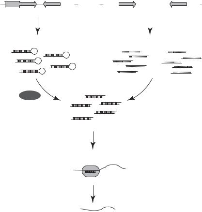
A siRNA molecule is generated under the action of an enzyme, known as “dicer,” which is an RNAse III molecule composed of a helicase domain, two RNAse III domains, a double-stranded RNA (dsRNA)-binding domain, and a PAZ domain (Fig. 10.13). The dicer enzyme can splice dsRNA into short siRNA. A typical siRNA is a double-stranded RNA about 21 nucleotide in length, and consists of a 5′ phosphate overhang at one end and a 3′ hydroxyl overhang at the other end. A siRNA fragment can form a complex with a multiprotein complex known as the RNA-induced silencing complex (RISC), which binds to a specific sequence of the siRNA and unwind the siRNA into single strands. The single-stranded siRNA can hybridize to complementary mRNA in the cytoplasm. The RISC complex is activated and can degrade the substrate mRNA bound by the siRNA, thus suppressing protein translation.
In mammalian cells, there is a RNA interference machinery, which has been evolved as a defense mechanism against the invasion of retroviruses. The RNA interference machinery can recognize and separate double-stranded RNA molecules into two single strands to form siRNAs. The siRNA molecules can recognize and recruit RNases, which degrade mRNA transcripts complementary to the siRNA molecules. On the invasion of retroviruses, siRNA sequences are generated and utilized for the destruction of the invaded viruses. This mechanism is also used for posttranscriptional gene regulation in mammalian cells. During gene transcription, certain RNA transcripts can fold to form hairpin-like double-stranded RNA structures, sometimes referred to as microRNA, which cannot be translated into proteins. The RNA interference machinery can detect and destroy these microRNA structures by generating siRNAs. Since the RNA interference machinery may not be able to completely remove target mRNAs, siRNA-mediated mRNA degradation is also referred to as gene knockdown.
 AAAAA
AAAAA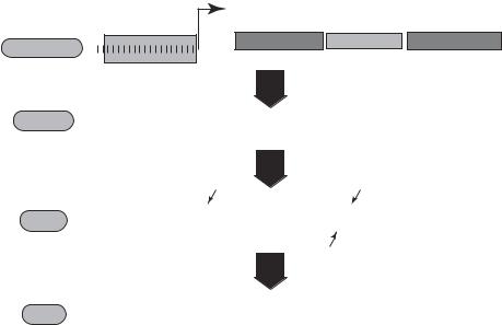
SMALL INTERFERING RNA-BASED THERAPY |
451 |
effective in knocking down gene expression, the effect is usually transient. It is necessary to conduct multiple transfection if long-term effect is required for experimental or therapeutic purposes.
To induce a long-term effect of RNA interference, a siRNA cloning vector can be constructed and used to transfect cells (Fig. 10.14). The transfected siRNA cloning vector contains a nucleotide sequence, which encodes a desired specific siRNA, and a gene expression vector, which induces gene expression when transfected into a cell. To construct a siRNA cloning vector, it is necessary to establish siRNA encoding gene sequence, which can be done by using a siRNA design program as described above. The siRNA-encoding gene sequence can be inserted into a cloning vector, such as the neomycin-resistant genecontaining psiSTRIKE cloning vector from Promega. The established siRNA cloning vector can be amplified by transfecting and growing E. coli cells. The amplified siRNA cloning vector can be purified and used for mammalian cell transfection. The following is an example of the siRNA cloning vector for the mRNA of the protein tyrosine phosphatase SH2 domain-containing protein tyrosine phosphatase 1 (SHP1).
The sequences for the SHP1 siRNA are as follows: 5′-ACCGAAAGGCCGGAACA AAT GTGTTTCAAGAGAACACATTTGTTCC GGCCTTTCTTTTTC-3′ and 5′- TGCAGA AAAAGAAAGGCCGGAACAAATGTGT TCTCTTGAAACACATTTGT TCCGGCC TTT-3′. In these sequences, the boldfaced fragments represent the target
|
+1 |
|
Transcription |
T7 class III promoter 3' |
Sense 25-mer Trimming loop Antlsense 25-mer |
AGCTAATACGACTCACTATA |
|
Template DNA A |
5' |
GCCATTATGCTGAGTGATATCCCNNNNNNNNNNNNNNNNNNNNNNNNNTCGTATCNNNNNNNNNNNNNNNNNNNNNNNNNTA
T7 RNA polymerase
Transcript PPPGGGNNNNNNNNNNNNNNNNNNNNNNNNNAGCAUAGNNNNNNNNNNNNNNNNNNNNNNNNNAUOH
Formation of hairpin RNA
PPP |
|
|
|
|
|
|
|
|
|
|
|
|
|
|
|
|
|
|
|
|
|
|
|
|
|
|
|
|
|
|
|
|
|
|
|
|
|
|
|
|
|
|
|
|
|
|
|
|
♥ |
GG |
|
|
|
|
|
|
|
|
|
|
|
|
|
|
|
|
|
|
|
|
|
|
|
|
|
|
|
|
|
|
|
|
|
|
|
|
|
|
|
|
|
|
|
|
|
|
|
|
|
♥ NNNNNNNNNNNNNNNNNNNNNNNNNA C |
|||||||||||||||||||||||||||||||||||||||||||||||||
shRNA |
|
|
|
|
|
|
|
|
|
|
|
|
|
|
|
|
|
|
|
|
|
|
|
|
|
|
|
|
|
|
|
|
|
|
|
|
|
|
|
|
|
|
|
|
|
|
|
|
A |
|
|
|
|
|
|
|
|
|
|
|
|
|
|
|
|
|
|
|
|
|
|
|
|
|
|
|
|
|
|
|
|
|
|
|
|
||||||||||||||
|
|
|
|
|
|
|
|
|
|
|
|
|
|
|
|
|
|
|
|
|
|
||||||||||||||||||||||||||||
UA |
NNNNNNNNNNNNNNNNNNNNNNNNN♥AU |
||||||||||||||||||||||||||||||||||||||||||||||||
HO |
|
|
|
|
|
|
|
|
|
|
|
|
|
|
|
|
|
|
|
|
|
|
|
|
|
|
|
|
|
|
|
|
|
|
|
|
|
|
|
|
|
|
|
|
|
|
|
|
|
Digestion by RNase T1
HONNNNNNNNNNNNNNNNNNNNNNNNNAGP siRNA 


HOUANNNNNNNNNNNNNNNNNNNNNNNNNOH
Walking across the entire gene, shifting one base at a time
Figure 10.14. Schematic representation of gene silencing by a shRNA expression vector. The shRNA is transcribed from a shRNA cloning vector and processed by Dicer to produce siRNA. The processed siRNA enters the RNA-induced silencing complex (RISC), where it targets mRNA for degradation. (Reprinted by permission of the Federation of the European Biochemical Societies from Itoa M et al: FEBS Lett 579:5988–95, copyright 2005.)
452 MOLECULAR ASPECTS OF BIOREGENERATIVE ENGINEERING
sequence, the italic boldfaced fragments represent the target reverse complement, the underlined fragments are for the mRNA loop, and the remainder are the overhang fragment (5′-ACC) and the U6 termination sequence (TTTTC-3′) for cloning purpose. The sequences for the control scrambled siRNA for SHP-1 are as follows: 5′-ACCGAAGAT- GCGAAGGGATAC TACTTCAAGAGAGTAGTATCCC T TCGCATCTTCTTTTTC-3′ and 5′-TGCAGAAAAAGAAGATGCGAAGGGATACTA TCTCTTGAA GTAGTATCCC TTCGCATCTT-3′. The SHP1 specific and scrambled siRNA sequences can be synthesized by a commercial carrier such as Proligo.
BIBLIOGRAPHY
10.1. Disorders Due to Gene Mutation
Kopp P, Jameson JL: Transmission of human genetic disease, in Principles of Molecular Medicine, Jameson JL, Collins FS, eds, Humana Press, Totowa, NJ, 1998, pp 43–58.
Schneider AS, Szanto PA: Pathology, 3rd ed, Lippincott Williams & Wilkins, Philadelphia, 2006.
Pasternak JJ: An Introduction to Human Molecular Genetics: Mechanisms of Inherited Diseases, 2nd ed, Wiley-Liss, Hoboken, NJ, 2005.
Killeen AA: Principles of Molecular Pathology, Humana Press, Totowa, NJ, 2004.
Watson JD, Berry A: DNA: The Secret of Life, Knopf, New York, 2003.
Pasternak JJ: An Introduction to Human Molecular Genetics: Mechanisms of Inherited Diseases, Fitzgerald Science Press, Bethesda, MD, 1999.
10.2. Principles of DNA Engineering
Schena M: Microarray Analysis, Wiley-Liss, Hoboken, NJ, 2003.
Grompe M, Johnson W, Jameson JL: Recombinant DNA and genetic techniques, in Principles of Molecular Medicine, Jameson JL, Collins FS, eds., Humana Press, Totowa, NJ, 1998, pp 9–24.
Watson JD, Gilman M, Witkowski J, Zoller M: Recombinant DNA, 2nd ed, Scientific American Books/Freeman, New York, 1992.
Kmiec EB: Gene targeting protocols, in Methods in Molecular Biology, Vol 133, Humana Press, Totowa, NJ, 2000.
Speicher DW, Reim DF: Microsequence analysis of electroblotted proteins, Anal Biochem 207:19– 23, 1992.
Jackson PJ: N-terminal protein sequencing for special application, in Methods in Molecular Biology, Vol 211: Protein Sequencing Protocols, 2nd ed, Smith BJ, ed, Humana Press, Totowa, NJ, 2003, pp 287–300.
Steinke L, Cook RG: Identification of phosphorylation sites by Edman degradation, in Methods in Molecular Biology, Vol 211: Protein Sequencing Protocols, 2nd ed, Smith BJ, ed, Humana Press, Totowa, NJ, 2003, pp 301–8.
Jackson PJ et al: Validation of protein sequencing in a regulated laboratory, in Methods in Molecular Biology, Vol 211: Protein Sequencing Protocols, 2nd ed, Smith BJ, ed, Humana Press, Totowa, NJ, 2003, pp 309–18.
Dupont DR, Yuen SW, Graham KS: Automated C-terminal protein sequence analysis using the alkylated-thiohydantoin method, in Methods in Molecular Biology, Vol 211: Protein Sequencing Protocols, 2nd ed, Smith BJ, ed, Humana Press, Totowa, NJ, 2003, pp 319–31.
BIBLIOGRAPHY 453
Harvey DJ: Identification of sites of glycosylation, in Methods in Molecular Biology, Vol 211: Protein Sequencing Protocols, 2nd ed, Smith B.J. ed, Humana Press, Totowa, NJ, 2003, pp 371–83.
Aitken A, Learmonth M: Analysis of sites of protein phosphorylation, in Methods in Molecular Biology, Vol 211: Protein Sequencing Protocols, 2nd ed, Smith BJ, ed, Humana Press, Totowa, NJ, 2003, pp 385–98.
Copley RR, Russell RB: Getting the most from your protein sequence, in Methods in Molecular Biology, Vol 211: Protein Sequencing Protocols, 2nd ed, Smith BJ, ed, Humana Press, Totowa, NJ, 2003, pp 411–30.
Shannon JD, Fox JW: Identification of phosphorylation sites by edman degradation, Tech Protein Chem VI:117–23, 1995.
Edman P: Method for determination of the amino acid sequence in peptides, Acta Chem Scand 4:283–93, 1950.
Burke DT, Carle GF, Olson MV: Cloning of large segments of exogenous DNA into yeast by means of artificial chromosome vectors, Science 236:806–12, 1987.
Cohen SN, Chang ACY, Boyer HW, Helling RB: Construction of biologically functional bacterial plasmids in vitro, Proc Natl Acad Sci USA 70:3240–4, 1973.
Nathans D, Smith HO: Restriction endonucleases in the analysis and restructuring of DNA molecules, Annu Rev Biochem 44:273–93, 1975.
Saiki RK, Gelfand DH, Stoffel S, Scharf SJ, Higuchi R, Horn GT et al: Primer-directed enzymatic amplification of DNA with a thermostable DNA polymerase, Science 239:487–91, 1988.
Sanger F, Nicklen S, Coulson AR: DNA sequencing with chain-terminating inhibitors, Proc Natl Acad Sci USA 74:5463–7, 1977.
Benton WD, Davis RW: Screening lambdagt recombinant clones by hybridization to single plaques in situ, Science 196:180–2, 1977.
Broome S, Gilbert W: Immunological screening method to detect specific translation products.
Proc Natl Acad Sci USA 75:2746–9, 1978.
Brown PO, Botstein D: Exploring the new world of the genome with DNA microarrays, Nat Genet 21(Suppl 1):33–7, 1999.
Gerhold DL, Jensen RV, Gullans SR: Better therapeutics through microarrays, Nat Genet 32(Suppl):547–51, 2002.
Grunstein M, Hogness DS: Colony hybridization: A method for the isolation of cloned DNAs that contain a specific gene, Proc Natl Acad Sci USA 72:3961–5, 1975.
Maniatis T, Hardison RC, Lacy E, Lauer J, O’Connell C, et al: The isolation of structural genes from libraries of eucaryotic DNA, Cell 15:687–701, 1978.
Southern EM: Detection of specific sequences among DNA fragments separated by gel electrophoresis, J Mol Biol 98:503–17, 1975.
Schildkraut CL, Marmur J, Doty P: The formation of hybrid DNA molecules and their use in studies of DNA homologies, J Mol Biol 3:595–617, 1961.
Ruitenberg MJ, Eggers R, Boer GJ, Verhaagen J: Adeno-associated viral vectors as agents for gene delivery: application in disorders and trauma of the central nervous system, Methods 28:182– 194, 2002.
Chan L, Fujimiya M, Kojima H: In vivo gene therapy for diabetes mellitus, Trends Mol Med 9:430–5, 2003.
Zhdanov R, Bogdanenko E, Moskovtsev A, Podobed O, Duzgunes N: Liposome-mediated gene delivery: Dependence on lipid structure, glycolipid-mediated targeting, and immunological properties, Meth Enzymol 373:433–65, 2003.
Bessis N, Doucet C, Cottard V, Douar AM, Firat H et al: Gene therapy for rheumatoid arthritis, J Gene Med 4:581–91, 2002.
454 MOLECULAR ASPECTS OF BIOREGENERATIVE ENGINEERING
Botstein D, Shortle D: Strategies and applications of in vitro mutagenesis, Science 229:1193–201, 1985.
Herskowitz I: Functional inactivation of genes by dominant negative mutations, Nature 329:219–22, 1987.
Robertson E, Bradley A, Kuehn M, Evans M: Germ-line transmission of genes introduced into cultured pluripotential cells by retroviral vector, Nature 323:445–8, 1986.
Jaenisch R: Transgenic animals, Science 240:1468–74, 1988.
10.3. Homologous Recombination
Bronson SK, Smithies O: Altering mice by homologous recombination using embryonic stem cells, J Biol Chem 269:27155–8, 1994.
Capecchi MR: Altering the genome by homologous recombination, Science 244:1288–92, 1989.
Gordon JW, Ruddle FH: Integration and stable germ line transmission of genes injected into mouse pronuclei, Science 214:1244–6, 1981.
Durai S, Mani M, Kandavelou K, Wu J, Porteus MH et al: Zinc finger nucleases: Custom-designed molecular scissors for genome engineering of plant and mammalian cells, Nucleic Acids Res 33:5978–90, 2005.
Gommans WM, Haisma HJ, Rots MG: Engineering zinc finger protein transcription factors: The therapeutic relevance of switching endogenous gene expression on or off at command, J Mol Biol 354:507–19, 2005.
Durai S, Mani M, Kandavelou K, Wu J, Porteus MH et al: Zinc finger nucleases: Custom-designed molecular scissors for genome engineering of plant and mammalian cells, Nucleic Acids Res 33:5978–90, 2005.
Vasileva A, Jessberger R. Precise hit: Adeno-associated virus in gene targeting, Nat Rev Microbiol 3:837–47, 2005.
Aplan PD: Causes of oncogenic chromosomal translocation, Trends Genet 22:46–55, 2006.
Glaser S, Anastassiadis K, Stewart AF: Current issues in mouse genome engineering, Nat Genet 37:1187–93, 2005.
Coghlan A, Eichler EE, Oliver SG, Paterson AH, Stein L: Chromosome evolution in eukaryotes: A multi-kingdom perspective, Trends Genet 21:673–82, 2005.
Porteus MH, Carroll D: Gene targeting using zinc finger nucleases, Nat Biotechnol 23:967–73, 2005.
Gerton JL, Hawley RS: Homologous chromosome interactions in meiosis: Diversity amidst conservation, Nat Rev Genet 6:477–87, 2005.
Klug A: Towards therapeutic applications of engineered zinc finger proteins, FEBS Lett 579:892–4, 2005.
Park Y, Gerson SL: DNA repair defects in stem cell function and aging, Annu Rev Med 56:495–508, 2005.
Pellegrini L, Venkitaraman A: Emerging functions of BRCA2 in DNA recombination, Trends Biochem Sci 29:310–6, 2004.
10.4. Antisense Oligonucleotide-Based Therapies
Izant JG, Weintraub H: Constitutive and conditional suppression of exogenous and endogenous genes by anti-sense RNA, Science 229:345–52, 1985.
Izant JG, Weintraub H: Inhibition of thymidine kinase gene expression by anti-sense RNA: A molecular approach to genetic analysis, Cell 36:1007–15, 1984.
BIBLIOGRAPHY 455
Wagner RW: Gene inhibition using antisense oligodeoxynucleotides, Nature 372:333–5, 1994.
Poulaki V, Mitsiades CS, Kotoula V, Tseleni-Balafouta S, Ashkenazi A et al: Regulation of Apo2L/ tumor necrosis factor-related apoptosis-inducing ligand-induced apoptosis in thyroid carcinoma cells, Am J Pathol 161:643–54, 2002.
Kawai H, Sato W, Yuzawa Y, Kosugi T, Matsuo S et al: Lack of the growth factor midkine enhances survival against cisplatin-induced renal damage, Am J Pathol 165(5):1603–12, Nov 2004.
10.5. Small Interfering RNA-Based Therapies
Hutvagner G, McLachlan J, Pasquinelli AE, Balint E, Tuschl T et al: A cellular function for the RNA-interference enzyme Dicer in the maturation of the let-7 small temporal RNA, Science 293:834–8, 2001.
Hutvagner G, Zamore PD: RNAi: Nature abhors a double-strand, Curr Opin Genet Dev 12:225–32, 2002.
Novina CD, Sharp PA: The RNAi revolution, Nature 430:161–4, 2004.
Sharp PA: RNA interference—2001, Genes Dev 15:485–90, 2001.
Hutvagner G, Zamore PD: A microRNA in a multiple-turnover RNAi enzyme complex, Science 297:2056–60, 2002.
Napoli C, Lemieux C, Jorgensen R: Introduction of a chalcone synthase gene into Petunia results in reversible co-suppression of homologous genes in trans, Plant Cell 2:279–89, 1990.
Dehio C, Schell J: Identification of plant genetic loci involved in a post transcriptional mechanism for meiotically reversible transgene silencing, Proc Acad Sci USA 91:5538–42, 1994.
Fire A, Xu S, Montgomery MK, Kostas SA, Driver SE, Mello CC: Potent and specific genetic interference by double–stranded RNA in Caenorhabditis elegans, Nature 391:806–11, 1998.
Aronin N: Target selectivity in mRNA silencing, Gene Ther 13:509–16, 2006.
Rodriguez-Lebron E, Paulson HL: Allele-specific RNA interference for neurological disease, Gene Ther 13:576–81, 2006.
Bonini NM, La Spada AR: Silencing polyglutamine degeneration with RNAi, Neuron 48:715–8, 2005.
Uprichard SL: The therapeutic potential of RNA interference, FEBS Lett 579:5996–6007, 2005.
Stecca B, Mas C, Ruiz i Altaba A: Interference with HH-GLI signaling inhibits prostate cancer, Trends Mol Med 11199–203, 2005.
Bennink JR, Palmore TN: The promise of siRNAs for the treatment of influenza, Trends Mol Med 10:571–4, 2004.
Ryther RC, Flynt AS, Phillips JA 3rd, Patton JG: siRNA therapeutics: Big potential from small RNAs, Gene Ther 12:5–11, 2005.
Hannon GJ, Rossi JJ: Unlocking the potential of the human genome with RNA interference, Nature 431:371–8, 2004.
Caplen NJ: Gene therapy progress and prospects. Downregulating gene expression: The impact of RNA interference, Gene Ther 11:1241–8, 2004.
Stevenson M: Dissecting HIV-1 through RNA interference, Nat Rev Immunol 3:851–8, 2003.