

Bioregenerative Engineering Principles and Applications - Shu Q. Liu
..pdfFas ligands
Fas
FADD
Caspase 8
Bid
Mitochondria
Releasing cytochrome C
Apaf1
Caspase 9
 Caspase3
Procaspase 3
Substrate proteins
Apoptosis
Figure 6.17. Schematic demonstration of the mechanisms of Fas-induced apoptosis. Based on bibliography 6.16.
APOPTOSIS 307
activated via autocatalytic cleavage. Caspase 8 can further activate a downstream protein known as Bid, a member of the Bcl2 family, by cleaving the C-terminal domain of the substrate. Following the cleavage by caspase 8, Bid can be translocated to the mitochondrial membrane. A major action of Bid is to release mitochondrial cytochrome c into the cytoplasm. Cytochrome c in turn activates a downstream protein named apoptosis proteinactivating factor-1 (Apaf-1), which binds ATP and interacts with procaspase 9, resulting in the formation of the active form of caspase 9 via autocatalytic cleavage. Caspase 9 can cleave procaspase 3, releasing the active form caspase 3. Caspase 3 is a terminal-stage protease, which cleaves and degrades a variety of signaling and structural proteins, including protein kinases, poly[A]polymerse, and actin filaments. Caspase 3 can also cleave DNA fragmentation factor (DFF), releasing a DFF subunit. This subunit can activate nucleases, which induces DNA degradation. These activities eventually lead to DNA fragmentation and cell degeneration. (See Table 6.10.)
Assessment of Cell Apoptosis [6.17]
There are several methods that can be used for assessing cell apoptosis. These methods have been developed on the basis of cell morphological and molecular changes in apoptotic cells and are classified into several groups: (1) methods based on changes in the structure of cell membrane, (2) methods based on changes in cell morphology, (3) methods based on DNA fragmentation, (4) methods based on cytochrome c translocation, and (5) methods based on caspase activities. These methods are briefly discussed here.
Assessing Changes in Cell Membrane Structure. A cell membrane contains asymmetrically distributed phospholipid species in the membrane bilayer. The cytoplasmic layer of the cell membrane is composed of phosphatidylserine, phosphatidylinositol, phosphatidylethanolamine, and phosphatidylcholine. The extracellular layer is composed of sphingomyelin, phosphatidylcholine, and phosphatidylethanolamine, but not phosphatidylserine. The asymmetrical distribution of phosphatidylserine is created and maintained by the activity of aminophospholipid translocase, which transports phosphatidylserine from the extracellular layer to the cytoplasmic layer of the cell membrane. In apoptosis, the activity of the aminophospholipid translocase is inhibited, and the content of phosphatidylserine in the extracellular layer of the cell membrane increases, even though the integrity of the cell membrane is uncompromised. Thus, the appearance of phosphatidylserine in the extracellular layer of the cell membrane is indicative of early cell apoptosis. Phosphatidylserine in the extracellular layer can be detected by using an assay for Annexin A5, which is a phosphatidylserine-binding protein. To detect phosphatidylserine, Annexin A5 can be tagged with a marker (e.g., biotin or fluorochrome) and incubated with cell samples. Positive labeling of cells with Annexin A5 suggests the translocation of phosphatidylserine to the extracellular layer of the cell membrane, which indicates the occurrence of apoptosis in the labeled cells.
In addition, the permeability of cell membrane is often increased in cell apoptosis because of the disorganization of phospholipids. In such a case, the cell membrane is permeable to certain types of fluorescent dyes, such as merocyanine (MC) 540 and 7- aminoactinomycin D (7-AAD), which can not pass through the plasma membrane of normal cells. These dyes can be incubated with cell samples and detected by fluorescence microscopy. The appearance of the dye within the cell suggests the occurrence of cell apoptosis. However, these fluorescent dyes are not specific to cell apoptosis. Any factors that cause an increase in cell membrane permeability can induce positive cell labeling.

308
TABLE 6.10. Characteristics of Selected Apoptosis Regulatory Proteins*
|
|
Amino |
Molecular |
|
|
Proteins |
Alternative Names |
Acids |
Weight (kDa) |
Expression |
Functions |
|
|
|
|
|
|
Fas ligand |
CD95 ligand, CD178, FAS, tumor |
281 |
31 |
Lymphocyte, testis |
A protein that interacts with the Fas receptor |
|
necrosis factor ligand superfamily |
|
|
|
and induces apoptosis |
|
member 6, TNFSF6, FASL, |
|
|
|
|
|
apoptosis antigen ligand 1, |
|
|
|
|
|
apoptosis (APO 1) antigen ligand 1 |
|
|
|
|
Fas receptor |
Tumor necrosis factor receptor |
335 |
38 |
Ubiquitous |
A transmembrane receptor that interacts with |
|
superfamily member 6, CD95, |
|
|
|
the Fas ligand and induces apoptosis |
|
FAS1, apoptosis-mediating |
|
|
|
|
|
surface antigen FAS, apoptosis |
|
|
|
|
|
antigen 1 |
|
|
|
|
FADD |
FAS-associated protein with death |
208 |
23 |
Ubiquitous |
An adaptor protein that interacts with the Fas |
|
domain |
|
|
|
receptor and TNF receptor; also mediates |
TNFα |
|
|
|
|
cell apoptosis |
Tumor necrosis factor, tumor necrosis |
233 |
26 |
Monocyte, |
Interacting with TNF receptor and regulating |
|
|
factor α, TNFA, cachectin |
|
|
macrophage |
cell apoptosis, proliferation, differentiation, |
|
|
|
|
|
and inflammatory reactions |
TNF receptor |
TNFR1α, TNFR1, tumor necrosis |
455 |
50 |
Heart, blood vessel, |
Interacting with TNFα, and mediating |
|
factor receptor superfamily |
|
|
leukocytes |
apoptosis and inflammatory reactions |
|
member 1A, p55 TNFR |
|
|
|
|
TRADD |
TNFR1-associated death-domain |
312 |
34 |
Ubiquitous |
Interacting with TNF receptor 1 and |
|
protein, tumor necrosisfactor |
|
|
|
regulating apoptosis and inflammation |
|
receptor 1-associated death- |
|
|
|
|
domain protein, tumor necrosis factor receptor 1-associated protein
Caspase 8 |
FADD homologous ICE/CED-3- |
496 |
58 |
Ubiquitous |
A cysteine–aspartic acid protease that |
|
ICE/CED-3-like protease, FADD- |
|
|
|
interacts with FADD and mediates the |
|
like ICE, MACH, MCH5, FLICE |
|
|
|
transduction of apoptotic signals |
Bid |
BH3 interacting death-domain |
195 |
22 |
Ubiquitous |
An apoptotic agonist activated by caspase 8, |
|
agonist, BID |
|
|
|
stimulating the release of cytochrome c, |
|
|
|
|
|
and regulating cell apoptosis |
Cytochrome c |
CYC |
105 |
12 |
Ubiquitous |
A mitochondrial electron transport chain |
|
|
|
|
|
component that mediates electron transfer |
|
|
|
|
|
and regulates apoptosis |
Apaf-1 |
Apoptotic protease-activating factor 1 |
1248 |
142 |
Ubiquitous |
Interacting with cytochrome c and forming |
|
|
|
|
|
an apoptosome, a structure that cleaves the |
|
|
|
|
|
preproprotein of caspase 9 and generates |
|
|
|
|
|
active caspase 9, resulting in apoptosis |
Caspase 9 |
CASP9, apoptotic protease MCH6, |
416 |
46 |
Ubiquitous |
A cysteine–aspartic acid protease that cleaves |
|
MCH6, ICE-like apoptotic |
|
|
|
procaspase 3, inducing the formation of |
|
protease 6, apoptotic protease- |
|
|
|
caspase 3 and apoptosis |
|
activating factor 3, APAF3 |
|
|
|
|
Caspase 3 |
CASP3, cysteine protease CPP32, |
277 |
32 |
Ubiquitous |
A downstream cysteine–aspartic acid |
|
CPP32, apoptosis-related cysteine |
|
|
|
protease that cleaves a variety of |
|
protease, APOPAIN |
|
|
|
cytoplasmic proteins, including protein |
|
|
|
|
|
kinases, nuclear proteins, and cytoskeletal |
|
|
|
|
|
proteins, and induces apoptosis |
|
|
|
|
|
|
*Based on bibliography 6.16.
309
310 FUNDAMENTAL CELLULAR FUNCTIONS
Assessing Changes in Cell Morphology. Apoptotic cells are associated with morphological changes, including cell membrane blebbing, DNA condensation with increased nucleus density, and nucleus disruption. The entire cell is eventually disintegrated into small pieces. Optical and electron microscopic approaches can be used to examine these morphological features. At the optical level, hematoxylin can be used to examine the morphology of cell nuclei. In addition, cell nucleus-binding fluorescent dyes, such as DAPI and Hoechst 33258, can be used for the same purpose. Usually, the fluorescent approach provides better images. At the electron microscopic level, cell membrane blebbing and DNA condensation (Fig. 6.16) can be observed with a much better resolution compared with the optical approach. Morphological examination is a key method for the identification of cell apoptosis and is often used as a standard for the confirmation of cell apoptosis detected by using other methods.
Assessing DNA Fragmentation. DNA fragmentation is a hallmark of cell apoptosis. Thus, apoptotic cells can be identified by assessing DNA fragmentation. Two approaches can be used for such purpose: DNA electrophoresis and terminal deoxynucleotidyl transferase-mediated dUTP nick end-labeling (TUNEL). For the DNA electrophoresis method, the pattern of DNA bands can be analyzed by comparing to DNA samples from normal control cells. An increase in the number of DNA bands in a large range of molecular size suggests the occurrence of cell apoptosis.
TUNEL is a method used for visualizing DNA fragments in situ. A key enzyme used for this assay is the terminal deoxynucleotidyl transferase, which catalyzes DNA synthesis at the ends of DNA fragments in the presence of deoxynucleotides (dNUPs). When a dNTP is tagged with a marker, such as a fluorescent molecule, the DNA fragments with the added dNTP can be visualized by fluorescence microscopy. This method can be used at the single cell level. However, the method is not apoptosis-specific. DNA fragmentation induced by other factors, such as cell injury, can be detected. Thus, the identification of morphological changes in apoptotic cells is often conducted together with the TUNEL method to confirm the results by TUNEL.
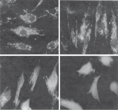
Assessing the Translocation of Cytochrome c. Cytochrome c is a protein component of the respiratory chain in the mitochondria. It is localized to the surface of the internal membrane of the mitochondria. Cytochrome c can be translocated from the mitochondria to the cytoplasm and contributes to the activation of apoptotic signaling pathways. The translocation of cytochrome c is a critical step in apoptosis. Thus, the detection of cytochrome c translocation from the mitochondria to the cytoplasm is indicative of apoptosis. An antibody can be used for examining the distribution of cytochrome c. Typical images of cytochrome c translocation are shown in Fig. 6.18.
Assessing the Activity of Caspases. Caspases are a group of proteinases that degrade proteins, ranging from signaling protein kinases to structural proteins, and play critical roles for the induction of cell apoptosis. The activation of caspases indicates the occurrence of apoptosis. Caspases are expressed in the form of inactive precursors, which can be activated by proteolytic cleavage at specific sites induced by proteinases. Thus, caspases cleavage is a sign of caspase activation. Immunoblotting is an effective method for the detection of caspase cleavage. The presence of reduced caspase subunits is indicative of caspase activation and the occurrence of apoptosis.
BIBLIOGRAPHY 311
A B
C D
Figure 6.18. Immunofluorescence detection of cytochrome c in fibroblasts. The micrographs show:
(A) control cells; (B–D) cells exposed to 0.5 μM naphthazarin (5,8-dihydroxy-1,4-naphthoquinone, an apoptosis inducer) for 1, 2, and 3 h, respectively. Note the translocation of cytochrome c from the mitochondria to the cytoplasm in panels B, C, and D. (Reprinted from Roberg K et al: Lysosomal release of cathepsin D precedes relocation of cytochrome c and loss of mitochondrial transmembrane potential during apoptosis induced by oxidative stress, Free Radical Biol Med 27:1228–37, 1999, with permission from Elsevier.)
BIBLIOGRAPHY
6.8. Classification and Structure of Selectins
E-Selectin
Bevilacqua MP, Stengelin S, Gimbrone MA Jr, Seed B: Endothelial leukocyte adhesion molecule 1: An inducible receptor for neutrophils related to complement regulatory proteins and lectins, Science 243:1160–5, 1989.
Collins T, Williams A, Johnston GI, Kim J, Eddy R et al: Structure and chromosomal location of the gene for endothelial-leukocyte adhesion molecule 1, J Biol Chem 266:2466–73, 1991.
Hidalgo A, Weiss LA, Frenette PS: Functional selectin ligands mediating human CD34+ cell interactions with bone marrow endothelium are enhanced postnatally, J Clin Invest 110:559–69, 2002.
Iida A, Nakamura Y: High-resolution SNP map in the 55-kb region containing the selectin gene family on chromosome 1q24-q25, J Hum Genet 48:150–4, 2003.
Wang N, Chintala SK, Fini ME, Schuman JS: Activation of a tissue-specific stress response in the aqueous outflow pathway of the eye defines the glaucoma disease phenotype, Nature Med 7:304–9, 2001.
312 FUNDAMENTAL CELLULAR FUNCTIONS
Watson ML, Kingsmore SF, Johnston GI, Siegelman MH, Le Beau MM et al: Genomic organization of the selectin family of leukocyte adhesion molecules on human and mouse chromosome 1, J Exp Med 172:263–72, 1990.
L-Selectin
Genbacev OD, Prakobphol A, Foulk RA, Krtolica AR, Ilic D et al: Trophoblast L-selectin-mediated adhesion at the maternal-fetal interface, Science 299:405–8, 2003.
Iida A, Nakamura Y: High-resolution SNP map in the 55-kb region containing the selectin gene family on chromosome 1q24-q25, J Hum Genet 48:150–4, 2003.
Lasky LA, Singer MS, Dowbenko D, Imai Y, Henzel WJ et al: An endothelial ligand for L-selectin is a novel mucin-like molecule, Cell 69:927–38, 1992.
Lasky LA, Singer MS, Yednock TA, Dowbenko D, Fennie C et al: Cloning of a lymphocyte homing receptor reveals a lectin domain, Cell 56:1045–55, 1989.
Ord DC, Ernst TJ, Zhou LJ, Rambaldi A, Spertini O et al: Structure of the gene encoding the human leukocyte adhesion molecule-1 (TQ1, Leu-8) of lymphocytes and neutrophils, J Biol Chem 265:7760–7, 1990.
Watson ML, Kingsmore SF, Johnston GI, Siegelman MH, Le Beau MM et al: Genomic organization of the selectin family of leukocyte adhesion molecules on human and mouse chromosome 1, J Exp Med 172:263–72, 1990.
P-Selectin
Burger PC, Wagner DD: Platelet P-selectin facilitates atherosclerotic lesion development, Blood 101:2661–6, 2003.
Hidalgo A, Weiss LA, Frenette PS: Functional selectin ligands mediating human CD34+ cell interactions with bone marrow endothelium are enhanced postnatally, J Clin Invest 110:559–69, 2002.
Hrachovinova I, Cambien B, Hafezi-Moghadam A, Kappelmayer J, Camphausen RT et al: Interaction of P-selectin and PSGL-1 generates microparticles that correct hemostasis in a mouse model of hemophilia A, Nature Med 9:1020–5, 2003.
Johnston GI, Bliss GA, Newman PJ, McEver RP: Structure of the human gene encoding granule membrane protein-140, a member of the selectin family of adhesion receptors for leukocytes, J Biol Chem 265:21381–5, 1990.
Johnston GI, Cook RG, McEver RP: Cloning of GMP-140, a granule membrane protein of platelets and endothelium: sequence similarity to proteins involved in cell adhesion and inflammation, Cell 56:1033–44, 1989.
Koyama H, Maeno T, Fukumoto S, Shoji T, Yamane T et al: Platelet P-selectin expression is associated with atherosclerotic wall thickness in carotid artery in humans, Circulation 108:524–9, 2003.
Marshall BT, Long M, Piper JW, Yago T, McEver RP et al: Direct observation of catch bonds involving cell-adhesion molecules, Nature 423:190–3, 2003.
Mayadas TN, Johnson RC, Rayburn H, Hynes RO, Wagner DD: Leukocyte rolling and extravasation are severely compromised in P selectin-deficient mice, Cell 74:541–54, 1993.
Rosenkranz AR, Mendrick DL, Cotran RS, Mayadas TN: P-selectin deficiency exacerbates experimental glomerulonephritis: A protective role for endothelial P-selectin in inflammation, J Clin Invest 103:649–59, 1999.
Human protein reference data base, Johns Hopkins University and the Institute of Bioinformatics, at http://www.hprd.org/protein.
6.9. Function of Selectins
Afshar-Kharghan V, Thiagarajan P: Leukocyte adhesion and thrombosis, Curr Opin Hematol 13:34–9, 2006.
BIBLIOGRAPHY 313
Simon SI, Green CE: Molecular mechanics and dynamics of leukocyte recruitment during inflammation, Annu Rev Biomed Eng 7:151–185, 2005.
Ley K, Kansas GS: Selectins in T-cell recruitment to non-lymphoid tissues and sites of inflammation, Nat Rev Immunol 4:325–35, 2004.
Kakkar AK, Lefer DJ: Leukocyte and endothelial adhesion molecule studies in knockout mice, Curr Opin Pharmacol 4(2):154–8, April 2004.
Rosen SD: Ligands for L-selectin: Homing, inflammation, and beyond, Annu Rev Immunol 22:129– 56, 2004.
Grunewald M, Griesshammer M: P-selectin modulation in haemostasis: One size fits all? Trends Mol Med 10:9–12, 2004.
Ulbrich H, Eriksson EE, Lindbom L: Leukocyte and endothelial cell adhesion molecules as targets for therapeutic interventions in inflammatory disease, Trends Pharmacol Sci 24:640–7, 2003.
6.10. Classification and Structure of Cadherins
E-Cadherin
Batlle E, Sancho E, Franci C, Dominguez D, Monfar M et al: The transition factor Snail is a repressor of E-cadherin gene expression in epithelial tumour cells, Nature Cell Biol 2:84–9, 2000.
Berx G, Staes K, van Hengel J, Molemans F, Bussemakers MJG et al: Cloning and characterization of the human invasion suppressor gene E-cadherin (CDH1), Genomics 26:281–9, 1995.
Boggon TJ, Murray J, Chappuis-Flament S, Wong E, Gumbiner BM et al: C-cadherin ectodomain structure and implications for cell adhesion mechanisms, Science 296:1308–3, 2002.
Cano A, Perez-Moreno MA, Rodrigo I, Locascio A, Blanco MJ et al: The transcription factor Snail controls epithelial-mesenchymal transitions by repressing E-cadherin expression, Nature Cell Biol 2:76–83, 2000.
Ceccherini I, Romeo G, Lawrence S, Breuning MH, Harris PC et al: Construction of a map of chromosome 16 by using radiation hybrids, Proc Natl Acad Sci USA 89:104–8, 1992.
Grady WM, Willis J, Guilford PJ, Dunbier AK, Toro TT et al: Methylation of the CDH1 promoter as the second genetic hit in hereditary diffuse gastric cancer, Nature Genet 26:16–7, 2000.
Guilford P, Hopkins J, Harraway J, McLeod M, McLeod N et al: E-cadherin germline mutations in familial gastric cancer, Nature 392:402–5, 1998.
Hayashi T, Carthew RW: Surface mechanics mediate pattern formation in the developing retina, Nature 431:647–52, 2004.
Huntsman DG, Carneiro F, Lewis FR, MacLeod PM, Hayashi A et al: Early gastric cancer in young, asymptomatic carriers of germ-line E-cadherin mutations, New Engl J Med 344:1904–9, 2001.
Jamora C, DasGupta R, Kocieniewski P, Fuchs E: Links between signal transduction, transcription and adhesion in epithelial bud development, Nature 422:317–22, 2003.
Kawasaki H, Taira K: Induction of DNA methylation and gene silencing by short interfering RNAs in human cells, Nature 431:211–7, 2004.
Maretzky T, Reiss K, Ludwig A, Buchholz J, Scholz F et al: ADAM10 mediates E-cadherin shedding and regulates epithelial cell–cell adhesion, migration, and beta-catenin translocation, Proc Natl Acad Sci USA 102:9182–7, 2005.
Natt E, Magenis RE, Zimmer J, Mansouri A, Scherer G: Regional assignment of the human loci for uvomorulin (UVO) and chymotrypsinogen B (CTRB) with the help of two overlapping deletions on the long arm of chromosome 16, Cytogenet Cell Genet 50:145–8, 1989.
Overduin M, Harvey TS, Bagby S, Tong KI, Yau P et al: Solution structure of the epithelial cadherin domain responsible for selective cell adhesion, Science 267:386–9, 1995.
Palmer HG, Larriba MJ, Garcia JM, Ordonez-Moran P, Pena C et al: The transcription factor SNAIL represses vitamin D receptor expression and responsiveness in human colon cancer, Nature Med 10:917–9, 2004.
314 FUNDAMENTAL CELLULAR FUNCTIONS
Perl AK, Wilgenbus P, Dahl U, Semb H, Christofori G: A causal role for E-cadherin in the transition from adenoma to carcinoma, Nature 392:190–3, 1998.
Risinger JI, Berchuck A, Kohler MF, Boyd J: Mutations of the E-cadherin gene in human gynecologic cancers, Nature Genet 7:98–102, 1994.
P-Cadherin
Hatta M, Miyatani S, Copeland NG, Gilbert DJ, Jenkins NA et al: Genomic organization and chromosomal mapping of the mouse P-cadherin gene, Nucleic Acids Res 19:4437–41, 1991.
Kremmidiotis G, Baker E, Crawford J, Eyre HJ, Nahmias J et al: Localization of human cadherin genes to chromosome regions exhibiting cancer-related loss of heterozygosity, Genomics 49:467–71, 1998.
Shimoyama Y, Yoshida T, Terada M, Shimosato Y, Abe O, Hirohashi S: Molecular cloning of a human Ca(2+)-dependent cell–cell adhesion molecule homologous to mouse placental cadherin: its low expression in human placental tissues, J Cell Biol 109:1787–94, 1989.
Sprecher E, Bergman R, Richard G, Lurie R, Shalev S et al: Hypotrichosis with juvenile macular dystrophy is caused by a mutation in CDH3, encoding P-cadherin, Nature Genet 29:134–6, 2001.
N-Cadherin
Garcia-Castro MI, Vielmetter E, Bronner-Fraser M: N-cadherin, a cell adhesion molecule involved in establishment of embryonic left-right asymmetry, Science 288:1047–51, 2000.
Hayashi T, Carthew RW: Surface mechanics mediate pattern formation in the developing retina, Nature 431:647–52, 2004.
Hermiston ML, Gordon JI: Inflammatory bowel disease and adenomas in mice expressing a dominant negative N-cadherin, Science 270:1203–6, 1995.
Miyatani S, Copeland NG, Gilbert DJ, Jenkins NA, Takeichi M: Genomic structure and chromosomal mapping of the mouse N-cadherin gene, Proc Natl Acad Sci USA 89:8443–7, 1992.
Reid RA, Hemperly JJ: Human N-cadherin: Nucleotide and deduced amino acid sequence, Nucleic Acids Res 18:5896 (only), 1990.
Tanaka H, Shan W, Phillips GR, Arndt K, Bozdagi O et al: Molecular modification of N-cadherin in response to synaptic activity, Neuron 25:93–107, 2000.
Wallis J, Fox MF, Walsh FS: Structure of the human N-cadherin gene: YAC analysis and fine chromosomal mapping to 18q11.2, Genomics 22:172–9, 1994.
Protocadherin 1
Del Mastro RG, Wang L, Simmons AD, Gallardo TD, Clines GA et al: Human chromosomespecific cDNA libraries: New tools for gene identification and genome annotation, Genome Res 5:185–94, 1995.
Sago H, Kitagawa M, Obata S, Mori N, Taketani S et al: Cloning, expression, and chromosomal localization of a novel cadherin-related protein, protocadherin-3, Genomics 29:631–40, 1995.
Sano K, Tanihara H, Heimark RL, Obata S, Davidson M et al: Protocadherins: A large family of cadherin-related molecules in central nervous system, EMBO J 12:2249–56, 1993.
Desmoglein 1
Allen E, Yu QC, Fuchs E: Mice expressing a mutant desmosomal cadherin exhibit abnormalities in desmosomes, proliferation, and epidermal differentiation, J Cell Biol 133:1367–82, 1996.
Amagai M, Klaus-Kovtun V, Stanley JR: Autoantibodies against a novel epithelial cadherin in pemphigus vulgaris, a disease of cell adhesion, Cell 67:869–77, 1991.
Arnemann J, Spurr NK, Wheeler GN, Parker AE, Buxton RS: Chromosomal assignment of the human genes coding for the major proteins of the desmosome junction, desmoglein DGI (DSG),
BIBLIOGRAPHY 315
desmocollins DGII/III (DSC), desmoplakins DPI/II (DSP), and plakoglobin DPIII (JUP), Genomics 10:640–5, 1991.
Buxton RS, Wheeler GN, Pidsley SC, Marsden MD, Adams MJ et al: Mouse desmocollin (Dsc3) and desmoglein (Dsg1) genes are closely linked in the proximal region of chromosome 18, Genomics 21:510–16, 1994.
Simrak D, Cowley CME, Buxton RS, Arnemann J: Tandem arrangement of the closely linked desmoglein genes on human chromosome 18, Genomics 25:591–4, 1995.
Wang Y, Amagai M, Minoshima S, Sakai K, Green KJ et al: The human genes for desmogleins (DSG1 and DSG3) are located in a small region on chromosome 18q12, Genomics 20:492–5, 1994.
Desmocollin
King IA, Arnemann J, Spurr NK, Buxton RS: Cloning of the cDNA (DSC1) coding for human type 1 desmocollin and its assignment to chromosome 18, Genomics 18:185–94, 1993.
Troyanovsky SM, Eshkind LG, Troyanovsky RB, Leube RE, Franke WW: Contributions of cytoplasmic domains of desmosomal cadherins to desmosome assembly and intermediate filament anchorage, Cell 72:561–74, 1993.
6.11. Function of Cadherins
Braga VM, Yap AS: The challenges of abundance: Epithelial junctions and small GTPase signaling,
Curr Opin Cell Biol 17:466–74, 2005.
Cowin P, Rowlands TM, Hatsell SJ: Cadherins and catenins in breast cancer, Curr Opin Cell Biol 17:499–508, 2005.
Junghans D, Haas IG, Kemler R: Mammalian cadherins and protocadherins: About cell death, synapses and processing, Curr Opin Cell Biol 17:446–52, 2005.
Lilien J, Balsamo J: The regulation of cadherin-mediated adhesion by tyrosine phosphorylation/ dephosphorylation of beta-catenin, Curr Opin Cell Biol 17:459–65, 2005.
Gumbiner BM: Regulation of cadherin-mediated adhesion in morphogenesis, Nat Rev Mol Cell Biol 6:622–34, 2005.
Carthew RW: Adhesion proteins and the control of cell shape, Curr Opin Genet Dev 15:358–63, 2005.
Takeichi M, Abe K: Synaptic contact dynamics controlled by cadherin and catenins, Trends Cell Biol 15:216–21, 2005.
Payne AS, Hanakawa Y, Amagai M, Stanley JR: Desmosomes and disease: Pemphigus and bullous impetigo, Curr Opin Cell Biol 16(5):536–43, Oct 2004.
Getsios S, Huen AC, Green KJ: Working out the strength and flexibility of desmosomes, Nat Rev Mol Cell Biol 5:271–81, 2004.
Nelson WJ, Nusse R: Convergence of Wnt, beta-catenin, and cadherin pathways, Science 303:1483– 7, 2004.
Cavallaro U, Christofori G: Cell adhesion and signalling by cadherins and Ig-CAMs in cancer, Nat Rev Cancer 4:118–32, 2004.
Human protein reference data base, Johns Hopkins University and the Institute of Bioinformatics, at http://www.hprd.org/protein.
6.12. Classification and Structure of Cell Surface Heparan Sulfate Proteoglycans
Syndecan 1
Alexander CM, Reichsman F, Hinkes MT, Lincecum J, Becker KA et al: Syndecan-1 is required for Wnt-1-induced mammary tumorigenesis in mice, Nature Genet 25:329–32, 2000.
Li Q, Park PW, Wilson CL, Parks WC: Matrilysin shedding of syndecan-1 regulates chemokine mobilization and transepithelial efflux of neutrophils in acute lung injury, Cell 111:635–46, 2002.