

Bioregenerative Engineering Principles and Applications - Shu Q. Liu
..pdfANATOMY AND PHYSIOLOGY |
507 |
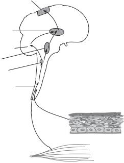
and spinal nerve fibers. The cranial nerve fibers contribute to the vagus nerve and regulate the contractility of smooth muscle cells. The spinal fibers control the movement of the neck and shoulder muscles. Cranial nerve XII (hypoglossal nerve) controls the contraction of the tongue and the geniohyoid muscles.
Cerebral Circulation. There is a rich network of blood vessels in the brain. The main arteries that supply blood to the brain are the left and right common carotid arteries, which originate from the aortic arch, and the left and right vertebral arteries, which originate from the subclavian arteries. Each common carotid artery is divided into the internal and external arteries before entering the cranial cavity. The internal carotid arteries enter the cranial cavity and supply blood to the brain, whereas the external carotid arteries supply blood to the facial tissues. The vertebral arteries enter the cranial cavity and join together to form the basilar artery. The internal carotid arteries and the basilar artery join together to form the cerebral arterial circle. Branches from the cerebral arterial circle supply blood to the brain. The cortex of each hemisphere is supplied by the anterior, middle, and posterior cerebral arteries from the cerebral arterial circle.
Arteries are eventually divided into numerous capillaries. The endothelium of the capillaries in the brain possesses well-developed tight junctions, constituting the blood– brain barrier. This barrier allows only lipid-soluble substances, such as CO2, CO, ethanol, and nicotine, to freely pass. Small water-soluble substances, such as amino acids and glucose, can be transported across the blood–brain barrier via active energy-consuming processes. Large molecules and particles, such as viruses and bacteria, cannot pass the blood–brain barrier under physiological conditions.
Blood from the brain is collected by small veins from the capillary network. The small veins conduct blood to a number of cranial venous sinuses. There are several small and medium-sized sinuses, including the superior sagittal, inferior sagittal, straight, cavernous, and occipital sinuses. Venous blood from these sinuses converges to larger sinuses, including the transverse and sigmoid sinuses, which drain blood into the internal jugular vein. Venous blood eventually returns to the right atrium via the subclavian and brachiocephalic veins and the superior vena cava.
Meninges and Cerebral Ventricles. Meninges are membranes of connective tissue, which cover and protect the brain and spinal cord. There are three meningeal layers: the dura mater/periosteum, arachnoid mater, and the pia mater. The dura mater is the outmost layer, which folds and extends into the brain fissues at three locations: the falx cerebri (between the two cerebral hemispheres), tentorium cerebelli (between the cerebrum and cerebellum), and falx cerebelli (between the two cerebellar hemispheres). The dura mater is attached to the periosteum of the cranial cavity, forming an integrated functional layer. The arachnoid mater is a thin membrane, which is separated from the dura mater by the subdural space filled with serous fluid. The pia mater is a membrane that is tightly attached to the brain. The pia mater is separated from the arachnoid mater by the subarachnoid space, which is filled with fluid and contains blood vessels.
The brain contains a number of cavities. These include the two lateral ventricles (one in each cerebral hemisphere), the third ventricle, and the fourth ventricle. The four ventricles are connected via two channels. The two lateral ventricles and the third ventricle are connected by a short channel known as the interventricular foramina. The third and fourth ventricles are connected by a long channel called the cerebral aqueduct. The fourth ventricle is continuous with the subarachnoid space and the spinal cord.
508 NERVOUS REGENERATIVE ENGINEERING
The brain ventricles, spinal cord, and subarachnoid space are filled with cerebrospinal fluid, which is similar in composition to the serum except that the cerebrospinal fluid contains a very low concentration of proteins. The cerebrospinal fluid is produced in the ventricles by a structure known as the choroid plexus, which contain capillaries and connective tissue covered by ependymal cells. The cerebrospinal fluid flows through the ventricles, leaves the fourth ventricle via several apertures, including the median aperture and two lateral apertures, flows through the subarachnoid space, and enters the dural venous sinuses, where cerebrospinal fluid joins the blood.
Spinal Cord. The spinal cord is a part of the central nervous system and extends from the brainstem to the vertebral column. At each transverse level, the spinal cord is composed of central gray matter and peripheral white matter. The gray matter consists of neurons, glial cells, and nerve axons, whereas the white matter consists of nerve axons. Spinal nerves arise from a large number of rootlets of the gray matter symmetrically at two dorsal and two ventral sites. Nerve fibers from several rootlets combine to form a nerve bundle, which leaves the spinal cord through the vertebral column. The spinal cord is vertically composed of several segments, known as the cervical, thoracic, lumbar, and sacral segments. The spinal cord gives rise to 31 pairs of spinal nerve bundles. Each pair of nerve bundles leaves the vertebral column at a corresponding vertebra. These nerves innervate peripheral tissues and organs.
At each level, the gray matter of the spinal cord is divided into two functional units: sensory and somatic motor control units. The sensory unit is located in the two dorsal sites symmetrically, whereas the somatic motor control unit is located in the two ventral sites. The dorsal nerve bundles contain input nerve fibers that transmit sensory signals from the peripheral receptors to the dorsal sensory ganglia, whereas the ventral nerve bundles contain output nerve fibers that transmit signals from the ventral motor control centers to the peripheral muscles. While the somatic motor control neurons are located within the gray matter, the sensory receiving and processing neurons are located in the dorsal root ganglia, which is located outside the spinal cord. The sensory neurons transmit signals from the ganglia to the dorsal gray matter of the spinal cord.
The Peripheral Nervous System. The peripheral nerve system is composed of neurons and nerve bundles outside the brain and spinal cord. A peripheral nerve bundle contains several structures: Schwann cell-enclosed nerve axons, blood vessels, and connective tissue. Each Schwann cell/axon bundle is enclosed with a connective tissue membrane known as endoneurium. A group of axons is enclosed within a perineurium membrane, forming a nerve fascicle. A number of axon fascicles are enclosed within an epineurium sheath, forming a nerve bundle. The connective tissue membranes protect the nerve fibers from injury.
The peripheral nerve fibers from the spinal cord are divided into various groups, based on the origin of the nerves and region of innervation. Each group is defined as a plexus, which contains several nerve bundles from a number of vertebrae. Major nerve plexuses include the cervical, brachial, thoracic, lumbosacral, coccygeal plexuses. The cervical plexus contains nerves from cervical vertebrae 1–4 and innervates the head and neck skin, muscle, and tissue. The brachial plexus originates from cervical vertebra 5 and thoracic vertebra 1, and innervates the upper limbs and part of the head. The thoracic plexus originates from thoracic vertebrae 1–12, and innervates the chest skin, muscle, and tissue. The lumbosacral plexus is from lumbar vertebrae 1–4 and sacral vertebrae 1–4, and innervates
510 NERVOUS REGENERATIVE ENGINEERING
mechanoreceptors respond to mechanical stimuli, such as tension, compression, and shearing, and are found in the epidermal tissue, muscle, inner ear, and internal organs. These receptors are responsible for the sensing of mechanical contacts and air vibrations (sounds). The chemoreceptors sense chemical stimuli, are responsible for the activities of smelling and tasting, and are found in the epidermal tissue. The thermoreceptors are responsible for the sensing of temperature and are found in the epidermal tissue. The photoreceptors sense light and are found in the retina. The pain receptors sense mechanical, chemical, and thermal stimuli and are found in almost all types of tissues.
The sensory signals sensed by the receptors are transmitted to the central nerve system via sensory nerves. There are various sensory pathways in the spinal cord and brainstem. These pathways are composed of neurons that are specific to sensory receptors. Each type of neuron is responsible for the transmission of a specific sensory signal. There are several ascending sensory pathways that transmit signals from the spinal cord to various parts of the brain. These include the spinothalamic pathway (transmitting pain, temperature, light, and mechanical signals from the spinal cord to the thalamus), the dorsal-column/medial– lemniscal pathway (transmitting proprioception, pressure, and vibration signals from the spinal cord to the brainstem), the spinocerebellar pathway (transmitting proprioception signals from the spinal cord to the cerebellum), and the spinoreticular pathway (transmitting tactile signals from the spinal cord to the reticular formation of the brainstem).
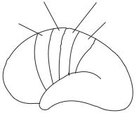
Sensory signals from the peripheral nerve system and the spinal cord are eventually converged to the primary sensory areas of the cerebral cortex. Each type of sensory input is projected to a designated cortical area. For instance, the somatic cortical area is located in the middle region of the cerebral cortex. Other regions are illustrated in Fig. 13.7. This area is organized in relation to the distribution of the peripheral tissues and organs. The sensory inputs from the head and face are transmitted to the inferior cortical area, whereas the sensory inputs from the lower limbs are projected to the superior cortical area. The sensory inputs from other organs are projected to corresponding areas between the head and the lower limbs.
Motor Control. The nervous motor system controls the contractile activities of the skeletal muscle cells by emitting action potentials and thus regulates the movement, posture, and balance of the body. There are two types of movements: involuntary and voluntary. An involuntary movement is controlled by the spinal cord motor centers and includes
Primary motor area |
Somatic sensory area |
Supplementary |
Somatic |
motor area |
association area |
Figure 13.7. Schematic representation of the distribution of the motor and sensory control regions in the cortex. Based on bibliography 13.1.
ANATOMY AND PHYSIOLOGY |
511 |
|
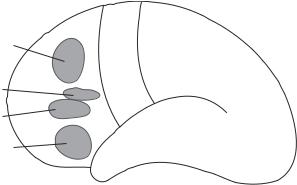
Legs |
|
|
Feet |
|
Hand activities |
Arms |
|
Fingers |
||
|
||
|
Neck |
|
Head movement |
Lips |
|
Tongue |
||
|
||
Eye movement |
|
|
Word formation |
|
Figure 13.8. Schematic representation of the distribution of the cortical motor control regions. Based on bibliography 13.1.
primarily reflexes of the skeletal muscle system. The reflex movement is not initiated through conscious judgment by the cortex. It should be noted that the heart beating, gastrointestinal motion, and blood vessel contraction are also involuntary movements, but these are controlled by the parasympathetic nerve system, not by the motor system. In contrast, a voluntary movement is consciously initiated and controlled by the cortex to achieve specified goals, such as running, walking, eating, writing, and talking.
Voluntary movements are induced in response to the action potentials initiated from the primary motor area located anterior to the primary sensory region of the cortex (Fig. 13.8). The primary motor area is organized in relation to the distribution of the peripheral tissues and organs. The action potentials initiated from the superior region of the primary motor area controls the muscles of the lower limbs, whereas the action potentials from the inferior area of the cortex controls the muscles of the head, face, and upper limbs. There is another cortical area, called premotor area, which is located anterior to the primary motor area and regulates the coordination of various muscles, ensuring accurate movements. The third important motor-control area is the prefrontal area, which is responsible for the regulation of moodand emotion-related movements.
The motor-control signals or action potentials are transmitted from the cortical motor control centers to the brainstem or spinal cord, and then to the peripheral skeletal muscles via motor nerves (Fig. 13.9). The motor nerves are divided into direct and indirect descending nerves. The direct nerves are responsible for the control of accurate muscle contraction, muscle tone, skilled movements, and emotion-related facial movements. The indirect nerves control less accurate muscle activities related to overall body movements. The direct nerves exist only in mammals.
Autonomic Control of Vital Activities. Vital activities include the heartbeat, regulation of bloodflow and pressure, and temperature regulation. These activities are primarily controlled by two nervous systems: the sympathetic and parasympathetic nervous systems.
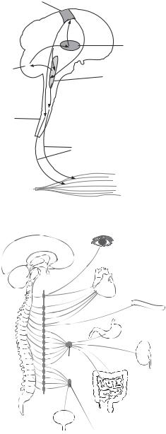
Sympathetic Nervous System. The sympathetic nerve system is composed of a number of control units. A typical sympathetic control unit consists of preganglionic neurons and postganglionic neurons (Fig. 13.10). The preganglionic neurons are located in the gray matter of the spinal cord in the region from thoracic vertebra 1 to lumbar vertebra 2. The postganglionic neurons are located in either a structure near the vertebrate column known
512 NERVOUS REGENERATIVE ENGINEERING
Cortical motor center
Thalamus
Cerebellum
Bulboreticular formation
Spinal cord
Nerves to skeletal muscle cells
Skeletal muscle
Figure 13.9. Schematic representation of the pathways of motor control signals. Based on bibliography 13.1.
Eye

 Heart
Heart
Blood vessel
Stomach
Celiac
ganglion
Kidney
|
|
Intestines |
|
Sympathetic |
Bladder |
Hypogastric plexus |
|
chain |
|||
|
|
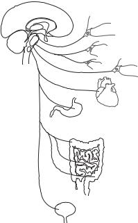
Figure 13.10. Schematic representation of the sympathetic nervous system. Based on bibliography 13.1.
as the sympathetic chain ganglia or a structure near the target tissues known as the collateral ganglia. The axons of preganglionic neurons join the spinal nerves, leave the spinal cord, and extend to the sympathetic chain ganglia. In the sympathetic chain ganglia, the sympathetic nerves are organized into four different groups: the spinal, sympathetic, splanchnic, and adrenal gland nerves.
ANATOMY AND PHYSIOLOGY |
513 |
For the spinal group, the preganglionic axons synapse with the postganglionic neurons in the sympathetic chain ganglia. The postganglionic axons leave the sympathetic chain ganglia, join the spinal nerves, and innervate peripheral tissues and organs. For the sympathetic group, the preganglionic axons synapse with the postganglionic neurons in the sympathetic chain ganglia. The postganglionic axons leave the sympathetic chain ganglia, form sympathetic nerves, and innervate peripheral tissues and organs. The heart is a major target of the sympathetic nerves. Activation of the sympathetic nerves induces an increase in cardiac contractility and the heartbeat. For the splanchnic group, the preganglionic axons enter the sympathetic chain ganglia, but do not form synapses there. They leave the sympathetic chain ganglia, form the splanchnic nerves, extend to the peripheral organs, and enter another sympathetic structure known as the collateral ganglia, where they synapse with postganglionic neurons. The postganglionic axons innervate target tissues, including the stomach and intestines. The adrenal gland nerves are different form others. The preganglionic axons do not synapse with postganglionic neurons, but directly extend to the adrenal gland and synapse with the adrenal medullar cells, stimulating the secretion of epinephrine and norepinephrine.
Parasympathetic Nervous System. This is another nervous system that controls the vital activities. As the sympathetic nervous system, the parasympathetic nervous system is composed of a number of control units. Each unit consists of preganglionic and postganglionic neurons (Fig. 13.11). The preganglionic neurons are located in the brainstem, and the postganglionic neurons are located in a structure near or within the target tissues or organs, known as the terminal ganglia. The preganglionic axons leave the brainstem, extend to and enter the terminal ganglia, where they synapse with the postganglionic
Ciliary ganglion
Sphenopalatine ganglion
Optic ganglion
Heart
Stomach
Intestine
Bladder
Figure 13.11. Schematic representation of the parasympathetic nervous system. Based on bibliography 13.1.
514 NERVOUS REGENERATIVE ENGINEERING
neurons. The postganglionic axons leave the terminal ganglia and innervate target tissues and organs.
Autonomic Regulation of Cardiovascular Functions. The activities of the heart and blood vessels are controlled by the sympathetic and parasympathetic nervous systems in a coordinated manner. The heart is innervated with both sympathetic and parasympathetic (vagus) nerves. Arteries and veins are primarily innervated with sympathetic nerves. Certain structures, such as the baroreceptors, which attach to the carotid arteries, are innervated with parasympathetic vagus nerves and play a critical role in regulating the cardiovascular activities.
The regulation of the cardiovascular functions is accomplished via autonomic reflexes, or nerve-controlled actions in response to peripheral inputs. In general, the sympathetic nerve system activates, whereas the parasympathetic system inhibits the cardiovascular activities. The two systems regulate the cardiovascular activities in coordination and counterbalance each other’s effects. An increase in the activity of the sympathetic system will activate the parasympathetic system via a feedback mechanism, and vice versa. Thus the cardiovascular activities are maintained within a relatively stable range, which is defined as the physiological range.
A typical example is baroreceptor-mediated regulation of arterial blood pressure. The baroreceptors detect changes in blood pressure. An increase in blood pressure beyond the physiological level stimulates the baroreceptors. Signals from the baroreceptors are transmitted to the brainstem via the vagus nerve and activate the parasympathetic cardiovascular control neurons. The parasympathetic signals are transmitted to the heart, inducing a decrease in the heartbeat and contractility. As a result, arterial blood pressure decreases. In contrast, a decrease in arterial blood pressure to a level below the physiological level will reduce stimulation to the baroreceptors as well as to the parasympathetic control neurons, resulting in a situation with relatively dominant sympathetic activities. Thus, heartbeat and cardiac contractility both increase simultaneously, leading to an increase in blood pressure.
NERVOUS DISORDERS
Nerve Injury
Etiology, Pathology, and Clinical Features [13.2]
Brain Injury. Brain injuries can be caused by head trauma and skull fractures resulting from physical impacts or penetrations. An injury beyond the tolerant level can cause changes in the structure and function of the brain at the injury site. Open trauma can cause hemorrhage, which significantly compromises the brain function at the hemorrhage site, often leading to dysfunction of the corresponding peripheral systems. Another common type of brain injury is concussion, which is defined as mild reversible brain injury with transient amnesia and loss of consciousness. This type of injury is often caused by blunt impacts on or sudden deceleration of the brain. Usually, concussion is diagnosed when no visible brain damage and hemorrhage are observed. The pathogenic mechanisms for the loss of consciousness are related to the dysfunction of the reticular system in the brainstem due to sudden rotation or movement of the brain. The mechanisms for amnesia are related to the injury of the cerebrum.
NERVOUS DISORDERS |
515 |
Severe head impact or deceleration causes brain contusion, which is often associated with various degrees of apparent brain injury, ranging from small superficial hemorrhage to large area necrotic destruction of the brain structure. The site and degree of brain injury can be detected by CT scan or MRI. This type of injury is often associated with prolonged loss of consciousness and amnesia. Clinical signs are dependent on the injury location and size. In certain cases, diffusive edema may occur within a short period following the trauma due to alterations in blood pressure (hypertension) and microcirculatory function. In addition, glial cells and leukocytes migrate to the injury and hemorrhage sites within hours to days and generate extracellular matrix components, eventually resulting in the formation of fibrous tissue or scars. The scars in the brain are often the causes of posttraumatic epilepsy.
Cranial Nerve Injury. Head trauma, especially basilar skull fractures, is often associated with injury of the cranial nerves, such as the optic, trochlear, olfactory, trigeminal, facial, and auditory nerves. Nerve injuries are often induced by shearing deformation of the skull. In severe cases, the nerve fibers can be completely severed. Nerve injury often induces degradation of the distal nerve fibers in association with biochemical changes, such as reduction in the expression of synaptophysin, which is an axonal marker (Fig. 13.12). Clinical signs are dependent on the type of nerve that is injured and the degree of the injury. For instance, minor injury of the optic nerves may cause blurring of vision, whereas bruising and transecting of the optic nerves will result in partial or complete blindness.
Spinal Cord Injury. Spinal cord injury can be induced by damage to, fracture, or dislocation of the vertebral column. The thoracic spinal cord is often injured by vertical compression. The cervical spinal cord can be injured by flexion. Spinal cord injury can be detected by imaging approaches such as X-ray and MRI. Spinal cord injuries result in functional changes in peripheral sensation and motor control. In severe cases, paralysis occurs in areas below the injured spinal cord.
Immediately following spinal cord injury, hemorrhage may occur, which significantly affect the function of the spinal cord neurons. Other changes include regional spinal cord edema and ischemia during the early period (about 4 h). Peripheral signs, such as demyelination of the peripheral nerves (Fig. 13.13) and regional loss of sensory input and movement, may be observed during this period. Spinal cord injury is usually reversible during the early period. Global infarction or necrosis occurs at the injury site about 8 h after spinal cord injury. Such injury is associated with complete paralysis below the injury site. Complete spinal cord transection or global spinal cord infarction is usually irreversible. At the injury site, glial cells and leukocytes are often activated. These cells can migrate to the damaged tissue and generate extracellular matrix, eventually forming fibrous tissue or scars.
EXPERIMENTAL MODELS OF SPINAL CORD INJURY. Spinal cord injury is often created in small rodents and used to investigate the mechanisms of nerve injury and regeneration. Several types of experimental models have been developed and reported in the literature. These include complete and partial spinal cord transection, blunt contusion, and compression. In the spinal cord transection model, the spinal cord is completely transected at a selected location, or selected nerve tracts in the spinal cord are transected. Neuronal growth and axonal regeneration can be observed after transection. In the blunt contusion model, a weight impactor can be dropped from a desired height to a segment of exposed