

Bioregenerative Engineering Principles and Applications - Shu Q. Liu
..pdf586 CARDIAC REGENERATIVE ENGINEERING
pulmonary valves. The mitral valves divide the left atrium and the left ventricle. They are open during the diastole, allowing blood entering the left ventricle. They are closed during the systole, preventing blood from regurgitation into the left atrium. The tricuspid valves separate the right atrium from the right ventricle, and have function similar to that of the mitral valves. Both mitral and tricuspid valves are referred to as atrioventricular valves. The aortic valves separate the left ventricle from the ascending aorta. These valves are open during systole, allowing blood to enter the aorta. They are closed during the diastole, preventing blood from flowing back to the left ventricle. The pulmonary valves separate the right ventricle from the pulmonary arterial trunk, and have function similar to that of the aortic valves. The aortic and pulmonary valves are referred to as arterial valves. The driving forces for the opening and closure of the cardiac valves are blood pressure gradients across the valves.
The heart is composed of a blood circulatory system, known as the coronary circulation. This system consists of coronary arteries, capillaries, and veins. Two major coronary arteries, the left and right coronary arteries, stem from the ascending aorta and supply blood to the left and right heart, respectively. The left coronary artery is divided into two major branches: the anterior descending artery and the left circumflex artery. The anterior descending artery supplies blood to the anterior wall of the left heart. The left circumflex artery supplies blood to the lateral and posterior wall of the left heart. The right coronary artery is divided into two major branches: the posterior descending artery and the right circumflex artery, which supply blood to the posterior and lateral wall of the right heart, respectively.
The heart consists of a rich capillary network to accommodate its high oxygen consumption activities. Capillaries are distributed within the cardiac muscular bundles. Deoxygenated blood is drained to the coronary venous system. Blood from the left heart is drained to a coronary vein known as the great cardiac vein, whereas blood from the right heart is drained to the small cardiac vein. These veins converge to the largest coronary vein, known as the coronary sinus, which empties blood into the right atrium.
Cardiac Cells
The heart is composed of a number of cell types, including cardiomyocytes, conducting cells, fibroblasts, epithelial cells, and vascular cells. Cardiomyocytes are elongated, multinucleated, striated, and involuntary contractile muscle cells. These cells consist of actin–myosin filament-containing sarcomeres, the fundamental units of cardiac contraction. Actin and myosin filaments are contractile structures, and their interactions induce sliding motions between the two types of filament, resulting in the contraction of the cardiomyocytes. The ordered arrangement of actin and myosin filaments gives the cardiomyocytes a striated pattern. Other subcellular structures of the cardiomyocytes are similar to those found in other cell types. Cardiomyocytes are organized into muscular bundles, which are further arranged with optimal alignments for the generation of contractile forces.
Cardiac conducting cells are specially differentiated cardiac muscle cells and are found in three structures: the sinoatrial (SA) node, the atrioventricular (AV) node, and the conducting bundles. The sinoatrial node is located at the right atrium near the root of the superior vena cava. The atrioventricular node is located at the right atrium near the intercept of the atrial and ventricular septa. The conducting bundle, known as the
ANATOMY AND PHYSIOLOGY OF THE HEART |
587 |
atrioventricular bundle, relays action potentials from the atrium to the ventricle. This bundle is divided into the left and right bundle branches. The left bundle branch is further divided into various generations of branches, which innervate the left ventricle. Similarly, the branches of the right bundle branch innervate the right ventricle. The terminal branches of the bundle branches are called Purkinje fibers, which conduct action potentials to the ventricular muscle cells. The conducting cells in the sinoatrial node, atrioventricular node, and conducting branches undergo automatic depolarization and initiate periodic action potentials, which induce cyclic cardiac contraction and relaxation. The conducting cells in these structures emit cyclic action potentials at different frequencies with the highest frequency found in the sinoatrial node (70–80 per minute). Thus, the action potentials from the sinoatrial node override those from other conducting structures and control the frequency of cardiac muscle contraction and heartbeat.
The heart also consists of fibroblasts. These cells are found in the connective tissue, which resides in extramuscular space, and are capable of producing and secreting extracellular matrix components, including collagen, elastin, and proteoglycans. Other cell types found in the heart include epithelial cells and vascular cells. Epithelial cells are the cells that line the internal and external surfaces of the atrial and ventricular wall and the surface of the valves. Vascular cells include endothelial cells and smooth muscle cells, which are found in the coronary blood vessels. Endothelial cells line the internal surface of blood vessels, including arteries, capillaries, and veins. Smooth muscle cells are found in the media of the arteries and veins. The structure and function of vascular cells are discussed further on page 660.
Cardiac Performance and Cycle
The heart conducts cyclic contractile activities and pumps blood into the arterial system. The cyclic activities include two mechanical events: contraction and relaxation (Fig. 14.2). Contraction occurs in a phase called systole, which lasts about 0.3–0.4 s in healthy humans, and relaxation occurs in another phase called diastole, which lasts about 0.4– 0.5 s. Systole is defined as the period from the beginning to the end of ventricular contraction, and the diastole is from the beginning to the end of ventricular relaxation. Systole and diastole constitute a cardiac cycle. A cardiac cycle is defined from the beginning of ventricular contraction to the ending of ventricular relaxation or the beginning of next ventricular contraction. Since action potential waves recorded by electrocardiography are consistent with cardiac mechanical events, these waves are usually used to identify cardiac cycles. The QRS wave complex, which reflects the spreading of action potentials within the ventricles and initiates ventricular contraction, is used to indicate the beginning of the systole and the ending of the diastole, although the beginning of the QRS wave complex is slightly earlier than the beginning of ventricular contraction.
A sequence of mechanical events occurs during the systole. When the ventricular muscle cells are excited by action potentials from the sinoatrial node, the ventricular muscle cells start contraction almost at the same time (owning to the fast-conducting activity of the Purkinje fibers). The contraction of the left ventricle rapidly increases ventricular blood pressure from about 5 to 120 mm Hg. The increased ventricular blood pressure rapidly closes the mitral valves due to the pressure gradient across the valves. There is a short period when the mitral valves are closed, but the aortic valves are not yet open. This period is known as the period of isometric contraction, since blood is not flowing from or to the ventricle. A continuous increase in the ventricular blood pressure results

588 CARDIAC REGENERATIVE ENGINEERING
Hg) |
|
Systole |
Diastole |
|
|
|
|
|
|
|
|
mm( |
120 |
|
|
Aortic pressure |
|
|
|
|
|||
|
|
|
|
||
Pressure |
80 |
|
|
|
|
40 |
|
LV pressure |
|
||
|
|
|
|||
|
|
|
|
|
|
|
0 |
LA pressure |
|
|
|
(ml) |
120 |
|
|
||
|
|
|
|
||
|
|
|
|
|
|
Volume |
|
|
|
LV volume |
|
80 |
|
|
|
|
|
|
|
|
|
|
|
|
40 |
|
|
|
|
ECG |
|
|
|
|
|
|
0 |
0.2 |
0.4 |
0.6 |
0.8 |
|
|
|
Time (Seconds) |
|
|
Figure 14.2. Electrical and mechanical events during a cardiac cycle. Based on bibliography 14.1.
in a pressure gradient across the aortic valves, leading to the opening of these valves and blood ejection from the left ventricle to the aorta. A similar sequence of mechanical events occurs in the right ventricle, except that blood pressure in the right ventricle is lower than that in the left ventricle. At the same time, the left and right atria are in the relaxing mode. The two atria collect blood from the pulmonary veins and vena cava, respectively. Approaching the end of ventricular contraction, the ventricles reduce contractile strength in association with a significant decrease in the ventricular blood pressure and volume. At the point when the ventricular blood pressure reaches a level just below the aortic blood pressure (for the left ventricle) and the pulmonary arterial blood pressure (for the right ventricle), the aortic and pulmonary valves are closed owning to the reversed pressure gradients across these valves. The closure of the aortic and pulmonary arterial valves indicates the ending of the systole and the beginning of the diastole.
During early diastole following the closure of the arterial valves, there is a period when the ventricular blood pressure is higher than that in the atria and the atrioventricular valves (mitral and tricuspid valves) are still closed. This period is called the period of isometric relaxation, during which blood is not flowing from or to the ventricles. Continuous ventricular relaxation results in a rapid decrease in the ventricular blood pressure. Soon, when the ventricular blood pressure drops below the atrial blood pressure, the atrioventricular valves are open, owing to the reversed pressure gradients across these valves, and blood flows from the left and right atria to the left and right ventricles, respectively. Two factors facilitate ventricular blood filling: continuous expansion of the ventricles and atrial contraction, which occurs in the late stage of diastole. The ventricles are completely filled before the next QRS-exciting wave complex arrives at the ventricle and initiates another cardiac cycle.
CARDIAC DISORDERS |
589 |
Regulation of Cardiac Performance
Cardiac activities undergo constant changes in response to various demands of bloodflow and oxygen consumption in the peripheral tissues and organs. A typical example is the increase in cardiac contraction and heartbeat in response to increased physical activities such as running and other forms of exercise. The change in cardiac activity is regulated by two mechanisms: intrinsic and extrinsic. The intrinsic mechanism is controlled by the natural properties of the cardiomyocytes, such as the length of the sarcomere or the fraction of the actin filaments that overlap with the myosin filaments. The contractile strength is proportional to the sarcomere length within a limited range. The muscle generates maximal forces at an optimal sarcomere length. With a further stretch or lengthening of the sarcomere, the contractile strength reduces. Such properties are described by Starling’s law, which is represented by a sarcomere length–tensile force curve. Under physiological conditions, the heart works in the ascending range of the sarcomere length–tensile force curve; that is, an increase in sarcomere length results in enhanced cardiac contraction. In a beating heart, the sarcomere length of the ventricular muscles is controlled by the diastolic blood volume of the ventricle. An increase in the diastolic blood volume results in an increase in the sarcomere length within a certain range.
The extrinsic mechanism is controlled by neural and hormonal factors. The heart is innervated with parasympathetic and sympathetic nerves. Activation of the parasympathetic nervous system suppresses the cardiac activity or contractility, whereas activation of the sympathetic nervous system exerts an opposite effect. The two nerve systems coordinately control the performance of the heart. In addition, the performance of the heart is regulated by hormones, including epinephrine and norepinephrine. Epinephrine and norepinephrine are produced by the adrenal medulla and released into the blood. The secretion of these hormones are controlled by the sympathetic nervous system and elevated in response to increased physical activities and emotional changes. The function of these hormones, especially epinephrine, is to stimulate cardiac contraction. These hormones can bind to and activate the β-adrenergic receptors in the membrane of the cardiomyocytes, inducing the activation of G-protein signaling pathways (see page 217). As a result, calcium is released into the cytoplasm. As calcium is required for the initiation of actin– myosin interactions, an increase in calcium concentration enhances actin–myosin interactions and the muscular contractile strength.
CARDIAC DISORDERS
Heart Failure and Cardiomyopathy
Pathogenesis, Pathology, and Clinical Features of Heart Failure [14.2]. Heart failure is a pathophysiological state in which the heart is unable to pump sufficient blood to the arterial system, resulting in the dysfunction of tissues and organs due to the lack of blood supply. Heart failure is an end event of cardiac and pulmonary disorders, which are among the most popular diseases. Each year about 960,000 patients are admitted to hospital because of heart failure in the United States. About 20% patients are associated with lifethreatening heart failure. Heart failure can be induced by a variety of disorders, including cardiac infarction, congenital heart disease, cardiomyopathy, myocarditis, anemia, hemorrhage, cardiac trauma, cardiac valve rupture, cardiac valvular diseases, arrhythmia, systemic hypertension, and excessive physical exercise. Although these disorders involve

590CARDIAC REGENERATIVE ENGINEERING
A.Ventricular remodeling after acute infarction
Initial infarct |
Expansion of |
Global remodeling |
|
infarct (hrs to days) |
(days to months) |
B. Ventricular remodeling in diastolic and systolic heart failure
Normal heart |
Hypertrophied heart |
Dilated heart |
|
(diastolic heart failure) |
(systolic heart failure) |
Figure 14.3. Ventricular remodeling after infarction (panel A) and in diastolic and systolic heart failure (panel B). At the time of an acute myocardial infarction, there is no clinically significant change in overall ventricular geometry (panel A). Within hours to days, the area of myocardium affected by the infarction begins to expand and become thinner. Within days to months, global remodeling can occur, resulting in overall ventricular dilatation, decreased systolic function, mitral valve dysfunction, and the formation of an aneurysm. The classic ventricular remodeling that occurs with hypertensive heart disease (middle of panel B) results in a normal-sized left ventricular cavity with thickened ventricular walls (concentric left ventricular hypertrophy) and preserved systolic function. There may be some thickening of the mitral valve apparatus. In contrast, the classic remodeling that occurs with dilated cardiomyopathy (right side of panel B) results in a globular shape of the heart, a thinning of the left ventricular walls, an overall decrease in systolic function, and distortion of the mitral valve apparatus, leading to mitral regurgitation. (Reprinted with permission from Jessup M, Brozena S: New Engl J Med 348:2007–18, copyright 2003 Massachusetts Medical Society. All rights reserved.)
different systems and are caused by different pathological factors, the consequence of these disorders is the impairment of cardiac contractility, eventually leading to cardiac failure (Fig. 14.3).
At the molecular and cellular levels, cardiac disorders may induce apoptosis of cardiomyocytes and/or reduction in cardiac contractility due to the impairment of calcium transport and handling. Cell apoptosis results in reduction in the density of cardiomyocytes, which impairs cardiac performance. Cardiac ischemia and infarction are typical cardiac disorders that induce cell apoptosis. The impairment of calcium transport is a primary cause for nonapoptotic heart failure. Calcium plays a critical role in the regulation of muscular contraction. Calcium is primarily stored in a subcellular structure known as
CARDIAC DISORDERS |
591 |
sarcoplasmic reticulum (SR). In patients with heart failure and experimental heart failure models, the load of calcium in the sarcoplasmic reticulum is often reduced. Thus, calcium released to the cytoplasm, in response to an action potential, is insufficient to induce optimal actin–myosin interaction, resulting in impaired cardiac performance. The reduction in the load of sarcoplasmic reticulum calcium is related to malfunction of several molecules, including sarcoplasmic reticulum calcium ATPase (SERCA2a), phospholamban (PLB), and/or the Na+ –Ca2+ -exchanger (NCX), which participate in the regulation of calcium handling and transport.
In response to the stimulation of an action potential, cardiomyocytes undergo depolarization, which stimulates the cell membrane voltage-dependent calcium channels to induce calcium influx. The increase in the cytoplasmic calcium concentration activates the ryanodine receptor and induces the transport of additional calcium from the sarcoplasmic reticulum to the cytoplasm. Released calcium binds to troponin C, which in turn initiates actin–myosin interaction and muscle cell contraction. Immediately after the contraction process, calcium must be removed from the cytoplasm, allowing the relaxation of the muscular cells. The transport of calcium from the cytoplasm to the sarcoplasmic reticulum is controlled primarily by the sarcoplasmic reticulum Ca2+ ATPase, while calcium transport from the cytoplasm to the extracellular space is controlled by the Na+ –Ca2+ exchanger. A reduction in the activity of sarcoplasmic reticulum calcium ATPase is thought to contribute to the reduction in the sarcoplasmic reticulum calcium load and the impairment of cardiac contractility, leading to the development of heart failure.
Cardiac contractility and performance are also regulated by the β-adrenergic receptor signaling system. These receptors interact with catecholamines, including epinephrine and norepinephrine, and stimulate the contraction of cardiac muscle cells. The binding of catecholamine ligands to the β-adrenergic receptor activates the Gs proteins, which induce the activation of a cascade of signaling molecules, including adenylyl cyclase, cAMP, and protein kinase A (PKA). Activated protein kinase A can phosphorylate downstream proteins that regulate the contractile activity of the cardiac muscle cells. One of the substrate molecules for protein kinase A is the cardiac ryanodine receptor, which controls the gating the calcium channels in the sarcoplasmic reticulum. The phosphorylation of the ryanodine receptor promotes diastolic calcium release from the sarcoplasmic reticulum, initiating cardiomyocyte contraction.
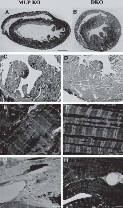
Furthermore, activated protein kinase A by the β-adrenergic receptor can phosphorylate a molecule known as phospholamban. In its dephosphorylated form, phospholamban inhibits the activity of sarcoplasmic reticulum Ca2+ ATPase. The PKA-induced phosphorylation of phospholamban results in the deinhibition of the sarcoplasmic reticulum Ca2+ ATPase activity. Thus, protein kinase A-induced phosphorylation of phospholamban removes the inhibitory effect of phospholamban on sarcoplasmic reticulum Ca2+ ATPase and enhances the cardiac contractility. The accumulation of dephosphorylated phosholamban or a decrease in the phosphorylation of the molecule reduces the cardiac contractility and promotes the development of heart failure. The knockout of phospholamban in mice with transgenic heart failure can reduce ventricular dilation and cardiac fibrosis, and improves the ventricular performance (Fig. 14.4). Thus, phospholamban is a potential target molecule for the treatment of cardiac failure.
There are various types of heart failure. In terms of the rate of progression, heart failure can be classified into acute and chronic heart failure. Acute heart failure occurs suddenly, often due to large cardiac infarction, cardiac trauma, sudden cardiac valve rupture, or severe hemorrhage, resulting in a rapid reduction in cardiac output and arterial blood
CARDIAC DISORDERS |
593 |
pressure. Acute heart failure is followed with the failure of all peripheral organs within a short period. Changes in the function of peripheral organs depend on the degree of heart failure. A complete loss of arterial blood pressure due to acute heart failure may result in cell death within minutes in vital organs such as the brain, heart, and kidney. Chronic heart failure progresses slowly over months, years, or decades, and are often a result of hypertension, cardiomyopathy, and valvular diseases, such as aortic valve stenosis and mitral valve regurgitation. Hypertension and valvular disorders induce a gradual increase in the cardiac workload. The heart may initially adapt to the workload by increasing its muscle size, a process known as hypertrophy. Within a certain period (months, years, or even decades), the heart is still capable of providing sufficient bloodflow to the peripheral systems. However, when the heart reaches the limitation of its adaptive capacity, additional workload may lead to heart failure. Cardiomyopathy is a pathological disorder that gradually deteriorates the structure and function of the cardiac muscle cells, resulting in cardiac malfunction and eventually cardiac failure.
According to the structure of the heart, heart failure can be classified into left and right heart failure. Left heart failure is defined as the failure of the left ventricle, whereas right heart failure is the failure of the right ventricle. Left heart failure can be induced by left coronary arteriosclerosis and left ventricular infarction, systemic hypertension, mitral and aortic valvular diseases, and cardiomyopathy. Right heart failure is often a result of pulmonary hypertension, hypoxia, and tricuspid and pulmonary valvular diseases.
Pathological changes in heart failure often include enlargement of the heart, left and right ventricular dilation for left and right heart failure, respectively, and ventricular wall hypertrophy. Usually, before the onset of heart failure, the heart intends to compensate for the loss of its function by increasing the mass of cardiac muscle via cellular hypertrophy. Such a compensatory remodeling process results in cardiac hypertrophy. Other pathological changes may be dependent on the disease that causes heart failure.
Patients with heart failure often express a number of clinical symptoms and signs. These include dyspnea, orthopnea, paroxysmal dyspnea, cyclic respiration, fatigue, headache, anxiety, insomnia, memory impairment, an increase in heartbeat, and a reduction in blood pressure. All these symptoms and signs are due to the lack of blood supply in various organs such as the lung (dyspnea, orthopnea, paroxysmal dyspnea, and cyclic respiration) and brain (headache, anxiety, insomnia, memory impairment). The increase in heartbeat is a sign of cardiac adaptation. A decrease in blood pressure or blood supply in the peripheral tissue stimulates the sympathetic nerve system, while it suppresses the parasympathetic system, resulting in an increase in the cardiac activity.
Experimental Models of Heart Failure [14.3]. Experimental heart failure can be established by using three methods: cardiac infarction, aortic constriction, and gene knockout. Cardiac infarction can be induced by coronary arterial ligation. In an open-chest surgery, a selected coronary artery can be ligated with a suture, inducing acute myocardial infarction distal to the ligated artery. The severity of heart failure is dependent on the size or generation of the ligated coronary artery. Usually, the ligation of a major branch of the left coronary artery can induce a severe left ventricular failure. Left heart failure can also be induced by increasing the afterload (or arterial blood pressure) of the left ventricle. Such a condition can be achieved by partial constriction of the ascending aorta. The degree of change in aortic blood pressure proximal to the constriction is dependent on the level of constriction. Following the constriction surgery, left ventricular hypertrophy can be developed within days. Severe hypertension and ventricular hypertrophy can lead to heart
594 CARDIAC REGENERATIVE ENGINEERING
failure. Another method for inducing heart failure is to knock out genes involved in the regulation of cardiac performance. A typical gene is the sarcoplasmic reticulum Ca2+ ATPase gene. As discussed above, sarcoplasmic reticulum Ca2+ ATPase plays a critical role in calcium handling and cardiac muscle contraction. A decrease in the expression of the sarcoplasmic reticulum Ca2+ ATPase gene induces heart failure. For these models, the degree of heart failure can be assessed by measuring the movement of the ventricular wall and the ejection fraction of the left ventricle by magnetic resonance imaging or echocardiography.
Pathogenesis, Pathology, and Clinical Features of Cardiomyopathy [14.4]. Cardiomyopathy is a disorder that originates from the intrinsic properties of the cardiomyocytes, but not as a result of other diseases, such as coronary arteriosclerosis, congenital heart disease, valvular disease, or hypertension. This disorder is characterized by ventricular dilation and/or hypertrophy and a progressive reduction in cardiac contractility and performance, eventually resulting in heart failure. Cardiomyopathy can be divided into two different types on the basis of etiology: primary and secondary cardiomyopathy. Primary cardiomyopathy is a myocardial disorder of unknown cause. Major forms of primary cardiomyopathy include the idiopathic and hereditary cardiomyopathy. Secondary cardiomyopathy is a myocardial disorder of known cause, often induced by diseases involving other organs. Typical examples include cardiomyopathy resulting from viral, bacterial, and fungal myocarditis, metabolic disorders, electrolyte and nutritional deficiency, systemic lupus erythematosus, rheumatoid arthritis, amyloidosis, muscular dystrophy, and toxic reactions resulting from alcohol and drugs. In either primary or secondary cardiomyopathy, molecular changes described in the section on heart failure may also occur.
Cardiomyopathy is associated with a number of characteristic pathological changes. These include enlargement of the heart and dilation of the ventricles, hypertrophy of the ventricular wall, interstitial fibrosis, leukocyte infiltration, and occasionally myocardial cell death. In dilated ventricles, thrombi may be found on the wall near the apex. Ventricular dilation is induced by a decrease in the cardiomyocyte contractility, which renders the ventricle unable to pump sufficient blood into the arterial system. Excessive residual blood contributes to the dilation of the ventricles. Ventricular dilation induces extension of the sarcomeres, which enhances the cardiac performance within a certain range (see page 589 of this chapter). Cell death may be attributed to the effects of toxic, metabolic wastes and infectious factors. Interstitial fibrosis develops as a result of enhanced fibroblast proliferation and excess extracellular matrix generation in response to cell death. Ventricular hypertrophy is often a result of adaptation to reduction in cardiac contractility and performance. Cardiomyocytes increase their size to compensate the loss of cardiac performance. Leukocyte infiltration is induced by infectious factors, if any, or cell death.
The clinical manifestations of cardiomyopathy are dependent on the level of changes in cardiac function, regardless the causes of the disorder. In the early stage, the patient may not experience any noticeable symptoms. With progressive deterioration of the cardiac function, patients may show heart failure symptoms and signs, such as dyspnea, fatigue, orthopnea, lower-limb edema, and palpitation. Patients may also feel chest pain. Cardiomyopathy eventually develops to heart failure. Strategies for the treatment of cardiomyopathy include the removal of primary causes, protection of cardiomyocytes from injury, and enhancement of myocardial contractility. In the end stage, the treatment of cardiomyopathy is similar to that of heart failure.
CARDIAC DISORDERS |
595 |
Experimental Model of Cardiomyopathy [14.4]. Experimental cardiomyopathy can be established by modulating or knocking out genes encoding proteins that regulate the integrity and contractility of cardiomyocytes. Candidate genes for such a purpose include the muscle-specific lim protein (MLP), dystrophin, sarcoglycans, desmin, myosin heavychain, myosin-binding protein C genes. These genes are involved in the regulation of cardiac contractility. The modulation or knockout of these genes exhibits the phenotype of cardiomyopathy. These models are usually established in the mouse. Transgenic mice with a desired disorder may be acquired from investigators who developed the model or from commercial carriers such as the Jackson Laboratory.
Conventional Treatment of Cardiac Failure and Cardiomyopathy [14.5]. Heart failure is usually treated with several approaches, including the removal of primary causes, reduction of cardiac workload, enhancement of cardiac contractility, and control of salt and water intake. The first approach (removal of primary causes) is dependent on the original disease. Each disease should be treated with distinct methods. The reduction of the cardiac workload is often a priority for the treatment of heart failure. There are several methods that can be used for such a purpose. These include reduction in blood volume and arterial blood pressure as well as reduction in physical activities. Blood volume can be reduced by administration of diuretics, and arterial blood pressure can be reduced by administration of vasodilators. In addition, salt and water intake should be carefully controlled to reduce circulating blood volume and the workload of the heart. The enhancement of cardiac contractility can be achieved by the administration of digitalis glycosides. These compounds can act on cardiac muscle cells, increase muscular contractility, and reduce the conduction of action potentials, thus slowing down the heartbeat. A commonly used compound is digoxin. Chemical compounds that augment the sympathetic nerve function by acting on the β-adrenergic receptors, such as epinephrine and dopamine, can be used to improve the cardiac contractility.
Molecular Therapy for Cardiac Failure and Cardiomyopathy [14.6]. Although numbers of conventional approaches have been established for the treatment of heart failure, these approaches are primarily used for the relief of cardiac symptoms, but not for longterm improvement of the cardiac function. Molecular and cellular studies have demonstrated that cardiac contractile dysfunction in heart failure may be related to intrinsic defects in molecular signaling mechanisms that control the cardiac contractility and/or cell apoptosis. Thus, therapeutic modulation of the contractility-controlling molecular mechanisms and prevention of cell apoptosis may provide new approaches for the treatment of heart failure. Available experimental evidence indicates that cardiac contractility is controlled by calcium handling and transport, which are regulated by several molecules localized to the cell membrane and sarcoplasmic reticulum, including sarcoplasmic reticulum Ca2+ ATPase, phospholamban, and Na+ –Ca2+ exchanger. The engineering manipulation of the genes that encode these proteins may exert therapeutic effects on heart failure.
In cardiac failure, the expression and activity of sarcoplasmic reticulum Ca2+ ATPase are often reduced. In animal models of heart failure, the overexpression of the sarcoplasmic reticulum Ca2+ ATPase gene by gene transfer enhances the contractility of cardiomyocytes and improves the cardiac performance of failing hearts. As discussed above, phospholamban is a molecule that regulates the activity of sarcoplasmic reticulum Ca2+ ATPase. In the dephosphorylated form, phospholamban inhibits the activity of