

Bioregenerative Engineering Principles and Applications - Shu Q. Liu
..pdf596 CARDIAC REGENERATIVE ENGINEERING
sarcoplasmic reticulum Ca2+ ATPase, suppressing the sarcoplasmic reticulum calcium pump function. The phosphorylation of phospholamban, induced by the β-adrenergic receptor-activated protein kinase A, results in the deinhibition of the sarcoplasmic reticulum Ca2+ ATPase activity. In mouse models of heart failure, the knockout of the phospholamban gene enhances the cardiac contractility and improves cardiac performance, suggesting that dephosphorylated phospholamban may be dominant in failing hearts. In vitro investigations have shown that the application of antisense mRNA for phospholamban to rat and human cardiomyocytes reduces the translation of the phospholamban protein and improves the contractile performance of these cells. The transfer of a domi- nant-negative mutant phospholamban gene to hamsters with experimental cardiomyopathy enhances the performance of failing hearts. These observations suggest that the suppression of phospholamban expression may be a potential approach for the treatment of heart failure. Since the activation of the β-adrenergic receptor–protein kinase A signaling pathway induces the phosphorylation of phospholamban, which removes the inhibitory effect of phospholamban on sarcoplasmic reticulum Ca2+ ATPase, enhancement of the β- adrenergic receptor may serve as an alternative approach (see below). The Na+ –Ca2+ exchanger regulates the transport of calcium from the cytoplasm to the extracellular space. The overexpression of the Na+ –Ca2+ exchanger gene reduces the contractility of cardiac myocytes, suggesting that the suppression of the Na+ –Ca2+ exchanger may exert therapeutic effects on a failing heart.
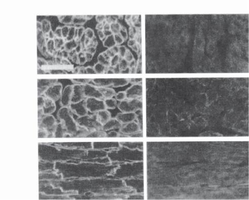
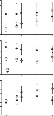
Myocardial β-adrenergic receptors play an important role in the regulation of cardiac contractility. Myocytes contain two dominant types of adrenergic receptor: β1-adrenergic receptor (accounting for 75–80%) and β2-adrenergic receptor (20–25%). The two types of receptor possess different functions. The activation of the β1-adrenergic receptors may induce cell apoptosis, whereas that of the β2-adrenergic receptors exerts a protective effect against cell apoptosis, leading to enhanced cardiac function. The different roles of the two types of receptor have been demonstrated in transgenic mice with targeted overexpression of β1- and β2-adrenergic receptors. In these mice, the overexpression of the β2-adrenergic receptor enhances the contractile performance of the cardiac muscle and prevents heart failure (Figs. 14.5 and 14.6), whereas the overexpression of the β1-adrenergic receptor facilitates heart failure. It has been suggested that the selective involvement of Gi proteins may be responsible for the different functions of the two receptors. In vivo transfer of the β2-adrenergic receptor to the animal heart has demonstrated an increase in the contractile activity of cardiac muscle cells in response to catecholamine and enhances the left ventricular function. Based on these investigations, it seems that the β2-adrenergic receptor gene may be used for the treatment of heart failure.
Tissue Regenerative Engineering for Cardiac Failure and Cardiomyopathy [14.7]. Based on the pathogenic mechanisms and clinical manifestations of cardiac failure, tissue regenerative engineering approaches have been developed for treating cardiac failure. The principle of cardiac tissue regenerative engineering is to enhance the cardiac performance. One method for such a purpose is to construct cardiomyocyte-containing tissue scaffolds, implant the constructed cardiac scaffold around the heart, and induce synchronized cardiomyocyte beating between the heart and the cardiac scaffold, thus enhancing the performance of the heart. Alternatively, the constructed cardiomyocyte-containing scaffold can be implanted around the abdominal aorta. The cyclic contractile activity of the cardiac scaffold can enhance the circulation by dynamically altering the diameter of the aorta.
CARDIAC DISORDERS |
597 |
Transgenic |
Control |
Atrium (cross)
Ventricle
(cross)
Ventricle
(long)
Figure 14.5. In situ demonstration of β2-adrenergic receptor transgene expression. Immunohistochemical labeling of the human β2-adrenergic receptor in TG4 (with transgenic overexpression of the β2-adrenergic receptor) and control myocardial frozen sections was done with rabbit antiserum to the COOH terminus of the human β2-adrenergic receptor. Atrium (cross), cross-sectioned atria; ventricle (cross), cross-sectioned ventricle; ventricle (long), longitudinal sectioned ventricle. Scale bar: 50 μm. (Reprinted with permission from Milano CA et al: Science 264:582–6, copyright 1994 AAAS.)
In an experimental model in the rat, researchers have constructed cardiac tissue scaffolds by seeding neonatal cardiomyocytes into a gel mixture containing collagen type I, matrigel, serum, and cell culture medium. The seeded cardiomyocytes can exhibit contractile activities. The matrix gel can be tailored into a desired shape according to the size and geometry of the host heart. The constructed cardiac tissue scaffold can be implanted around to host heart to cover a desired area. Following the implantation surgery, the implanted cardiac tissue scaffold can develop into a structure with organized cardiac muscle cells, which express mature cardiac protein markers such as actinin, connexin 43, and cadherins. More importantly, the implanted cardiac tissue scaffold demonstrates periodic contractile activities. Such activities can enhance the contractile performance of the host heart and reduce the pathological effect of heart failure. These investigations demonstrate that cell-based tissue regenerative engineering approaches can be used to improve the cardiac function in heart failure.
CARDIAC DISORDERS |
599 |
There are a number of vascular disorders that contribute to the pathogenesis of cardiac ischemia and infarction. These vascular disorders include coronary arteriosclerosis, thrombosis, and embolism. All these disorders block the coronary arterial bloodflow, resulting in blood and oxygen deficiency. The most common cause is coronary arteriosclerosis, a vascular disorder characterized by the presence of intimal atheroma, which grows continuously and partially or completely blocks bloodflow (see page 674 for details). Coronary arterial thrombosis is an acute, complex process involving endothelial cell injury, activation of the blood coagulation system, adhesion of platelets and leukocytes to injured endothelium, and formation of thrombi, which partially or completely block the lumen of an artery. Embolism is a condition with the arterial lumen blocked with loose thrombi detached from an upstream artery. All these vascular disorders cause similar changes in the heart.
Cardiac infarction is associated with apparent structural changes in the heart, while cardiac ischemia may not exhibit noticeable structural changes. When specimens are collected from an infracted area, one may find characteristic structural changes at different stages. In the acute stage, major structural changes include local edema, massive death of cardiac muscle cells, and leukocyte infiltration, in association with locally reduced contractile activities. If the patient survives, the dead cardiac muscles cannot self-regenerate, but are gradually replaced with proliferating fibroblasts and fibrous tissue composed of primarily collagen matrix and proteoglycans. In the late stage of cardiac infarction, a pathological examination often reveals changes seen in a typical scar tissue, including scattered fibroblasts and fibrous extracellular matrix.
The clinical manifestations of ischemic heart disease are dependent on the degree and location of arterial obstruction. When a coronary artery is partially blocked, the distal cardiac muscles may experience transient ischemia, but are not completely devoid of bloodflow. Such a condition often gives rise to a clinical syndrome known as angina pectoris. A major symptom of this syndrome is transient chest pain or discomfort. Often, pain is radiated to the left shoulder and both arms or to the back, neck, jaw, and teeth. Angina usually occurs after physical activities or emotional changes. An electrocardiographic examination often reveals ST-segment depression. In particular, a change in the ST segment after a defined level of exercise in a “stress test” provides further evidence for cardiac ischemia. A coronary angiographic test can provide convincing evidence ensuring the presence of partial arterial obstruction and cardiac ischemia.
In the case of acute cardiac infarction, patients often experience deep, heavy, crushing pain in the chest. Although the pain occurs at locations similar to those in angina pectoris, it is often more severe and lasts longer. The pain may radiate to the shoulder, neck, back, and jaw. The pain is usually associated with other symptoms and signs, such as sweating, weakness, nausea, vomiting, and sudden drop in arterial blood pressure. However, about 15% of patients with cardiac infarction may not experience any pain. Such infarction is referred to as silent infarction. This is a more dangerous situation, because patients with severe cardiac infarction can be easily ignored without prompt medical attention.
Conventional Treatment of Ischemic Heart Disease [14.8]. Acute cardiac infarction is often associated with two types of life-threatening disorder: electrical rhythmic disorder (arrhythmia) and mechanical pump failure. The principle of treating acute cardiac infarction is thus to prevent these disorders and minimize the size of cardiac infarction. Patients are always given additional oxygen to maintain an optimal level of blood oxygen to minimize the spread of cardiac infarction, in association with the administration of analgesics,
600 CARDIAC REGENERATIVE ENGINEERING
which keeps the patients calm, lowers physical activities and emotional stress, and reduces the heartbeat and oxygen consumption.
An electrocardiographic examination may reveal various forms of atrial and ventricular arrhythmia. The most serous life-threatening arrhythmias are ventricular tachycardia (heart rate >100 beats/min) and ventricular fibrillation. These forms of arrhythmia occur during the first 24 h following the onset of cardiac infarction. Often, patients are given a preventive treatment with an antiarrhythmia drug such as lidocaine. Ventricular tachycardia and fibrillation, if any, should be treated immediately by defibrillation. Another form of severe arrhythmia is sinus bradycardia (heart rate <45 beats/min). Patients may be administrated with atropine (note that atropine increases heart rate and should be used with caution). In a life-threatening case, patients should be given electrical pacing when blood pressure drops rapidly.
The impairment of the mechanical or contractile performance of an infracted heart is dependent on the size of the infract. Cardiac infarcts with a size larger than a critical level may result in heart failure. In the absence of heart failure, patients are often associated with tachycardia and an increase in arterial blood pressure as the heart intends to compensate for the lost function due to infarction. In such a case, α-adrenergic blocker should be administrated to lower the heart rate and arterial blood pressure. In the presence of heart failure, inotropic agents such as digitalis glycosides or catecholamines may be administrated to raise arterial blood pressure. However, these agents are not given for preventive purposes because they increase heart rate, cardiac contractility, and oxygen consumption, which may facilitate the spread of cardiac infarction. Other treatments described above should be applied in the presence of acute heart failure.
In a large fraction of patients with cardiac infarction, thrombus formation is a major cause of acute arterial obstruction near an atheromatic lesion. In such a case, thrombus dissolution should be carried out promptly with thrombolytic agents to reduce the arterial obstruction, introduce reperfusion, and minimize the size of cardiac infarcts. Common thrombolytic agents include streptokinase and tissue plasminogen activator. These agents can be used to effectively lyse freshly formed thrombi.
Molecular Regenerative Engineering for Ischemic Heart Disease. Molecular engineering approaches can be used to treat cardiac infarction and to improve cardiac function. The principle of cardiac molecular engineering for cardiac infarction is to prevent acute cell death, promote cell survival, protect cells from reperfusion injury, enhance angiogenesis, and improve the contractility of impaired myocardial cells. A number of molecules can be used to prevent cell death and promote cell survival. These include growth factors, such as fibroblast growth factor (FGF), platelet-derived growth factor (PDGF), epidermal growth factor (EGF), and vascular endothelial growth factor or (VEGF), antiapoptotic proteins, such as Bcl2, protein kinase B, and Akt, and inhibitors of proinflammatory cytokines. Experimental investigations have demonstrated that these factors can be used effectively to protect impaired cells from death.
Growth Factors as Therapeutic Agents for Cardiac Regenerative Engineering
fiBROBLAST GROWTH FACTORS. Fibroblast growth factors (FGFs) are a family of growth factors, including about 22 known members, including FGF1–22. Among these members, FGF1 and FGF2, also known as acidic and basic FGF, respectively, have been intensively
CARDIAC DISORDERS |
601 |
studied and characterized. Other members are discovered more recently and have become the targets of increasing investigations. The members of the FGF family share about 30– 70% identical amino acid sequence. Fibroblast growth factors play a critical role in the regulation of multiple biological processes, including cell proliferation, cell differentiation, cell migration, tissue morphogenesis, and organ formation during development and remodeling. Different FGFs exhibit distinct functions, depending on the structure of the individual members and the types of target cells. In this section, the two wellcharacterized FGFs, including FGF1 and FGF2, are discussed.
Fibroblast Growth Factor 1 (FGF1, Acidic FGF, or aFGF) [14.9]. Fibroblast growth factor 1 is a 155-amino acid protein with molecular weight of 17 kDa. This growth factor is also known as heparin-binding growth factor 1 (HBGF1), since heparin sulfate glycosaminoglycans can bind to FGF1 and facilitate the interaction of FGF1 with FGF receptor (FGFR). Fibroblast growth factor 1 shares the same gene with two growth factors known as endothelial cell growth factors α and β (ECGF α and ECGF β, respectively). The distinct forms of FGF1, ECGFα, and ECGFβ, result from different processes of posttranslational splicing. Fibroblast growth factor 1 is 14 amino acids shorter and ECGFα is 20 amino acid shorter at the N-terminus than ECGFβ. The gene for these growth factors is localized to chromosome 5 at gene locus 5q31. The amino acid sequence is highly conserved among mammals. Fibroblast growth factor 1 is expressed in several tissue types, including the central nervous system (primarily the cortex), kidney, pancreas, spleen, and skeletal muscle. This growth factor can interact with all four types of FGF receptors, including FGFR1, FGFR2, FGFR3, and FGFR4.
Fibroblast growth factor 1 exerts a mitogenic effect via the interaction with the fibroblast growth factor receptor (FGFR, see section on fibroblast growth factor receptors below for characterization of FGFR). Investigations by crystallography have demonstrated that FGF1 can interact with the extracellular immunoglobulin-like ligand-binding domain 2 and the linker between domains 2 and 3 of the FGFR. Indeed, this domain and the linker are general binding sites for all FGFs. The binding specificity of distinct FGFs is achieved via the interaction of the N-terminus of the FGFs and the immunoglobulin-like ligand-binding domain 3. This structural analysis provides a basis for understanding the interaction of FGF1 with its receptor.
Fibroblast growth factor 1 plays an important role in the regulation of cell proliferation, differentiation, and morphogenesis in a variety of tissue types during embryonic development and adult remodeling. FGF1 is expressed in the neuronal cells of the central nerve system and is responsible for the regeneration of neuronal cells in nerve injury. There exist three of the four types of FGF receptors in the central nerve system. These receptors can interact with FGF1 to induce mitogenic responses. Selective heparan proteoglycans (HSPGs) may mediate the interaction of FGF1 with its receptor. FGF1 also contributes to the formation of the liver from the foregut endoderm. During the embryonic stage, FGF1 is expressed in the mesoderm, which is close to the foregut endoderm, and released to the endoderm, where it stimulates the differentiation of stem and progenitor cells into liver cells. A treatment with FGF1 can induce the foregut endoderm to express genes that are required for liver generation.
Fibroblast Growth Factor 2 (FGF2, Basic FGF, or bFGF) [14.10]. Fibroblast growth factor 2 is protein of 288 amino acids with molecular weight approximately 23 kDa. This
602 CARDIAC REGENERATIVE ENGINEERING
growth factor is expressed widely and participates in the regulation of tissue and organ development, angiogenesis, wound healing, cell regeneration, and tumorigenesis. The level of FGF2 expression in the central nervous system is considerably higher than that in other systems, suggesting a critical role for FGF2 in regulating the development and survival of the neurons. Fibroblast growth factor 2 is encoded by a single gene, which is localized to chromosome 4 at locus 4q25-q27. The length of the human FGF2 is 288 amino acids, whereas that of the mouse FGF2 is 154 amino acids.
Fibroblast growth factor 2 can interact with all four types of FGF receptor, initiating intracellular signaling events. Studies with crystallography have demonstrated that FGF2 can bind to the extracellular immunoglobulin-like domains 2 and 3 of the FGF receptors. The binding of FGF2 induces dimerization of the receptors. The interaction of FGF2 with the immunoglobulin domain 2 is critical for stabilizing the dimerization of the FGF receptor. The interaction of FGF2’s N-terminus with the immunoglobulin domain 3 of the FGF receptor plays a critical role in the specificity of the ligand–receptor interaction. These observations provide a structural basis for understanding the function of FGF2.
Fibroblast growth factor 2 plays a critical role in the regulation of multiple biological processes, including cell proliferation, differentiation, and morphogenesis during embryonic development and adult remodeling. This growth factor is highly expressed in the central nervous system during embryonic development. The interaction of FGF2 with FGF receptors activates intracellular signaling events, which control the differentiation of neuronal stem cells and the pattern formation of neurons. In the transgenic mouse model of FGF2 knockout induced by homologous recombination, histological abnormalities can be found in the frontal motor sensory area of the cortex with a significant reduction in the neuronal density, although the genetic modulation does not significantly influence the lifespan and fertility of the mouse. These observations suggest a role for FGF2 in regulating neurogenesis. Fibroblast growth factor 2 also participates in regulating the formation of other systems, including the limb, liver, and the Langerhans islets of the pancreas by controlling cell differentiation and proliferation.
Fibroblast growth factor 2 is known as a factor that regulates angiogenesis. In animal models of arterial ligation, a treatment with FGF2 significantly promotes arteriogenesis in regions distal to the ligation site. The application of platelet-derived growth factor (PDGF), together with FGF2, to the arterial ligation model exerts a synergistic effect on arteriogenesis. These growth factors upregulate the expression of PDGF receptors α and β, and activate intracellular mitogenic signaling pathways, leading to enhanced activity of angiogenesis. Furthermore, FGF2 has been shown to induce lymphangiogenesis in the mouse cornea.
Another function of FGF2 is the regulation of wound healing and cell regeneration in tissue and organ injury. For instance, in animal models of bone fracture, a treatment with FGF2 can significantly enhance the union of fractured bones. Such a treatment can also stimulate the mineralization and enhance the mechanical stiffness of the fractured bones. When the FGF2 gene is disrupted by genetic modulation, the rate of mineral deposition and bone formation are significantly reduced. Furthermore, FGF2 promotes cell survival and protects cells from apoptosis. In particular, FGF2 enhances the differentiation of transplanted cardiomyocyte progenitor cells into mature cardiomyocytes. FGF2-deficient progenitor cells exhibit reduced capability of differentiating to cardiomyocytes. These observations suggest that FGF2 is a critical factor for the regulation of wound healing and cell regeneration.
CARDIAC DISORDERS |
603 |
fiBROBLAST GROWTH FACTOR RECEPTORS. Fibroblast growth factor receptors (FGFRs) are a group of single-pass membrane proteins that interact with FGFs and can activate corresponding intracellular signaling pathways, resulting in the activation of mitogenic cellular processes such as cell proliferation, differentiation, migration, and pattern formation. There exist four known types of FGFR, designated as FGFR1, FGFR2, FGFR3, and FGFR4. These receptor types exhibit differential ligand binding affinity and tissuespecific expression. In general, each FGFR is composed of an extracellular region, a transmembrane region, and a cytoplasmic region. In the extracellular region, there are three distinct domains known as immunoglobulin-like domains, which are responsible for the binding of FGFs. A catalytic tyrosine kinase domain is found in the cytoplasmic region. This domain serves as a protein tyrosine kinase that phosphorylates downstream signaling molecules. Upon the binding of a FGF ligand, which is mediated by heparan sulfate glycosaminoglycans, the FGFRs are stimulated to form heteroor homodimers, which induce autophosphorylation of the cytoplasmic domains of the receptors. This process induces the recruitment of adaptor and linker proteins to the receptor cytoplasmic domains, resulting in the activation of corresponding signaling pathways, such as the ras–mitogen-activated protein kinase pathway (see page 151), and activation of mitogenic cellular processes, such as cell proliferation and migration.
Fibroblast Growth Factor Receptor 1 [14.11]. Fibroblast growth factor receptor 1 (FGFR1) is also known as FMS-like tyrosine kinase 2 or FLT2 (note that FMS is macrophage colony-stimulating factor receptor encoded by the fms oncogene). The human FGFR1 is composed of 820 amino acids with molecular weight 92 kDa. The FGFR1 gene is localized to chromosome 8 at locus 8p11.1 and 8p11.2. Fibroblast growth factor receptor 1 is expressed in a variety of tissue types, including the central nerve system, kidney, lung, mammary gland, blood vessels, stomach, pancreas, thymus, uterus, and cornea. This growth factor receptor primarily interacts with FGF1, FGF2, and FGF5, resulting in the activation of the receptor. Activated FGFR1 can interact with adapter proteins, including growth factor receptor-bound protein 2 (Grb2) and Sos, which in turn activate intracellular signaling pathways and regulate mitogenic processes such as cell proliferation, differentiation, migration, and pattern formation. Mutation of FGFR1 contributes to the development of several disorders, including Pfeiffer syndrome (an anautosomal dominant craniosynostosis syndrome characterized by craniofacial anomalies and broad thumbs and large toes) and Kallmann syndrome (characterized by craniosynostosis).
Fibroblast Growth Factor Receptor 2 [14.12]. Fibroblast growth factor receptor 2 (FGFR2) is a 758-amino acid transmembrane receptor protein tyrosine kinase with molecular weight of approximately 92 kDa. It is also known as a keratinocyte growth factor receptor, fibroblast growth factor receptor BEK, protein tyrosine kinase receptor-like 14
(TK14), or BEK. The receptor is composed of several domains, including three extracellular immunoglobulin-like domains, a transmembrane domain, and a tyrosine kinase domain. The FGFR2 gene is localized to chromosome 10 at locus 10q26. This receptor is expressed in a variety of tissues, including the brain, thymus, cornea, skin, pancreas, stomach, prostate gland and uterus. This receptor can interact with numbers of ligands, including FGF1, FGF2, FGF5, FGF7, FGF9, FGF10, and phospholipase C/γ1. The primary function of the FGFR2 is to regulate cell proliferation, differentiation, and pattern formation during embryonic development and adult remodeling. Mutation of the FGFR2 gene may induce several disorders, such as Pfeiffer syndrome, colorectal carcinoma, and gastric cancer.
604 CARDIAC REGENERATIVE ENGINEERING
Fibroblast Growth Factor Receptor 3 [14.13]. Fibroblast growth factor receptor 3 (FGFR3) is an 806 amino acid transmembrane protein tyrosine kinase with molecular weight 88 kDa. This receptor is also known as human tyrosine kinase JTK4. It is expressed in the central nerve system, liver, intestine, cartilage, lung, and thymus. Fibroblast growth factor receptor 3 can interact with FGF1, FGF8, and FGF9, inducing the activation of the receptor. FGFR3 can activate intracellular adapter proteins, including Grb2 and SH2 domain-containing protein tyrosine phosphatase 2 (SHP2). These adaptor proteins induce activation of intracellular mitogenic signaling pathways. The mutation of FGFR3 induces several disorders, including thanatophoric dwarfism (sporadic lethal skeletal dysplasia with limb shortening, macrocephaly, platyspondyly, and reduced thoracic cavity), gastric and colorectal cancers, and hypochondroplasia (chondrodystrophy or abnormal development of cartilage).
Fibroblast Growth Factor Receptor 4 [14.14]. Fibroblast growth factor receptor 4 (FGFR4), also known as TKF, is a 802-amino acid transmembrane protein tyrosine kinase with molecular weight 88 kDa. This receptor is expressed in the central nervous system, heart, lung, liver, intestine, adrenal gland, pancreas, spleen, thymus, retina, and cornea. Fibroblast growth factor receptor 4 can interact with FGF1, FGF2, FGF6, FGF8, and FGF19, inducing mitogenic cellular activities. The mutation of FGFR4 induces breast cancer and ovarian cancer.
EPIDERMAL GROWTH FACTOR [14.15]. Epidermal growth factor (EGF), also known as urogastrone, is synthesized first as a precursor of 1168 amino acids with molecular weight128 kDa. The EGF precursor os converted to mature EGF by protein cleavage. The mature EGF is a 53 polypeptide with molecular weight 6 kDa. The EGF gene is localized to chromosome 4 at locus 4q25. Epidermal growth factor is expressed in the skin, intestine, ovary, pancreas, prostate gland, uterus, and blood vessels. Epidermal growth factor interacts with and activates EGF receptor (EGFR), leading to the activation of intracellular signaling pathways involving Grb2, PI3 kinase, Ras, and mitogen-activated protein kinase (see page 151). The activation of these signaling molecules results in cellular activities, such as cell proliferation and migration. The mutation of EGF gene may contribute to the development sporadic malignant melanoma.
EPIDERMAL GROWTH FACTOR RECEPTOR [14.16]. Epidermal growth factor receptor (EGFR) is a transmembrane receptor protein kinase of 1210 amino acids with molecular weight134 kDa. The gene of EGFR is localized to chromosome 7 at locus 7p12.3–p12.1. This receptor is expressed ubiquitously. The EGFR is composed of two cheY-homologous receiver (REC) domains and three furin-like repeats in the extracellular region, a transmembrane domain, and a cytoplasmic protein tyrosine kinase. Note that the REC domain is a sequence homologous to that of the cheY protein, which regulates the rotation of E. coli flagellate motors, and the furin-like repeat is a cysteine-rich sequence found in receptors involved in signal transduction mediated by receptor tyrosine kinases. Epidermal growth factor receptor can interact with EGF, inducing autophosphorylation of the receptor. Phosphorylated EGFR can activate intracellular signaling molecules, including protein kinase A, Ras, focal adhesion kinase, integrins, Sos, protein kinase Cα, STAT, and SH2 domain-containing protein tyrosine kinase 2 (SHP2). The activation of these signaling molecules leads to mitogenic cellular processes such as cell proliferation, migration, and adhesion.
CARDIAC DISORDERS |
605 |
PLATELET-DERIVED GROWTH FACTOR A CHAIN [14.17]. Platelet-derived growth factor A chain (PDGF A), also known as platelet-derived growth factor α peptide, is a protein of 211 amino acids with molecular weight 24 kDa. The PDGF A gene is localized to gene locus 7p22. Platelet-derived growth factor A is expressed in several tissues, including the uterus, lung, and blood vessels. Platelet-derived growth factor A chain can form a homodimer, known as PDGF AA, with another PDGF A molecule. PDGF A chain can interact with PDGF receptor α to induce mitogenic cellular activities, such as cell proliferation and migration. Platelet-derived growth factor A can bind to several extracellular matrix proteins, including collagen, laminin 1, and perlecan. The binding of PDGF A to collagen molecules requires the presence of calcium, whereas the binding to perlecan is not calcium-dependent. The formation of PDGF A–matrix protein complexes is an effective mechanism for the storage of PDGF A in the extracellular space. Platelet-derived growth factor A can be rapidly released in the case of cell injury, facilitating cell regeneration. The expression of PDGF A is upregulated in the vascular smooth muscle cells and endothelial cells in vascular disorders such as atherosclerosis, inducing smooth muscle cell proliferation and migration from the media to the intima, critical processes that contribute to atherogenesis. Furthermore, PDGF A plays a role in regulating spermatogenesis. The deficiency of PDGF A in a transgenic mouse model induces the arrest of spermatogenesis.
PLATELET-DERIVED GROWTH FACTOR B CHAIN [14.18]. Platelet-derived growth factor B chain (PDGF B) is also known as PDGFβ polypeptide, V-SIS platelet-derived growth factor β polypeptide, and Simian sarcoma viral oncogene homolog. It is a 241-amino acid protein with molecular weight 27 kDa. The PDGF B gene, also known as the sis oncogene, is localized to chromosome 22 at locus 22q12.3–q13.1. Platelet-derived growth factor B is expressed in the heart, blood vessels, testis, kidney, eye, and ovary. This growth factor often forms a dimer with another PDGF B chain, known as PDGF BB. The dimeric complex can interact with PDGF receptor α and PDGF receptor β. The binding of PDGF BB to PDGFR induces dimerization of the receptors and autophosphorylation of the receptor cytoplasmic tyrosine kinase domains, a critical process for the action of mitogenic signaling pathways involving Ras, Raf1, and mitogen-activated protein kinases. The activation of these signaling molecules enhances cell proliferation and migration. PDGF B can bind to extracellular matrix proteins, including collagen and perlecan, a process for the storage of PDGF B.
PLATELET-DERIVED GROWTH FACTOR RECEPTOR α [14.19]. Platelet-derived growth factor receptor α (PDGFRα) is a transmembrane receptor protein tyrosine kinase (1089 amino acids, 123 kDa). It is also known as PDGFR A or PDGFR2. The gene of PDGFRα is localized to chromosome 4 at locus 4q12. The receptor is composed of an extracellular region, a transmembrane region, and a cytoplasmic region. The extracellular region contains three immunoglobulin (Ig)-like domains and an immunoglobulin constant 2-type (IGC2) domain. PDGFRα is expressed in the brain, liver, pancreas, bone, platelets, and B cells. The extracellular region of the PDGFRα can interact with PDGF A and PDGF B, inducing dimerization of the receptors and autophosphorylation of the receptor cytoplasmic tyrosine kinase domains. Phosphorylated PDGFRα can activate several intracellular signaling molecules, including guanine nucleotide-releasing factor 2, growth factor receptor-bound protein 2 (Grb2), phospholipase Cγ and SH2 domain-containing protein tyrosine phosphatase 2 (SHP2). The activation of these signaling molecules results in