

Bioregenerative Engineering Principles and Applications - Shu Q. Liu
..pdf566 NERVOUS REGENERATIVE ENGINEERING
Parkinson’s disease is associated with several distinct pathological changes. These include the progressive loss of pigmented neurons in the substantia nigra and dorsal motor nucleus, the presence of Lewy bodies (eosinophilic cytoplasmic contents with halos) (Fig. 13.28), and reduction in nonpigmented neurons. The pathogenesis of the disease may be related to the disorder of the dopamine metabolic system, resulting in a reduction in the production of dopamine. It has been observed that the level of tyrosine hydroxylase, a key enzyme for the production of dopamine, is reduced significantly in patients with Parkinson’s disease. This is consistent with the reduction in the level of dopamine. Thus, reduced dopamine in the central motor control system may contribute to the degeneration of the pigmented neurons. Although it is not certain whether hereditary factors play a role, there are patients with a family history of Parkinson’s disease.
A |
α-syn |
B |
α-syn |
C |
α-syn |
|
|
|
|
100μm |
100μm |
D |
HLA E |
HLA |
50μm
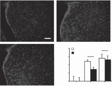
Figure 13.28. Neuron loss and Lewy body formation in Parkinson’s disease. (A) Control. (B–E) Parkinson’s disease. (A–C) Neuromelanin pigmented neurons immunohistochemically stained with an anti-α synuclein antibody. Scale in B is equivalent to that for A. There is an obvious loss of pigmented dopamine neurons in Parkinson’s disease (B) compared with the control (A). Some remaining pigmented dopamine neurons in Parkinson’s disease contain α-synuclein-immunoreactive Lewy bodies (arrowheads in C). (D,E) Specimens immunohistochemically stained with antibodies to HLA-DP/DQ/DR (HLA), a marker for the major histocompatibility complex class II protein. Scale in E is equivalent to that for D. HLA-immunoreactive upregulated microglia (arrows) near nonimmunoreactive pigmented neurons (asterisks) in thick (D) and thin (E) midbrain sections from patients with Parkinson’s disease (D and E). The inserts in D and E were from Parkinson’s disease patients with α-synuclein and parkin gene mutation, respectively. (Reprinted from Orr CF et al: A possible role for humoral immunity in the pathogenesis of Parkinson’s disease, Brain 128:2665–74, copyright 2005 by permission of Oxford University Press.)
DEGENERATIVE NEURAL DISEASES |
567 |
Conventional Treatment [13.35]. Parkinson’s disease is induced predominantly by reduction or depletion of cerebral dopamine, the neurotransmitter of dopaminergic neurons. Thus, the principle of treating Parkinson’s disease is to maintain or restore the level of dopamine in the central nervous system. Several types of pharmacological substances have bee used to achieve such a goal. These include dopamine precursors, such as L-dihy- droxyphenylalanine (L-dopa; also known as levodapa), dopamine agonists, such as bromocriptine, pergolide, lisuride, ropinirole, and pramipexole, and substances that inhibit the degradation of dopamine, such as selegiline. L-Dopa is the most effective drug for the treatment of Parkinson’s disease. L-Dopa can be used alone or in combination with other agents. One type of such agents is decarboxylase inhibitors such as carbidopa and benserizide. Decarboxylase is an enzyme that catalyzes L-dopa breakdown and the conversion of L-dopa to dopamine. Carbidopa reduced the activity of decarboxylase. Because carbidopa cannot pass through the blood–brain barrier, the administration of carbidopa together with L-dopa reduces the rate of L-dopa breakdown in the peripheral system, allowing a large fraction of L-dopa to reach the brain. This strategy also reduces the side effect of dopamine, such as nausea and hypotension, in the peripheral systems.
Dopamine agonists, such as bromocriptine, pergolide, lisuride, ropinirole, and pramipexole, can interact with the dopamine receptor, mimicking the activity of dopamine. These agents can be administrated before L-dopa is used or as a supplement to L-dopa treatment. Selegiline is an inhibitor for the monoamine oxidase, which degrades dopamine, and thus can be used to reduce the degradation of dopamine in the brain. It should be noted that treatment with all these agents may only be effective for treating the symptoms of Parkinson’s disease, but not be effective for preventing the progression of the disease.
Molecular Regenerative Engineering for Parkinson’s Disease [13.36]. Given the pathogenic mechanisms of Parkinson’s disease as discussed on page 565, strategies for molecular engineering treatment of Parkinson’s disease include the promotion of the survival of dopaminergic neurons, maintenance of the dopamine level in the brain, and protection of dopaminergic neurons from apoptosis. These strategies are tested primarily in rodent and nonhuman primate models of Parkinson’s disease. These models are induced by administration of chemical toxins, such as 6-hydroxydopamine (6-OHDA) and 1-methyl-4- phenyl-1,2,3,6-tetrahydropyridine (MPTP), which reduce the production of dopamine in neurons and promote neuronal apoptosis. It is important to note that these animal models may not completely assemble the human Parkinson’s disease. Results form these models should be interpreted with caution.
PROMOTION OF THE SURVIVAL OF DOPAMINERGIC NEURONS. For promoting the survival of dopaminergic neurons, neurotrophic factors, such as nervous growth factor (NGF), brainderived neurotrophic factor (BDNF), glial cell line-derived neurotrophic factor (GDNF), and platelet-derived growth factor (PDGF) or their genes can be delivered to the brain by using approaches of direct injection or spinal cavity injection (see page 521 of this chapter for these neurotrophic factors) (Fig. 13.29). In animal models of Parkinson’s disease, the overexpression of neurotrophic factors can exert several effects that benefit the treatment of the disease, including an increase in the synthesis of dopamine, protection of dopaminergic neurons from toxic injury and apoptosis, and enhancement of the sprouting of dopaminergic neurons. A large number of investigations have demonstrated the effectiveness of neurotrophic factor gene transfer in mitigating the symptoms of Parkinson’s
DEGENERATIVE NEURAL DISEASES |
569 |
converts L-dopa to dopamine. The deficiency of this enzyme may contribute to the reduction in the dopamine level and the pathogenesis of Parkinson’s disease. The transfer of the amino acid decarboxylase gene into the striatum of nonhuman primates with induced Parkinson’s disease results in increased conversion of L-dopa to dopamine and reduction in Parkinson’s symptoms. The cotransfer of the genes encoding multiple enzymes for the synthesis of dopamine has been demonstrated to provide a synergistic effect and exhibit significant improvement in the dopamine level and motor control in Parkinson’s animal models. Similar results have also been found in studies with rodent models. The tyrosine hydroxylase and amino acid decarboxylase genes are considered potential candidate genes for the treatment of human Parkinson’s disease.
PROTECTION OF DOPAMINERGIC NEURONS FROM APOPTOSIS. Another molecular approach for treating Parkinson’s disease is to protect neurons from apoptosis. Parkinson’s disease is associated with an increase in neuronal apoptosis, which is thought to contribute to the pathogenesis of Parkinson’s disease. Several factors, including oxidative stress and free radicals resulting from dopamine metabolism and ion toxicity, may induce neuronal apoptosis. An enzyme, known as Cu/Zn superoxide dismutase, plays a critical role in detoxifying superoxide, which forms toxic peroxynitrite with nitric oxide. The overexpression of the Cu/Zn superoxide dismutase gene in cultured neurons by gene transfection has been shown to protect the cells from 6-OHDA-induced toxic effects. Animals with overexpressed Cu/Zn superoxide dismutase gene exhibit increased resistance to oxidative stress and apoptosis. These observations suggest that the Cu/Zn superoxide dismutase gene can potentially serve as a therapeutic gene for the treatment of Parkinson’s disease.
Apoptosis is regulated by the Bcl2 family of proteins. While some of these proteins are proapoptotic, the Bcl2 protein exerts an antiapoptotic effect (see page 304 for signaling mechanisms of apoptosis). Experimental investigations have shown that overexpression of the Bcl2 protein in the striatum by gene transfection protects neurons from apoptosis and induces an increase in the number of tyrosine hydroxylse-positive neurons. In transgenic mice with overexpressed Bcl2 gene, neurons in the central nervous system are protected from the toxic effect of 1-methyl-4-phenyl-1,2,3,6-tetrahydropyridine (MPTP), which induces a reduction in dopamine and degeneration of dopaminergic neurons. Cotransfer of the glial cell line-derived neurotrophic factor gene with the Bcl2 gene significantly augments the beneficial effect of glial cell line-derived neurotrophic factor gene. Both factors synergistically prevent neurons from apoptosis. These investigations suggest that the antiapoptosis protein genes can be used to as therapeutic genes for enhancing the survival of dopaminergic neurons.
Cell apoptosis is regulated by a protein kinase known as C-Jun N-terminal kinase (JNK). The phosphorylation of C-Jun N-terminal kinase has been shown to activate c-Jun and promote cell apoptosis. A protein called JNK-interacting protein 1 (JIP1) can inhibit the phosphorylation of C-Jun N-terminal kinase. The transfer of the JIP1 gene into the striatum induces the overexpression of the gene, which is associated with reduced phosphorylation of C-Jun N-terminal kinase and reduced activity of caspase 3, a proteinase that induces cell apoptosis. Furthermore, the transfer of a dominant negative c-jun gene protects neurons from the toxic effect of 1-methyl-4-phenyl-1,2,3,6-tetrahydropyridine (MPTP) and promotes cell survival. Thus, the negative regulators of JNK may serve as therapeutic factors for the molecular treatment of Parkinson’s disease.
Caspase 3 is a critical downstream proteinase that cleaves a variety of intracellular proteins, such as actin filaments, signaling protein kinases, and nuclear proteins, and
570 NERVOUS REGENERATIVE ENGINEERING
induces cell apoptosis. Caspase inhibitor genes can be used and transferred into the brain to suppress cell apoptosis. Such inhibitor genes include the p35 gene and the family of the inhibitor of apoptosis protein genes. The transfer of these genes into the striatum in the model of 6-OHDA-induced Parkinson’s disease significantly inhibits the activity of caspases and delays the occurrence of cell apoptosis. Transgenic mice with p35 overexpression exhibit increased neuronal tolerance to the toxic effect of 1-methyl-4-phenyl- 1,2,3,6-tetrahydropyridine (MPTP). Caspase inhibitors also augment the mitogenic effect of neurotrophic factors. In general, all caspase inhibitor genes can be used for molecular treatment of Parkinson’s disease.
Cell Regenerative Engineering for Parkinson’s Disease [13.37]. In cell regenerative engineering for Parkinson’s disease, the primary goal is to transplant stem or other types of cell into the brain to restore the structure and function of impaired dopaminergic neurons. Typical cell types for such a purpose include embryonic stem cells, fetal neuronal stem and progenitor cells, adult neuronal stem cells, and cell lines with transferred genes that enhance desired functions. To date, cell regenerative engineering approaches have been tested primarily in animal models. Since these models may not completely assemble the human Parkinson’s disease, results from animal models should be interpreted with caution.
As discussed on page 381, embryonic stem cells from the blastocyst are capable of differentiating into all specified cell types, including neurons, under appropriate conditions. Fetal neuronal precursor cells can differentiate into dopamine-synthesizing neurons. These stem cells can be used and transplanted into the central nervous system for neuronal regeneration. It is expected that the embryonic and fetal stem cells can differentiate into dopaminergic neurons, thus improving the motor control capability. A number of investigations have demonstrated that dopamine-synthesizing neurons from embryonic and fetal stem cells can be used for transplantation into the central nervous system. The transplanted cells can be integrated into the host brain (Fig. 13.30). Such a treatment significantly improves the level of dopamine production and the survival of dopaminergic neurons in association with reduced Parkinson’s symptoms. However, the application of embryonic or fetal stem cells to human patients remains an issue of ethical debate. To date, most investigations in this area are conducted by using animal models.
Given the controversial issues for the use of embryonic and fetal stem cells, many investigators have searched for neuronal stem cells in the central nervous system. Indeed, the adult brain contains cells that exhibit stem cell characteristics such as self-renewing and differentiation. These cells are considered adult neural stem cells and can differentiate into neurons, astrocytes, and oligodendrocytes. The adult neural stem cells can be found in the wall of the ventricular system, the olfactory system, and the hippocampus of the brain (see page 395 for details). These cells can be potentially used for the treatment of Parkinson’s disease.
Although the transplantation of stem and progenitor cells demonstrates beneficial effects on the treatment of Parkinson’s disease, natural wildtype cells may not be able to sufficiently boost the production and release of the neurotransmitter dopamine and neurotrophic factors necessary for the survival of neurons. To resolve such an issue, stem and progenitor cells have been transfected with genes encoding proteins that promote the differentiation of stem and progenitor cells to dopaminergic neurons. Two types of gene, including the Nurr1 and von Hippel–Lindau (VHL) protein genes, have been used for such a purpose.
|
DEGENERATIVE NEURAL DISEASES |
571 |
|
|
A |
B |
|
0 |
CC |
|
|
A 200 |
|
|
|
|
|
|
|
|
B 750 LV |
|
|
|
G |
|
|
|
C 1,500 |
|
|
|
2,500 |
|
C |
D |
|
D 3,000 |
d |
|
|
|
|
|
||
|
E 3,500 |
v |
|
|
|
|
|
|
|
4,000 |
F |
AC |
|
|
G |
|
|
||
4,300 (μm) |
|
|
|
|
|
|
|
|
|
|
|
E |
F |
G |
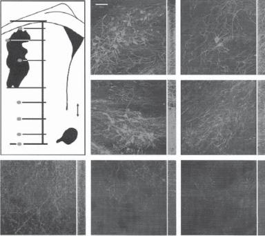
Figure 13.30. Integration of Nurr1 embryonic stem cells into the striatum of hemiparkinsonian rats. The diagram shows a drawing of a single section through a graft (G) in the striatum (LV, lateral ventricle; AC, anterior commissure). Single confocal images after immunohistochemistry for tyrosine hydroxylase (TH) are shown (A–G) from regions marked by red dots in the diagram. The distribution of cells and processes through the thickness of the section (35 μm) is shown by the z series displayed in green on the right. Note the many TH+ processes that extend away from the graft into the parenchyma of the host striatum (D–F). Scale bar: 50 μm. (Reprinted by permission from Macmillan Publishers Ltd.: Kim JH et al: Dopamine neurons derived from embryonic stem cells function in an animal model of Parkinson’s disease, Nature 418:50–6, copyright 2002.)
Nurr1 is a protein that belongs to the nuclear receptor superfamily of transcription factors. This protein is expressed predominantly in the developing and mature dopaminergic neurons of the central nervous system, and is essential for the differentiation of the mesencephalic precursors to dopaminergic neurons and for the survival of mature dopaminergic neurons. The von Hippel–Lindau protein is a tumor suppressor that downregulates the transcriptional activity of mitogenic genes. This protein is primarily expressed in the central nervous system and plays a role in regulating the differentiation of the mesencephalic precursors to dopaminergic neurons. Thus, genes encoding the Nurr1 and von Hippel–Lindau proteins can be potentially used to boost the differentiation of stem and progenitor cells into dopamine-synthesizing neurons. Several investigations have demonstrated that the transfer of these genes into stem or progenitor cells enhances the synthesis of dopamine, increases the density of tyrosine hydroxylase-positive neurons, improves the survival of dopaminergic cells, and reestablishes dopamine-dependent motor behaviors in animal models of Parkinson’s disease. Other potential genes that can be used
572 NERVOUS REGENERATIVE ENGINEERING
to boost the function of transplanted neuronal stem cells include neurotrophic factor genes, dopamine synthesis-promoting genes, and Cu/Zn superoxide dismutase genes.
Nonneuronal cell types have also been used to create dopamine-synthesizing cell lines by gene transfer. These cells include fibroblasts, astrocytes, Schwann cells, myoblasts, and marrow stromal cells. These cell types are readily available and easy to collect in comparison to embryonic and fetal stem cells. Although these cell types may not be able to differentiate into neurons, they can carry and deliver necessary proteins such as neurotrophic factors and dopamine synthesis enzymes for enhancing the survival of neurons.
Multiple Sclerosis
Etiology, Pathogenesis, and Clinical Manifestations [13.38]. Multiple sclerosis is a neural disorder that is characterized by myelin degradation, demyelination, and the generation of insoluble precipitates or plaques in various locations, in association with dysfunction of multiple cerebral regions and the spinal cord. Clinical signs include motor weakness, partial paralysis, abnormal sensation, abnormal vision, tremor, and dysarthria. The insoluble plaques can be detected by X-ray and magnetic resonance imaging. The disorder progresses slowly with a latent period of 1–10 years starting with a minor symptom. One of the distinct features of the disorder is the intermittent occurrence of the symptoms and signs. Multiple sclerosis is found in about 0.001% of the population. The disorder is often detected in patients at the age of 20–40. Hereditary factors may influence the occurrence of the disease. About 15% of the patients with multiple sclerosis have relatives with the same disorder. Siblings of patients with multiple sclerosis have a risk factor significantly greater than that of the general population.
Multiple sclerosis is associated with the destruction of myelin sheaths. However, this process does not apparently affect the structure of neurons and axons. A pathological examination often reveals an uneven surface of the spinal cord, while the brain surface may appear normal. Scattered lesions or insoluble plaques may be found in the nervous system with a size ranging from millimeters to centimeters with pink/gray-colored white matter due to demyelination. The lesion is found primarily in the white matter near the cerebral ventricles, in the brainstem, and in the spinal cord, but not in peripheral nerves. The optic nerves are often affected by the disorder. Structural alterations are dependent on the progression of the disorder. Fresh lesions exhibit partial destruction of myelin sheaths and infiltration of lymphocytes and mononuclear cells. With the progression of the disorder, there appears increased infiltration of microglial cells (macrophages) and increased size of sclerotic lesions. In the late stage, few myelin sheaths can be found. Affected regions often exhibit increased fibrous tissue with reduced lymphocytes and macrophages.
A number of factors may contribute to the pathogenesis of multiple sclerosis. These factors include viral infection, autoimmune reactions, and synergistic effects of both viral and autoimmune factors. Patients with multiple sclerosis exhibit immune reactions against viral products. Herpes viruses have been implicated in the initiation and development of multiple sclerosis, since DNA from these viruses is found in multiple sclerosis plaques. However, little direct evidence has been obtained for the role of viruses. Another factor that potentially contributes to the induction and development is autoimmune reaction. In such a case, the host immune cells may not recognize myelin proteins as the body’s own components and may initiate immune reactions to attack and destroy the myelin structure
MULTIPLE SCLEROSIS |
573 |
and sometimes the nerve axon. A major line of supporting evidence for the autoimmune mechanism is the existence of antibodies against the myelin components in the serum of patients with multiple sclerosis. Viral infection and autoimmune reactions may coordinately influence the process of demyelination. Several types of virus, such as rubella and rubeola, may contain protein components that are similar to some proteins in the myelin sheath of nerve axons. The exposure of T and B lymphocytes to these viruses may induce lymphocyte immunization. Immunized lymphocytes in turn recognize and attack the host myelin.
Further investigations have demonstrated that multiple sclerosis is associated with the infiltration of mononuclear cells in the area of demyelination, axonal loss, and glial fibrosis. Given the involvement of T lymphocytes, it has been hypothesized that activated antigen-specific T cells initiate specific immune reactions and induce the infiltration of non-antigen-specific mononuclear cells into the brain. The mononuclear cells in turn interact with and destroy oligodendrocytes by releasing toxic substances in association with the degradation of myelin sheaths. In the early stage of the disease, oligodendrocytes are capable of surviving and remyelinating axons. However, in the late stage, these cells are gradually committed to apoptosis.
Multiple sclerosis causes changes in the physiological function of nervous axons. A major change is delayed transmission of action potentials. This is due to the destruction of the axon myelin sheath. Under physiological conditions, the transmission of action potentials in the myelinated axons (as high as 100 m/s) is much faster than that in unmyelinated axons (as low as 0.25 m/s). The destruction of the myelin sheath influences the electrical conduction of the axons. In severe cases, the transmission of electrical signals can be completely blocked. The impairment and blockade of electrical signals ultimately influence the function of peripheral tissues and organs.
Conventional Treatment [13.39]. Multiple sclerosis is a disease possibly induced by viral infection and autoimmune reactions. The principle of treating multiple sclerosis is administration of antiinflammatory and immunosuppressor agents. Corticosteroids are commonly used as antiinflammatory agents. These agents usually give noticeable results within about 2 weeks. Immunosuppressor agents, such as azathioprine and cyclophosphamide, have been used for the treatment of multiple sclerosis with some positive results. However, these substances compromise with the normal immune function and impose toxic effects. Such harmful influences may preclude the widespread use of the immunosuppressor agents. In addition, appropriate physical exercise is necessary to stimulate impaired motor control systems. Bacterial infection should be prevented and treated promptly, if any. Other disorders associated with multiple sclerosis should be treated properly.
Molecular Regenerative Engineering for Multiple Sclerosis [13.40]. As the pathogenesis of multiple sclerosis is attributed to the destruction of the myelin sheath and oligodendrocytes by inflammatory reactions involving activated mononuclear cells, a potential approach for this disease is to suppress inflammatory reactions and inhibit the infiltration of bloodborne mononuclear cells into the brain. The enhancement of oligodendrocyte proliferation and migration into the area of demyelinated axons may also provide therapeutic effects. Gene therapeutic approaches have been developed to achieve these goals. These approaches have been tested primarily in the animal model of multiple sclerosis: autoimmune encephalomyelitis (EAE). It should be noted that, since the animal model
574 NERVOUS REGENERATIVE ENGINEERING
may not completely assemble the human disease, information from experimental observations may not be directly applied to the human disease.
Several therapeutic strategies have been developed and used to suppress inflammatory reactions. These include the enhancement of antiinflammatory cytokines and induction of B-lymphocyte tolerance to myelin-related antigens. Since mononuclear cells and B- lymphocytes are bloodborne cells, genes encoding antiinflammatory proteins can be delivered into the bloodstream. Potential genes for antiinflammatory purposes include the interleukin (IL)1β, IL2, IL4, IL6, IL10, tumor necrosis factor (TNF)α, and transforming growth factor (TGF)β1 genes. The transfer of these genes into animals with autoimmune encephalomyelitis reduces pathological signs and clinical symptoms of the disorder, although controversial results are observed for some IL molecules, such as IL4 and IL10. These antiinflammatory protein genes can be delivered to target tissues by three approaches: injection into the bloodstream, directly into the brain, and into the cerebrospinal fluid cavities.
The enhancement of B-lymphocyte tolerance to myelin-related antigens is another potential approach for the treatment of multiple sclerosis. Certain viruses may contain components that partially assemble the structure of myelin proteins. Viral infection may expose B lymphocytes to the myelin-like viral components and induce immunization of the lymphocytes, producing antimyelin protein antibodies. These antibodies may interact with host myelin proteins and contribute to autoimmune processes, potentially inducing multiple sclerosis. In an experimental study, a recombinant IgG–myelin basic protein (MBP) gene is inserted into a retroviral vector and transferred into B lymphocytes. These B cells were introduced into the bloodstream of mice with autoimmune encephalomyelitisinduced multiple sclerosis. Compared to control mice without B cell transplantation, the B cell-transplanted mice exhibit reduced pathological signs and clinical symptoms of multiple sclerosis. It is thought that the presence of myelin basic protein in the B cells increases the tolerance of these cells to the myelin basic protein, thus reducing the production and secretion of antimyelin protein antibodies and mitigating autoimmune reactions.
Gene therapeutic approaches have also been developed to enhance the proliferation and migration of oligodendrocytes and to promote the remyelination of impaired axons, as demyelination results in axonal loss and neurological impairment. Candidate genes for such a purpose include neurotrophic factor and nerve growth factor genes. Since the therapeutic targets of these genes are the oligodendrocytes in the central nervous system, direct brain gene delivery usually gives satisfactory results. Although neurotrophic factors and their genes can be injected into the bloodstream, the therapeutic efficiency is usually low, as it is difficult for protein and DNA molecules to pass through the blood–brain barrier. Another delivery route is the cerebrospinal fluid. In an experimental model of autoimmune encephalomyelitis-induced multiple sclerosis, a herpes virus-derived vector containing the fibroblast growth factor gene was transferred into the cerebrospinal fluid. Fibroblast growth factor is known to promote the proliferation and differentiation of oligodendrocytes. The introduction of this growth factor to the cerebrospinal fluid enhances remyelination of impaired axons and reduces pathological signs and clinical symptoms of multiple sclerosis.
Cell Regenerative Engineering [13.41]. Cellular engineering approaches have been developed for the treatment of experimental multiple sclerosis induced by autoimmune encephalomyelitis. These include the transplantation of oligodendrocytes, oligodendrocyte
BIBLIOGRAPHY 575
precursors, or genetically modified memory T lymphocytes with enhanced secretion of growth factors or antiinflammatory factors. Oligodendrocytes or their precursors can be directly delivered to the lesion sites of multiple sclerosis. A fraction of these cells can integrate into the native system and generate myelin proteins and sheaths. These cells can also be transfected with growth factor genes to enhance their capability of proliferation and migration. T lymphocytes can be transfected with antiinflammatory protein genes, such as the interleukin (IL)1β, IL2, IL4, IL6, IL10, tumor necrosis factor (TNF)α genes, enhancing the production and secretion of antiinflammatory factors. These cells can be transplanted to the bloodstream, from where they can migrate into the lesion sites of the brain. Alternatively, T cells can be directly delivered to the central nervous system. T lymphocytes can also be transfected with growth factor genes to promote their capability of producing growth factors.
BIBLIOGRAPHY
13.27. Etiology, Pathology, and Clinical Manifestations for Alzheimer’s Disease
Wilquet V, De Strooper B: Amyloid-beta precursor protein processing in neurodegeneration, Curr Opin Neurobiol 14:582–8, 2004.
Reinhard C, Hebert SS, De Strooper B: The amyloid-beta precursor protein: Integrating structure with biological function, EMBO J 24:3996–4006, 2005.
Annaert W, De Strooper B: A cell biological perspective on Alzheimer’s disease, Annu Rev Cell Dev Biol 18:25–51, 2002.
Dominguez DI, De Strooper B: Novel therapeutic strategies provide the real test for the amyloid hypothesis of Alzheimer’s disease, Trends Pharmacol Sci 23:324–30, 2002.
Lu PJ, Wulf G, Zhou XZ, Davies P, Lu KP: The prolyl isomerase Pin1 restores the function of Alzheimer-associated phosphorylated tau protein, Nature 399:784–8, 1999.
Panda D, Samuel JC, Massie M, Feinstein SC, Wilson L: Differential regulation of microtubule dynamics by threeand four-repeat tau: Implications for the onset of neurodegenerative disease,
Proc Natl Acad Sci USA 100:9548–53, 2003.
Rapoport M, Dawson HN, Binder LI, Vitek MP, Ferreira A: Tau is essential to beta-amyloid- induced neurotoxicity, Proc Natl Acad Sci USA 99:6364–9, 2002.
Ros R, Thobois S, Streichenberger N, Kopp N, Sanchez MP et al: A new mutation of the tau gene, G303V, in early-onset familial progressive supranuclear palsy, Arch Neurol 62:1444–50, 2005.
SantaCruz K, Lewis J, Spires T, Paulson J, Kotilinek L et al: Tau suppression in a neurodegenerative mouse model improves memory function, Science 309:476–81, 2005.
Skipper L, Wilkes K, Toft M, Baker M, Lincoln S et al: Linkage disequilibrium and association of MAPT H1 in Parkinson disease, Am J Hum Genet 75:669–77, 2004.
Spillantini MG, Murrell JR, Goedert M, Farlow MR, Klug A et al: Mutation in the tau gene in familial multiple system tauopathy with presenile dementia, Proc Natl Acad Sci USA 95:7737– 41, 1998.
Spittaels K, van den Haute C, van Dorpe J, Geerts H, Mercken M et al: Glycogen synthase kinase- 3-beta phosphorylates protein tau and rescues the axonopathy in the central nervous system of human four-repeat tau transgenic mice, J Biol Chem 275:41340–9, 2000.
Stamer K, Vogel R, Thies E, Mandelkow E, Mandelkow EM: Tau blocks traffic of organelles, neurofilaments, and APP vesicles in neurons and enhances oxidative stress, J Cell Biol 156:1051– 63, 2002.