

Bioregenerative Engineering Principles and Applications - Shu Q. Liu
..pdf536 NERVOUS REGENERATIVE ENGINEERING
as nerve growth factor, neurotrophin 3, brain-derived neurotrophic factor, and basic fibroblast growth factor genes. The transgenic cells can be used for transplantation into injured nerve tissues. These cells can express and release the transfected neurotrophic genes at a high rate, rapidly stimulating the regeneration of injured neurons and axons. Candidate cell types for such a purpose include embryonic stem cells, glial cells, Schwann cells, olfactory ensheathing cells, and fibroblasts. Preliminary investigations have demonstrated promising results for the use of genetically modified cells for the regeneration of injured neurons and axons.
Tissue Regenerative Engineering for Nerve Injury. Tissue regenerative engineering is to construct and provide nerve matrix scaffolds, which serve to enhance and guide neuronal regeneration and axonal outgrowth. Compared to molecular and cell regenerative engineering, which can be applied to both the central and peripheral nerve systems, tissue regenerative engineering has been primarily used for the treatment of peripheral nerve injury. This is attributed to the difficulty of accessing the central nerve system and the possibility of inducing nerve injury by tissue scaffold implantation. For peripheral nerve engineering, several strategies have been developed and used, including guidance of axonal regeneration and outgrowth, stimulation of neuronal proliferation by graft-based assistance, and improvement of neuronal function.
Stimulation of Neuronal Regeneration and Guidance of Axonal Outgrowth by GraftBased Assistance [13.20]. As discussed on page 517 of this Chapter, severed nerve axons can regenerate and establish reconnection between the severed segments. However, such reconnection requires appropriate guidance from the Schwann cells, which form a sheath along the severed axons. When the severed proximal axon is relocated away from the distal axon, it is difficult for the proximal axon to reconnect to the severed distal axon. In conventional treatment, severed nerve bundles are reconnected surgically. However, when a segment of a nerve bundle is severely damaged or removed, it is impossible to reconnect the severed proximal to the distal nerve bundle. Under such as circumstance, it is necessary to provide guidance to the injured axons, allowing axonal extension along a desired path. For the past decade, a number of biological and synthetic materials have been developed and used to construct nerve scaffolds for the stimulation of axonal sprouting and the guidance of axonal extension. These materials include autologous nerve tissue grafts, allogenic (from the same animal species) and xenogeneic nerve tissue grafts (from different animal species), extracellular matrix components (laminin, fibronectin, collagen, and fibrin), and synthetic polymeric materials [poly (L-lactic acid), poly-L-lactide-ε- caprolactone, polyurethane, poly(2-hydroxyethyl methacrylate-co-methyl methacrylate), and polypyrrole-hyaluronic acid]. Nerve grafts based on these materials are briefly discussed here.
AUTOLOGOUS NERVE TISSUE GRAFTS [13.21]. Autologous nerve tissue specimens can be collected from the patient who receives grafts. Autologous nerve tissue is an ideal material type for constructing nerve grafts. Certain subcutaneous nerves, such as the branches of the saphenous nerve, can be used as nerve grafts. A nerve branch can be isolated and grafted into a severed nerve bundle. Such a graft usually provides effective support and guidance to the injured nerve and significantly enhances neuronal regeneration and axonal outgrowth. Autologous nerve tissue is usually considered the gold standard for constructing nerve grafts. Other types of natural tissue, such as autologous blood vessels, skeletal
NERVOUS DISORDERS |
537 |
muscle, and soft connective tissues, have also been used as nerve grafts. These tissues can be tailored into the shape of nerve bundles and grafted into severed nerves. However, the effectiveness of these tissues is limited compared to the autologous nerve grafts.
ALLOGENIC AND XENOGENEIC TISSUE GRAFTS [13.22]. Allogenic grafts are those collected from different individuals of the same animal species. Xenogeneic grafts are from individuals of different animal species. Various types of allogenic and xenogeneic tissue, such as nerve tissue, soft connective tissue, intestinal submucosa, and amnion, have been used as nerve grafts. However, the cellular components of allogenic and xenogeneic tissues, especially living cells, induce acute immune rejection reactions. Thus, the cellular components must be removed from the graft tissue. Decellularized matrix scaffolds can be produced by appropriate thermal, detergent, or NaOH (or KOH) treatment. The cell-free grafts can be used as scaffolds that guide neuronal regeneration and axonal outgrowth. These grafts, however, are not as effective as the autologous nerve grafts.
EXTRACELLULAR MATRIX COMPONENTS [13.23]. Extracellular components, including collagen, fibronectin, laminin, and fibrin, have been used to construct scaffolds for stimulating and guiding neuronal regeneration and axonal outgrowth. These molecules play an important role in the regulation of neuronal proliferation and migration as well as axonal extension and innervation during development. Some of these matrix components, such as collagen, fibronectin, and laminin, serve as substrates for cell attachment and migration, which are critical processes for the morphogenesis of the nerve system during development and regeneration of injured nerve axons. Other extracellular matrix molecules, such as fibrinogen, fibrin gels, peptide scaffolds, alginate, agarose, and chitosan, have been applied to injured nerves for enhancing axonal outgrowth in experimental models. It is important to point out that not all extracellular matrix components are neuronal growth promoters. Certain molecules of the proteoglycan family, such as chondroitin sulfate proteoglycan (CSPG) and proteoglycan phosphocan, are typical inhibitory factors for neuronal regeneration and axonal outgrowth. The inhibitory effect of these molecules may play a role in the prevention of glial scar formation, a hindrance for the regeneration and extension of nerve axons.
SYNTHETIC MATERIALS [13.24]. A number of synthetic polymeric materials have been developed and used for the regeneration of injured nerves in experimental models. These materials include poly(glycolic acid) (PGA), poly(lactic acid) (PLA), poly(lactic-co- glycolic acid) (PLGA), poly(caprolactones), biodegradable poly(urethane), polyorganophosphazene, methacrylate-based hydrogels, and poly(3-hydroxybutyrate). These polymeric materials are biodegradable, relatively low in toxicity, and easy to process. Scaffolds made of these materials have been tested in animal models for nerve regeneration. Results from these investigations are promising. Nonbiodegradable synthetic materials, such as polytetrafluoroethylene (PTFE), have also been used as scaffolds for nerve regeneration with satisfactory results.
Polymeric materials can be used in combination with nerve regeneration stimulators, such as neurotrophic factors and antiapoptotic factors or their genes. A polymeric scaffold may serve not only as a guide for nerve regeneration but also as a drug delivery device. The polymeric scaffold and the nerve regeneration stimulators may synergistically enhance the regeneration of injured nerves. However, compared to autogenous nerve grafts, the effectiveness of the synthetic polymeric materials remains limited. A critical problem with
538 NERVOUS REGENERATIVE ENGINEERING
the synthetic materials is their properties of inducing host inflammatory reactions. Improving biocompatibility of polymers is a major challenge in nerve regeneration with synthetic materials.
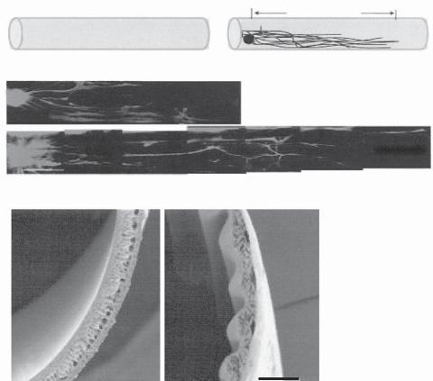
ROLE OF GEOMETRIC FACTORS [13.25]. While the material properties are critical to the modulation of neuronal regeneration and axonal outgrowth, geometric factors, such as the shape, orientation, curvature, and dimension of matrix substrate and scaffolds, may influence the extension and direction of regenerating axons. For instance, when neurons are cultured on a polymeric matrix surface, the direction of axonal outgrowth is dependent on the matrix structure and the orientation of the matrix fibers (Fig. 13.21). The curvature of
A |
Outgrowth Length |
|
|
0 days |
7 days |
B |
~5.4 mm |
|
|
C |
~9.6 mm |
|
|
1 mm |
|
D |
E |
Figure 13.21. Biomaterial implantation for guiding axonal outgrowth. (A) Schematic illustration of neurite outgrowth evaluation in a dorsal root ganglion (DRG) explant model. DRG explant was seeded at one end of a cylinder of polyurethane hollow fiber membrane (HFM) and allowed to grow for 7 days. Neurite outgrowth was assessed based on neurite length measurement. (B) Neurite outgrowth on a smooth HFM surface for 7 days. (C) Neurite outgrowth on a HFM surface with aligned grooves for 7 days. (D,E) Scanning electron micrographs showing the morphology of HFM membrane with smooth and grooved surfaces, respectively. (Reprinted with permission of John Wiley & Sons, Inc. from Zhang N et al: Fabrication of semipermeable hollow fiber membranes with highly aligned texture for nervous guidance, J Biomed Mater Rese Pt A 75:941–9, copyright 2005.)
BIBLIOGRAPHY 539
the matrix fibers influences the direction of axonal extension. Furthermore, the geometry of the matrix substrate regulates the rate of neuronal regeneration. As shown in several studies, magnetically aligned collagen fibers elicit a more significant effect on neuronal regeneration than randomly aligned collagen fibers. These observations demonstrate that geometric factors may serve as cues for the neuronal regeneration and axonal outgrowth.
IMPROVEMENT OF NERVE FUNCTION [13.26]. For the treatment of nerve injury, in addition to the promotion of nerve regeneration, another important strategy is to improve the function of regenerated axons. A major approach for such a purpose is to electrically stimulate impaired and regenerated neurons and axons. Several types of devices have been developed and tested. These include cochlear, retinal, spinal cord, and brain stimulators. These devices can be used to improve nerve functions such as hearing, vision, and muscular movement control. It becomes now clear that a successful treatment of nerve injury requires both anatomical nerve regeneration and functional recovery.
BIBLIOGRAPHY
13.1. Anatomy and Physiology of the Nervous System
Lai C, Peripheral glia: Schwann cells in motion, Curr Biol 15(9):R332–4, May 2005.
Guyton AC, Hall JE: Textbook of Medical Physiology, 11th ed, Saunders, Philadelphia, 2006.
McArdle WD, Katch FI, Katch VL: Essentials of Exercise Physiology, 3rd ed, Lippincott Williams & Wilkins, Baltimore, 2006.
Germann WJ, Stanfield CL (with contributors Niles MJ, Cannon JG): Principles of Human Physiology, 2nd ed, Pearson Benjamin Cummings, San Francisco, 2005.
Thibodeau GA, Patton KT: Anatomy & Physiology, 5th ed, Mosby, St Louis, 2003.
Boron WF, Boulpaep EL: Medical Physiology: A Cellular and Molecular Approach, Saunders, Philadelphia, 2003.
Ganong WF: Review of Medical Physiology, 21st ed, McGraw-Hill, New York, 2003.
13.2. Nerve Injury
Schneider AS, Szanto PA: Pathology, 3rd ed, Lippincott Williams & Wilkins, Philadelphia, 2006.
McCance KL, Huether SE: Pathophysiology: The Biologic Basis for Disease in Adults & Children, 5th ed, Elsevier Mosby, St Louis, 2006.
Porth CM: Pathophysiology: Concepts of Altered Health States, 7th ed, Lippincott Williams & Wilkins, Philadelphia, 2005.
Frazier MS, Drzymkowski JW: Essentials of Human Diseases and Conditions, 3rd ed, Elsevier Saunders, St Louis, 2004.
Kwon BK, Tetzlaff W: Spinal cord regeneration: From gene to transplants, Spine 26(Suppl):S13–22, 2001.
Feringa ER, McBride RL, Pruitt JN: Loss of neurons in the red nucleus after spinal cord transection, Exp Neurol 100:112–20, 1988.
Goshgarian HG, Koistinen JM, Schmidt ER: Cell death and changes in the retrograde transport of horseradish peroxidase in rubrospinal neurons following spinal cord hemisection in the adult rat, J Compar Neurol 214:251–7, 1983.
540 NERVOUS REGENERATIVE ENGINEERING
McDonald JW, Sadowsky C: Spinal cord injury, Lancet 359:417–25, 2002.
Schwab ME: Repairing the injured spinal cord, Science 295:1029–31, 2002.
13.3. Molecular Regenerative Engineering for the Nervous System
Koprivica V, Cho KS, Park JB, Yiu G, Atwal J et al: EGFR activation mediates inhibition of axon regeneration by myelin and chondroitin sulfate proteoglycans, Science 310:106–10, 2005.
Houle JD, Ye JH: Survival of chronically-injured neurons can be prolonged by treatment with neurotrophic factors, Neuroscience 94:929–36, 1999.
Yamada M, Natsume A, Mata M, Oligino T, Goss J et al: Herpes simplex virus vector-mediated expression of Bcl-2 protects spinal motor neurons from degeneration following root avulsion, Exp Neurol 168:225–30, 2001.
Chen DF, Schneider GE, Martinou JC, Tonegawa S: Bcl-2 promotes regeneration of severed axons in mammalian CNS, Nature 385:434–39, 1997.
Chierzi S, Strettoi E, Cenni MC, Maffei L: Optic nerve crush: axonal responses in wild-type and bcl-2 transgenic mice, J Neurosci 19:8367–76, 1999.
Holm K, Isacson O: Factors intrinsic to the neuron can induce and maintain its ability to promote axonal outgrowth: A role for BCL2? Trends Neurosci 22:269–73, 1999.
Kobayashi NR, Fan DP, Giehl KM et al: BDNF and NT-4/5 prevent atrophy of rat rubrospinal neurons after cervical axotomy, stimulate GAP-43 and Talpha1-tubulin mRNA expression, and promote axonal regeneration, J Neurosci 17:9583–95, 1997.
Schumacher PA, Siman RG, Fehlings MG: Pretreatment with calpain inhibitor CEP-4143 inhibits calpain I activation and cytoskeletal degradation, improves neurological function, and enhances axonal survival after traumatic spinal cord injury, J Neurochem 74:1646–55, 2000.
Beattie MS, Farooqui AA, Bresnahan JC: Review of current evidence for apoptosis after spinal cord injury, J Neurotrauma 17:915–25, 2000.
Crowe MJ, Bresnahan JC, Shuman SL et al: Apoptosis and delayed degeneration after spinal cord injury in rats and monkeys, Nat Med 3:73–6, 1997.
Emery E, Aldana P, Bunge MB et al: Apoptosis after traumatic human spinal cord injury, J Neurosurg 89:911–20, 1998.
Martin LJ, Al Abdulla NA, Brambrink AM et al: Neurodegeneration in excitotoxicity, global cerebral ischemia, and target deprivation: A perspective on the contributions of apoptosis and necrosis, Brain Res Bull 46:281–309, 1998.
Liu XZ, Xu XM, Hu R et al: Neuronal and glial apoptosis after traumatic spinal cord injury, J Neurosci 17:5395–406, 1997.
13.4. Brain-Derived Neurotrophic Factor (BDNF)
Jones KR, Reichardt LF: Molecular cloning of a human gene that is a member of the nerve growth factor family, Proc Natl Acad Sci USA 87:8060–4, 1990.
Maisonpierre PC, Le Beau MM, Espinosa R III, Ip NY, Belluscio L et al: Human and rat brainderived neurotrophic factor and neurotrophin-3: Gene structures, distributions and chromosomal localizations, Genomics 10:558–68, 1991.
Lee R, Kermani P, Teng KK, Hempstead BL: Regulation of cell survival by secreted proneurotrophins, Science 294:1945–8, 2001.
Pang PT, Teng HK, Zaitsev E, Woo NT, Sakata K et al: Cleavage of proBDNF by tPA/plasmin is essential for long-term hippocampal plasticity, Science 306:487–91, 2004.
BIBLIOGRAPHY 541
Chen H, Weber AJ: BDNF enhances retinal ganglion cell survival in cats with optic nerve damage,
Invest Ophthalm Vis Sci 42:966–74, 2001.
Kawamura K, Kawamura N, Mulders SM, Sollewijn Gelpke MD et al: Ovarian brain-derived neurotrophic factor (BDNF) promotes the development of oocytes into preimplantation embryos,
Proc Natl Acad Sci USA 102:9206–11, 2005.
Conover JC, Erickson JT, Katz DM, Bianchi LM, Poueymirou WT et al: Neuronal deficits, not involving motor neurons, in mice lacking BDNF and/or NT4, Nature 375:235–8, 1995.
Liu X, Ernfors P, Wu H, Jaenisch R: Sensory but not motor neuron deficits in mice lacking NT4 and BDNF, Nature 375:238–41, 1995.
Guillin O, Diaz J, Carroll P, Griffon N, Schwartz J-C et al: BDNF controls dopamine D3 receptor expression and triggers behavioural sensitization, Nature 411:86–9, 2001.
Lyons WE, Mamounas LA, Ricaurte GA, Coppola V, Reid SW et al: Brain-derived neurotrophic factor-deficient mice develop aggressiveness and hyperphagia in conjunction with brain serotonergic abnormalities, Proc Natl Acad Sci USA 96:15239–44, 1999.
Peng S, Wuu J, Mufson EJ, Fahnestock M: Precursor form of brain-derived neurotrophic factor and mature brain-derived neurotrophic factor are decreased in the pre-clinical stages of Alzheimer’s disease, J Neurochem 93(6):1412–21, 2005.
Jones KR, Reichardt LF: Molecular cloning of a human gene that is a member of the nerve growth factor family, Proc Natl Acad Sci USA 87(20):8060–4, 1990.
Baker-Herman TL, Fuller DD, Bavis RW, Zabka AG, Golder FJ et al: BDNF is necessary and sufficient for spinal respiratory plasticity following intermittent hypoxia, Nature Neurosci 7:48–55, 2004.
Chen H, Weber AJ: BDNF enhances retinal ganglion cell survival in cats with optic nerve damage,
Invest Ophthalm Vis Sci 42:966–74, 2001.
Conover JC, Erickson JT, Katz DM, Bianchi LM, Poueymirou WT et al: Neuronal deficits, not involving motor neurons, in mice lacking BDNF and/or NT4, Nature 375:235–8, 1995.
Du J, Poo M: Rapid BDNF-induced retrograde synaptic modification in a developing retinotectal system, Nature 429:878–83, 2004.
Guillin O, Diaz J, Carroll P, Griffon N, Schwartz J-C et al: BDNF controls dopamine D3 receptor expression and triggers behavioural sensitization, Nature 411:86–9, 2001.
Hofer MM, Barde Y-A: Brain-derived neurotrophic factor prevents neuronal death in vivo, Nature 331:261–2, 1988.
Huang ZJ, Kirkwood A, Pizzorusso T, Porciatti V, Morales B et al: BDNF regulates the maturation of inhibition and the critical period of plasticity in mouse visual cortex, Cell 98:739–55, 1999.
Jones KR, Reichardt LF: Molecular cloning of a human gene that is a member of the nerve growth factor family, Proc Natl Acad Sci USA 87:8060–4, 1990.
Kawamura K, Kawamura N, Mulders SM, Sollewijn Gelpke MD, Hsueh AJW: Ovarian brainderived neurotrophic factor (BDNF) promotes the development of oocytes into preimplantation embryos, Proc Natl Acad Sci USA 102:9206–11, 2005.
Kernie SG, Liebl DJ, Parada LF: BDNF regulates eating behavior and locomotor activity in mice, EMBO J 19:1290–1300, 2000.
Kovalchuk Y, Hanse E, Kafitz KW, Konnerth A: Postsynaptic induction of BDNF-mediated longterm potentiation, Science 295:1729–34, 2002.
Lawrence JM, Keegan DJ, Muir EM, Coffey PJ, Rogers JH et al: Transplantation of Schwann cell line clones secreting GDNF or BDNF into the retinas of dystrophic Royal College of Surgeons rats, Invest Ophthalm Vis Sci 45:267–74, 2004.
Lee R, Kermani P, Teng KK, Hempstead BL: Regulation of cell survival by secreted proneurotrophins, Science 294:1945–8, 2001.
542 NERVOUS REGENERATIVE ENGINEERING
Li Y, Jian J-C, Cui K, Li N, Zheng Z-Y et al: Essential role of TRPC channels in the guidance of nerve growth cones by brain-derived neurotrophic factor, Nature 434:894–8, 2005.
Liu X, Ernfors P, Wu H, Jaenisch R: Sensory but not motor neuron deficits in mice lacking NT4 and BDNF, Nature 375:238–41, 1995.
Lyons WE, Mamounas LA, Ricaurte GA, Coppola V, Reid SW et al: Brain-derived neurotrophic factor-deficient mice develop aggressiveness and hyperphagia in conjunction with brain serotonergic abnormalities, Proc Natl Acad Sci USA 96:15239–44, 1999.
Ming G, Wong ST, Henley J, Yuan X, Song H et al: Adaptation in the chemotactic guidance of nerve growth cones, Nature 417:411–8, 2002.
Pang PT, Teng HK, Zaitsev E, Woo NT, Sakata K et al: Cleavage of proBDNF by tPA/plasmin is essential for long-term hippocampal plasticity, Science 306:487–91, 2004.
Human protein reference data base, Johns Hopkins University and the Institute of Bioinformatics, at http://www.hprd.org/protein.
13.5. Neurotrophin 3
Jones KR, Reichardt LF: Molecular cloning of a human gene that is a member of the nerve growth factor family, Proc Natl Acad Sci USA 87:8060–4, 1990.
Maisonpierre PC, Le Beau MM, Espinosa R III, Ip NY, Belluscio L et al: Human and rat brainderived neurotrophic factor and neurotrophin-3: Gene structures, distributions, and chromosomal localizations, Genomics 10:558–68, 1991.
Kalcheim C, Carmeli C, Rosenthal A: Neurotrophin 3 is a mitogen for cultured neural crest cells,
Proc Natl Acad Sci USA 89:1661–5, 1992.
Ramer MS, Priestley JV, McMahon SB: Functional regeneration of sensory axons into the adult spinal cord, Nature 403:312–6, 2000.
Cosgaya JM, Chan JR, Shooter EM: The neurotrophin receptor p75(NTR) as a positive modulator of myelination, Science 298:1245–8, 2002.
Kuruvilla R, Zweifel LS, Glebova NO, Lonze BE, Valdez G et al: A neurotrophin signaling cascade coordinates sympathetic neuron development through differential control of TrkA trafficking and retrograde signaling, Cell 118:243–55, 2004.
Ernfors P, Lee K-F, Kucera J, Jaenisch R: Lack of neurotrophin-3 leads to deficiencies in the peripheral nervous system and loss of limb proprioceptive afferents, Cell 77:503–12, 1994.
Tessarollo L, Vogel KS, Palko ME, Reid SW, Parada LF: Targeted mutation in the neurotrophin-3 gene results in loss of muscle sensory neurons, Proc Natl Acad Sci USA 91:11844–8, 1994.
Donovan MJ, Hahn R, Tessarollo L, Hempstead BL: Identification of an essential nonneuronal function of neurotrophin 3 in mammalian cardiac development, Nature Genet 14:210–3, 1996.
13.6. Neurotrophin (NT)4
Ip NY, Ibanez CF, Nye SH, McClain J, Jones PF et al: Mammalian neurotrophin-4: structure, chromosomal localization, tissue distribution, and receptor specificity, Proc Natl Acad Sci USA 89:3060–4, 1992.
Ibanez CF: Neurotrophin-4: the odd one out in the neurotrophin family, Neurochem Res 21:787–93, 1996.
Robinson LLL, Townsend J, Anderson RA: The human fetal testis is a site of expression of neurotrophins and their receptors: regulation of the germ cell and peritubular cell population, J Clin Endocr Metab 88:3943–51, 2003.
Xie C-W, Sayah D, Chen Q-S, Wei W-Z, Smith D et al: Deficient long-term memory and long-lasting long-term potentiation in mice with a targeted deletion of neurotrophin-4 gene, Proc Natl Acad Sci USA 97:8116–21, 2000.
BIBLIOGRAPHY 543
13.7. Nerve Growth Factor (NGF)
Ramer MS, Priestley JV, McMahon SB: Functional regeneration of sensory axons into the adult spinal cord, Nature 403:312–6, 2000.
Ieda M, Fukuda K, Hisaka Y, Kimura K, Kawaguchi H et al: Endothelin-1 regulates cardiac sympathetic innervation in the rodent heart by controlling nerve growth factor expression, J Clin Invest 113:876–84, 2004.
Zabel BU, Eddy RL, Scott J, Shows TB: The human nerve growth factor gene (NGF) is located on the short arm of chromosome 1 [abstract], Cytogenet Cell Genet 37:614, 1984.
Ullrich A, Gray A, Berman C, Dull TJ: Human beta-nerve growth factor gene sequence highly homologous to that of mouse, Nature 303:821–5, 1983.
Lee R, Kermani P, Teng KK, Hempstead BL: Regulation of cell survival by secreted proneurotrophins, Science 294:1945–8, 2001.
Harrington AW, Leiner B, Blechschmitt C, Arevalo JC, Lee R et al: Secreted proNGF is a pathophysiological death-inducing ligand after adult CNS injury, Proc Natl Acad Sci USA 101:6226– 30, 2004.
13.8. Glial Cell-Derived Neurotrophic Factor
Lin LF, Doherty DH, Lile JD, Bektesh S, Collins F: GDNF: A glial cell line-derived neurotrophic factor for midbrain dopaminergic neurons, Science 260(5111):1130–2, May 1993.
Quintero EM, Willis LM, Zaman V, Lee J, Boger HA et al: Glial cell line-derived neurotrophic factor is essential for neuronal survival in the locus coeruleus-hippocampal noradrenergic pathway, Neuroscience 124(1):137–46, 2004.
Gianino S, Grider JR, Cresswell J, Enomoto H, Heuckeroth RO: GDNF availability determines enteric neuron number by controlling precursor proliferation, Development 130(10):2187–98, May 2003.
Haase G, Dessaud E, Garces A, de Bovis B, Birling M et al: GDNF acts through PEA3 to regulate cell body positioning and muscle innervation of specific motor neuron pools, Neuron 35(5):893– 905, Aug 2002.
Shakya R, Jho EH, Kotka P, Wu Z, Kholodilov N et al: The role of GDNF in patterning the excretory system, Dev Biol 283(1):70–84, July 2005.
Whitehead J, Keller-Peck C, Kucera J, Tourtellotte WG: Glial cell-line derived neurotrophic factor-dependent fusimotor neuron survival during development, Mech Dev 122(1):27–41, Jan 2005.
Beck KD, Valverde J, Alexi T, Poulsen K, Moffat B et al: Mesencephalic dopaminergic neurons protected by GDNF from axotomy-induced degeneration in the adult brain, Nature 373:339–41, 1995.
Boucher TJ, Okuse K, Bennett DLH, Munson JB, Wood JN et al: Potent analgesic effects of GDNF in neuropathic pain states, Science 290:124–7, 2000.
Durbec P, Marcos-Gutierrez CV, Kilkenny C, Grigoriou M, Wartiowaara K et al: GDNF signalling through the Ret receptor tyrosine kinase, Nature 381:789–93, 1996.
Gash DM, Zhang Z, Ovadia A, Cass WA, Yi A et al: Functional recovery in parkinsonian monkeys treated with GDNF, Nature 380:252–5, 1996.
Gill SS, Patel NK, Hotton GR, O’Sullivan K, McCarter R et al: Direct brain infusion of glial cell line-derived neurotrophic factor in Parkinson disease, Nature Med 9:589–95, 2003.
Kordower JH, Emborg ME, Bloch J, Ma SY, Chu Y et al: Neurodegeneration prevented by lentiviral vector delivery of GDNF in primate models of Parkinson’s disease, Science 290:767–73, 2000.
544 NERVOUS REGENERATIVE ENGINEERING
Lin L-FH, Doherty DH, Lile JD, Bektesh S, Collins F: GDNF: A glial cell line-derived neurotrophic factor for midbrain dopaminergic neurons, Science 260:1130–2, 1993.
Meng X, Lindahl M, Hyvonen ME, Parvinen M, de Rooij DG et al: Regulation of cell fate decision of undifferentiated spermatogonia by GDNF, Science 287:1489–93, 2000.
Moore MW, Klein RD, Farinas I, Sauer H, Armanini M et al: Renal and neuronal abnormalities in mice lacking GDNF, Nature 382:76–9, 1996.
Nguyen QT, Parsadanian AS, Snider WD, Lichtman JW: Hyperinnervation of neuromuscular junctions caused by GDNF overexpression in muscle, Science 279:1725–9, 1998.
Oppenheim RW, Houenou LJ, Johnson JE, Lin L-FH, Li L et al: Developing motor neurons rescued from programmed and axotomy-induced cell death by GDNF, Nature 373:344–6, 1995.
Pichel JG, Shen L, Shang HZ, Granholm AC, Drago J et al: Defects in enteric innervation and kidney development in mice lacking GDNF, Nature 382:73–6, 1996.
Sanchez MP, Silos-Santiago I, Frisen J, He B, Lira SA et al: Renal agenesis and the absence of enteric neurons in mice lacking GDNF, Nature 382:70–3, 1996.
Tomac A, Lindqvist E, Lin L-FH, Ogren SO, Young D et al: Protection and repair of the nigrostriatal dopaminergic system by GDNF in vivo, Nature 373:335–9, 1995.
Treanor JJS, Goodman L, de Sauvage F, Stone DM, Poulsen KT et al: Characterization of a multicomponent receptor for GDNF, Nature 382:80–3, 1996.
Human protein reference data base, Johns Hopkins University and the Institute of Bioinformatics, at http://www.hprd.org/protein.
13.9. Prevention of Ion Flux
Agrawal SK, Fehlings MG: Mechanisms of secondary injury to spinal cord axons in vitro: Role of Na+, Na(+)-K(+)-ATPase, the Na(+)-H+ exchanger, and the Na(+)-Ca2+ exchanger, J Neurosci 16:545–52, 1996.
Agrawal SK, Fehlings MG: Role of NMDA and non-NMDA ionotropic glutamate receptors in traumatic spinal cord axonal injury, J Neurosci 17:1055–63, 1997.
Agrawal SK, Fehlings MG: The effect of the sodium channel blocker QX-314 on recovery after acute spinal cord injury, J Neurotrauma 14:81–8, 1997.
Agrawal SK, Nashmi R, Fehlings MG: Role of L- and N-type calcium channels in the pathophysiology of traumatic spinal cord white matter injury, Neuroscience 99:179–88, 2000.
Li S, Jiang Q, Stys PK: Important role of reverse Na(+)-Ca(2+) exchange in spinal cord white matter injury at physiological temperature, J Neurophysiol 84:1116–9, 2000.
Nashmi R, Jones OT, Fehlings MG: Abnormal axonal physiology is associated with altered expression and distribution of Kv1.1 and Kv1.2 K+ channels after chronic spinal cord injury, Eur J Neurosci 12:491–506, 2000.
Agrawal SK, Fehlings MG: Mechanisms of secondary injury to spinal cord axons in vitro: Role of Na+, Na(+)-K(+)-ATPase, the Na(+)-H+ exchanger, and the Na(+)-Ca2+ exchanger, J Neurosci 16(2):545–52, Jan 1996.
Rosenberg LJ, Teng YD, Wrathall JR: 2,3-Dihydroxy-6-nitro-7-sulfamoyl-benzo(f)quinoxaline reduces glial loss and acute white matter pathology after experimental spinal cord contusion, J Neurosci 19:464–75, 1999.
Shi R, Blight AR: Differential effects of low and high concentrations of 4-aminopyridine on axonal conduction in normal and injured spinal cord, Neuroscience 77:553–62, 1997.
Stys PK, Steffensen I: Na(+)-Ca2+ exchange in anoxic/ischemic injury of CNS myelinated axons, Ann NY Acad Sci 779:366–78, 1996.
Stys PK: General mechanisms of axonal damage and its prevention, J Neurol Sci 233:3–13, 2005.
BIBLIOGRAPHY 545
13.10. Prevention of Excitotoxicity
Li S, Stys PK: Mechanisms of ionotropic glutamate receptor-mediated excitotoxicity in isolated spinal cord white matter, J Neurosci 20:1190–8, 2000.
Wada S, Yone K, Ishidou Y et al: Apoptosis following spinal cord injury in rats and preventative effect of N-methyl-D-aspartate receptor antagonist, J Neurosurg 91:98–104, 1999.
Li S, Stys PK: Na(+)-K(+)-ATPase inhibition and depolarization induce glutamate release via reverse Na(+)-dependent transport in spinal cord white matter, Neuroscience 107(4):675–83, 2001.
Stys PK: General mechanisms of axonal damage and its prevention, J Neurol Sci 233:3–13, 2005.
Stys PK: White matter injury mechanisms, Curr Mol Med 4(2):113–30, March 2004.
13.11. Stimulation of Stem Cell Differentiation
Kobayashi NR, Fan DP, Giehl KM et al: BDNF and NT-4/5 prevent atrophy of rat rubrospinal neurons after cervical axotomy, stimulate GAP-43 and Talpha1-tubulin mRNA expression, and promote axonal regeneration, J Neurosci 17:9583–95, 1997.
Ramer MS, Priestley JV, McMahon SB: Functional regeneration of sensory axons into the adult spinal cord, Nature 403:312–6, 2000.
Houweling DA, Bar PR, Gispen WH, Joosten EA: Spinal cord injury: Bridging the lesion and the role of neurotrophic factors in repair, Prog Brain Res 117:455–71, 1998.
Terenghi G: Peripheral nerve regeneration and neurotrophic factors, J Anat 194:1–14, 1999.
Blesch A, Lu P, Tuszynski MH: Neurotrophic factors, gene therapy, and neural stem cells for spinal cord repair, Brain Res Bull 57:833–8, 2002.
Jones LL, Oudega M, Bunge MB, Tuszynski MH: Neurotrophic factors, cellular bridges and gene therapy for spinal cord injury, J Physiol 533:83–9, 2001.
Murakami Y, Furukawa S, Nitta A, Furukawa Y: Accumulation of nerve growth factor protein at both rostral and caudal stumps in the transected rat spinal cord, J Neurol Sci 198:63–9, 2002.
Whitworth IH, Brown RA, Dore CJ, Anand P, Green CJ et al: Nerve growth factor enhances nerve regeneration through fibronectin grafts, J Hand Surg 21:514–22, 1996.
Bloch J, Fine EG, Bouche N, Zurn AD, Aebischer P: Nerve growth factorand neurotrophin-3- releasing guidance channels promote regeneration of the transected rat dorsal root, Exp Neurol 172:425–32, 2001.
Ramer MS, Priestley JV, McMahon SB: Functional regeneration of sensory axons into the adult spinal cord, Nature 403:312–16, 2000.
Sendtner M, Holtmann B, Kolbeck R, Thoenen H, Barde YA: Brain-derived neurotrophic factor prevents the death of motoneurons in newborn rats after nerve section, Nature 360:757–9, 1992.
Yan Q, Elliott J, Snider WD: Brain-derived neurotrophic factor rescues spinal motor neurons from axotomy-induced cell death, Nature 360:753–5, 1992.
Henderson CE, Camu W, Mettling C, Gouin A, Poulsen K et al: Neurotrophins promote motor neuron survival and are present in embryonic limb bud, Nature 363:266–70, 1993.
Oudega M, Hagg T: Neurotrophins promote regeneration of sensory axons in the adult rat spinal cord, Brain Res 818:431–8, 1999.
Bloch J, Fine EG, Bouche N, Zurn AD, Aebischer P: Nerve growth factorand neurotrophin-3- releasing guidance channels promote regeneration of the transected rat dorsal root, Exp Neurol 172:425–32, 2001.