

Bioregenerative Engineering Principles and Applications - Shu Q. Liu
..pdf526 NERVOUS REGENERATIVE ENGINEERING
Figure 13.18. Neurotrophin-3 (NT3) enhances axonal growth of injured neurons into neural stem cell (NSC) grafts in injured spinal cord. Neural stem cell (mouse clone C17.2) grafts were transplanted to the lesion site of injured rat spinal cord. The growth of the dorsal column ascending sensory axons to the NSC graft was measured. (A) The ascending sensory axons rarely grow into the grafts of C17.2 NSCs without NT3 gene transfer. (B) Image from panel A with a higher magnification. (C) The ascending sensory axons could penetrate the graft of NSCs with NT3 gene transfer. (D) Image from panel C with a higher magnification. g: graft; h: host; dashed lines indicate host–graft interface. Scale bars: 177 μm in A,C; 44 μm in B,D. (E) Measurements of axon density within C17.2 grafts and C17.2–NT3 grafts (*P < 0.05). (Reprinted from Lu P et al: Neural stem cells constitutively secrete neurotrophic factors and promote extensive host axonal growth after spinal cord injury, Exp Neurol 181:115–29, copyright 2003, with permission from Elsevier.)
tiation of neurons. This factor also acts on the hippocampal neurons and contributes to the development of long-term memory. Furthermore, neurotrophin 4 is expressed in the fetal testis, contributing to the regulation of germ cell development and morphogenesis.
NERVE GROWTH FACTOR [13.7]. Nerve growth factor is a protein complex composed of four subunits, including one α unit, two β units, and one γ unit. The molecular weight of the complex is about 130 kDa. The genes that encode these subunits are mapped to chromosome 1 and locus 1p13.1. The gene sequence is similar between the human and mouse. Nerve growth factor subunits are produced at first as precursor proteins. The precursors are released into the extracellular matrix and cleaved by matrix metalloproteinases (MMP)7, MMP3, and serine protease plasmin. The cleavage activates the protein subunits. It is interesting to note that the precursor of nerve growth factor may exert an effect that is opposite to that of the mature form. In the rat and mouse models of brain injury, the precursor of nerve growth factor, produced and released by injured neurons, can
NERVOUS DISORDERS |
527 |
bind to the neurotrophin receptor p75 and induce apoptosis. The blockade of the interaction of the nerve growth factor precursor with the neurotrophin receptor reduces apoptosis.
The expression of nerve growth factor is regulated by several factors, including endothelin-1 (EDN1), endothelin receptor, protein kinase C, the Src family protein tyrosine kinases, mitogen-activated protein kinases, and activator protein 1 (AP1). Upregulated endothelin 1 can interact with and activate its receptor, which in turn induces the activation of the intracellular signaling protein kinases. The deficiency of endothelin 1 in transgenic mice results in a reduction in the expression of nerve growth factor, which is associated with the loss of neurons during the late embryonic stage and a reduction in the level of cardiac norepinephrine and sympathetic innervation. The overexpression of nerve growth factor in endothelin-1-deficient mice enhances the production of norepinephrine and sympathetic innervation.
Nerve growth factor is produced and released by neurons and plays a critical role in the regulation of growth and differentiation of sympathetic and sensory neurons of the nerve system. For instance, in a rat model of spinal cord dorsal root injury, a treatment with nerve growth factor induces the regeneration of injured dorsal axons and functional reconnection of injured synapses.
GLIAL CELL LINE-DERIVED NEUROTROPHIC FACTOR [13.8]. Glial cell line-derived neurotrophic factor (GDNF) is a member of the transforming growth factor-β superfamily. This is a 211 amino acid protein produced by the neural glial cells. The gene of glial cell linederived neurotrophic factor is localized to chromosome 5p13.1–p12. Glial cell line-derived neurotrophic factor exists in the form of disulfide-bonded homodimer, which is usually glycosylated.
The primary function of glial cell line-derived neurotrophic factor is to promote the survival, proliferation, and differentiation of neurons in the central nervous system. In particular, this neurotrophic factor is present in the midbrain and enhances the survival and proliferation of dopaminergic neurons. In the embryonic midbrain, glial cell linederived neurotrophic factor plays a critical role in regulating the differentiation and morphogenesis of neuronal cells as well as the uptake of dopamine. It is important to note that glial cell line-derived neurotrophic factor exerts a specific effect on the dopaminergic neuron. It does not significantly influence the mitogenic activity of other types of neurons such as γ-aminobutyric-containing and serotonergic neurons. The dopaminergic neuronspecific features render this factor a potential therapeutic agent for the treatment of Parkinson’s disease, which is possibly caused by progressive degeneration of the midbrain dopaminergic neurons.
Glial cell line-derived neurotrophic factor has been shown to mediate the survival and innervation of the motor neurons during development. The deficiency of glial cell line-derived neurotrophic factor in transgenic mice induces a reduction in motor axonal innervation to skeletal muscle cells. The overexpression of glial cell line-derived neurotrophic factor enhances the innervation of the motor axons. Furthermore, glial cell linederived neurotrophic factor may be involved in the regulation of motor neuron pattern formation. In the deficiency of glial cell line-derived neurotrophic factor, motor neurons are mispositioned within the spinal cord in association with reduced innervation into the skeletal muscle tissue. These observations suggest the importance of glial cell linederived neurotrophic factor in regulating the development of the peripheral nervous system.
528 NERVOUS REGENERATIVE ENGINEERING
Glial cell line-derived neurotrophic factor also plays a role in regulating the development of other systems. For instance, during the embryonic development, glial cell linederived neurotrophic factor is expressed in the metanephric mesenchyme and acts on the ureteric bud epithelium, contributing to the regulation of ureter outgrowth and epithelial branching of the excretory system of the embryo (see Chapter 8 for the excretory system of the embryo).
Prevention of Cell Death. Nerve cell death is often induced by injury and injuryassociated complications, such as ischemia and hypoxia, and is the most critical pathological event that deteriorates the function of the nervous system. In central nerve injury, most cells die in the injury site because of injury-associated complications. As reported in several studies, about 25–50% of the neurons die near the site of spinal cord axotomy within 4 weeks following the occurrence of the injury. The survival cells are highly atrophic with reduced capability of survival and proliferation.
Conventional approaches, as described on page 519 of this chapter, can be used to improve blood and oxygen supply in nerve injury, the most important step for the prevention of continuous cell death. However, nerve cells, especially injured nerve cells, may continue to commit to death even after the reestablishment of bloodflow. Thus, it is necessary to apply therapeutic approaches to injured nerve cells. Recent work has demonstrated that molecular engineering approaches can be used for such a purpose. It is now well established that cell death is mediated by extracellular ligands, including tumor necrosis factor α and Fas ligand, and intracellular death signaling molecules and caspases (see page 304). Extracellular stimuli, such as ischemia and hypoxia, may activate these factors and induce cell death. Molecular engineering approaches can be used to reduce cell death by suppressing the activity of cell death-promoting factors and by enhancing the activity of cell death-inhibiting factors. For instance, antibodies may be developed and used to block the extracellular cell death inducers TNF-α and Fas ligand. Genes encoding cell death inhibitors, such as Bcl2, can be used to transfect injured neuronal cells to prevent cell death. Furthermore, the nerve growth-stimulating factors, such as brain-derived neurotrophic factor, neurotrophin-4/5, nerve growth factor, and glial-derived neurotrophic factor, can prevent cell injury and death. These factors can be directly delivered to the site of nerve injury. The genes that encode these neurotrophic factors can be used to transfect injured nerve cells. These approaches have been shown to be effective for the prevention of neuronal death.
Prevention of Secondary Nerve Injury. Secondary injury is defined as disorders induced by pathological alterations initiated by the original nerve injury. Injury-induced pathophysiological alterations include, but are not limited to, hemorrhage (open wound), hypoxia (reduction in oxygen supply), inflammation (bacterial or nonbacterial), release of harmful enzymes (proteinases), and disorder of ion fluxes. These alterations significantly worsen the original injury and induce secondary injury of the nerve system, resulting in cell death and tissue disintegration. Thus, immediately after the initial nerve injury, it is critical to control these pathophysiological changes and protect the nerve system from secondary injury. Conventional approaches for the treatment of secondary injury have been described on page 519. Several molecular engineering approaches have been developed and tested for the prevention of secondary nerve injury. Strategies for preventing secondary injury include prevention of the disorder of ion transport and excitotoxicity.
NERVOUS DISORDERS |
529 |
PREVENTION OF ION flUX [13.9]. The flux of ions, including sodium and calcium, is increased in nerve injury and contributes to secondary nerve injury (Fig. 13.19). Under physiological conditions, sodium is concentrated in the extracellular fluid. The concentration of sodium in the extracellular matrix is significantly higher than that in the cytoplasm. The concentration gradient for the sodium across the cell membrane is maintained by the
EXTRACELLULAR SPACE |
|
|
|
|
|
|
|
|
|
||
|
|
10 |
|
|
|
|
|
|
10 |
|
|
|
|
AMPA-R |
|
O2 + |
|
|
|
AMPA-R |
|
||
|
|
|
glucose |
|
|
|
|
? |
|||
14 |
|
|
|
|
|
|
|
|
|||
|
|
|
|
|
|
|
|
|
P2X 11 |
||
K |
Cl |
mGluR1 |
|
|
1a |
3a |
|
4 |
|
|
|
|
|
|
|
|
|
|
|||||
|
|
|
|
|
|
|
|
|
|||
KCC |
|
|
|
2K |
3Na Na |
3Na |
1Ca |
|
|
|
|
|
lac |
|
|
GluR4 |
|
||||||
|
|
MY |
|
|
|
|
|
||||
|
|
|
|
Na-K |
|
|
NCX |
|
|||
|
|
|
|
|
ATPase |
|
|
|
K Ca |
||
GABA-B ADENOSINE |
|
|
|
|
|
|
|
? |
|
||
|
|
|
|
NaV1.6 |
|
|
|
||||
|
|
|
|
|
|
|
|
|
|
||
G |
|
3b |
|
|
|
|
|
8 |
mGluR1 |
|
|
|
|
|
|
ATP |
|
|
|
|
|||
CaV1.x |
K |
|
|
|
|
|
|
glut Na |
CaV2.2 |
||
PKC |
2a |
|
|
|
|
|
5 |
Na |
K PLC |
9 |
12 |
RyR1 |
|
|
|
|
Ca |
2b |
|
|
|||
|
|
|
|
|
|
|
|
||||
|
|
|
|
|
|
|
IP3R |
|
|
||
|
|
|
|
|
|
|
|
|
|
|
|
? |
“AR” |
|
|
|
Na |
|
|
|
|
|
|
|
Ca-ATPase |
? |
|
|
“AR” |
|
|
||||
13 |
|
|
|
|
Ca-ATPase |
||||||
|
|
Ca |
|
|
|
|
|
|
|
||
|
|
|
|
|
|
|
|
|
Ca |
|
|
|
|
1b |
|
|
|
|
6 |
calpain |
spectrin, NF... |
1b |
|
|
|
|
|
|
|
|
phospholipase C |
|
|
||
 phospholipase A2 AXOPLASM
  protein kinase C
nitric oxide synthase
NO
7
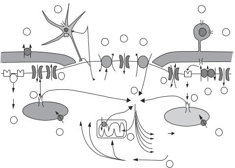
Figure 13.19. Mechanisms of ion-induced nerve injury. An energy deficit and/or excess demand impairs ATP-dependent pumps such as the Na–K ATPase (1a) and Ca ATPase (1b). Internal stores of Ca may contribute significantly to axonal Ca accumulation, triggered by depolarization via L- type Ca channels (2a) and/or generation of IP3 (2b). The rise in flux through noninactivating Na+ channels (3a) will increase [Na]i and, together with depolarization caused by K efflux through a variety of K channels (3b), will stimulate the Na–Ca exchanger to operate in the reverse Ca import mode (4). This Ca accumulation (5) promotes destructive events including mitochondrial Ca overload (especially during reoxygenation) (6), and overactivation of several Ca-dependent enzyme systems (7). NO will inhibit mitochondrial respiration and alter other cellular proteins. Some Na influx may occur through Na/K-permeable inward rectifier channels (8). Glutamate is also released through reversal of Na-dependent glutamate transport (9), causing cellular injury from activation of ionotropic glutamate receptors (10). ATP-activated P2X purinergic receptors may cause Cadependent oligodendroglial injury (11). A component of Ca influx into damaged axons directly through voltage-gated Ca channels is also likely (12). GABA and adenosine release may play an “autoprotective” role (13). Anion transporters such as the K—Cl cotransporter participate in volume dysregulation in glia and the myelin sheath, contributing to conduction abnormalities (14). The locations of the various channels and transporters are drawn for convenience and do not necessarily reflect actual distributions. (Reprinted from Stys PK: General mechanisms of axonal damage and its prevention, J Neurol Sci 233:3–13, copyright 2005, with permission from Elsevier.)
530 NERVOUS REGENERATIVE ENGINEERING
continuous action of the sodium pumps in the cell membrane. Calcium is stored in the endoplasmic reticulum (ER) system in normal cells. The concentration of calcium in the cytoplasm is much lower than that within the ER. The calcium concentration gradient across the ER membrane is established and maintained by the calcium pumps in the ER membrane.
In nerve injury, especially in the presence of ischemia, cell membrane exhibits enhanced permeability to ions, resulting in an increase in ion flux. Such a change influences the level of membrane potential as well as the initiation and transmission of the action potential of the neurons, thus reducing the function of the nerve system. Ion channel blockers, including sodium channel blockers (QX-314, tetrodotoxin, and procaine) and calcium channel blockers (diltiazem, verapamil, and ω-conotoxin GVIA), have been developed and used to reduce ion flux. Such an approach has been shown to be effective for the prevention of secondary nerve injury. The ion channel blockers can be directly delivered into the spinal fluid. Alternatively, the blockers can be injected into the blood. Since the blockers are relatively small molecules, they can pass the blood–brain barrier and reach the injured nerve cells and axons.
PREVENTION OF EXCITOTOXICITY [13.10]. Central nerve injury is associated with excitotoxic activities mediated by glutamate, which contributes to secondary nerve injury. Glutamate-mediated secondary injury is often found in the nerve axon-concentrated white matter of the spinal cord. As discussed above, nerve injury induces disorder of sodium influx, which in turn influences the efflux of potassium. These alterations induce the disruption of the concentration gradient for sodium and potassium. Such a disruption activates the sodium-dependent glutamate transporter, resulting in the release of endogenous glutamate. Released glutamate can interact with and activate the α-amino-3- hydroxy-5-methyl-isoxazole-4-propionate (AMPA)/kainate receptor. This receptor mediates calcium release and transport, resulting in cytosolic accumulation of calcium. Calcium is known to activate various enzymes, such as calpains and phospholipases. These enzymes can cause irreversible injury of the axons. A treatment with a glutamate antagonist (e.g., kynurenic acid) or a selective AMPA antagonist (e.g., GYKI52466) significantly reduces secondary axonal injury. The inhibition of the sodium-dependent glutamate transport with L-trans-pyrrolidine-2,4-dicarboxylic acid can also protect the axons from injury. These substances may be potentially used for the prevention of secondary nerve injury.
Stimulation of Stem Cell Differentiation [13.11]. One of the strategies for inducing neuronal regeneration is to enhance the differentiation of the neural stem cells to neurons. Neurons have long been considered well-differentiated cells that exhibit a very low level of regenerative activity. However, recent work has demonstrated that there exist neuronal stem cells in a number of locations in the adult central nerve system, including the caudal portion of the subventricular zone, olfactory bulb, hippocampus, striatum, optic nerve, corpus callosum, spinal cord, cortex, retina, and hypothalamus. A typical type of stem cell is the neuroepithelial cells at these locations. The neuroepithelial cells are able to differentiate into neurons or glial cells to replace injured and dead cells. Thus, a conceivable therapeutic approach in nerve regenerative engineering is to promote the differentiation of the stem cells. Furthermore, neurons can extend their axons and replace injured and severed axonal segments. The enhancement of axonal growth is another approach for the treatment of nerve injury.
NERVOUS DISORDERS |
531 |
An effective method for stimulating the differentiation of neural stem cells is to deliver neurotrophic factors to injured nerve tissues. Neurotrophic factors have long been known to stimulate neural stem cell proliferation and differentiation. In the nerve system, there are a number of growth factors, also known as neurotrophic factors. These include nerve growth factor (NGF), brain-derived neurotrophic factor (BDNF), neurotrophin 3 (NT3), and neurotrophin-4/5 (NT4/5). These neurotrophic factors are required for the expression of the nerve regeneration-associated genes and stimulate neuronal regeneration. The neurotrophic factors also directly stimulate axonal regeneration and enhance structural and functional reconnection between injured neurons. The functions of these regenerationstimulating factors are discussed on page 521 of this chapter.
It is important to address that neuronal regeneration is regulated not only by stimulatory neurotrophic factors but also by inhibitory factors. The inhibitory factors suppress the regeneration of neuronal cells. The understanding of the inhibitory effect helps to clarify the mechanisms of controlling neuronal regeneration. A typical inhibitory factor is growth and differentiation factor 11 (GDF11), a member of the transforming growth factor (TGF)β protein superfamily. This factor is expressed in the olfactory neuroepithelial cells, which are thought to include neural stem cells. When upregulated, GDF11 exerts an inhibitory effect on olfactory neurogenesis by activating an inhibitory factor p27, which is implicated in the induction of cell cycle arrest. The knockout of the GDF11 gene results in an increase in neurogenesis. The inhibitory effect of GDF11 represents a mechanism by which the density of neurons is checked and maintained at a certain level. GDF11 may be activated when excessive neurogenesis is induced, whereas GDF11 is suppressed when neurogenesis is reduced. This is a typical example of the feedback regulatory mechanism.
Neuronal regeneration is also inhibited by bone morphogenetic protein 2 (BMP2), a member of the BMP family, which are involved in the development of the nervous system. A treatment with BMP2 in cultured neural stem cells induces a significant reduction in the formation of a certain type of nerve cell that expresses the neuronal marker microtubule-associated protein 2, but enhances the formation of other type of nerve cells that express a glial cell marker S100β. These observations suggest that BMP2 may stimulate the transformation of neural stem cells to glial cells, while inhibiting the formation of neuronal cells. Further investigations have demonstrated that BMP7, another member of the BMP family, is upregulated in spinal cord injury. This factor may stimulate the regeneration of glial cells in injured spinal cord. Taken together, these investigations suggest that, in nerve regenerative engineering, both the stimulatory and inhibitory mechanisms should be taken into account.
Enhancement of Axonal Extension, Adhesion, and Reconnection [13.12]. Under appropriate conditions, the neuron is capable of regenerating its axon when the axon is severed. It is interesting to note that axon transection can serve as a stimulus for the initiation of axon regeneration. Such an injury can induce the expression of regeneration-associated genes, including c-jun and c-fos. The protein products of these genes enhance the expression of several factors that are involved in axon extension, including the Tα1-tubulin and nerve cell adhesion molecule (NCAM). The Tα1-tubulin is a cytoplasmic growth cone protein and is involved in growth cone guidance. The nerve cell adhesion molecule is a factor that regulates cell attachment and migration. These molecules promote axonal regeneration. Nerve regeneration fails in the absence of these factors. For therapeutic purposes, the genes of these factors can be used to construct recombinant genes, which
532 NERVOUS REGENERATIVE ENGINEERING
can be transferred to the injury site of the central nerve system by using methods described on page 436. The enhancement of gene expression by gene transfer facilitates the extension of injured axon.
Cell adhesion and attachment to substrate are critical processes for nerve axonal extension and regeneration. A number of adhesion molecules, such as growth-associated protein 43 (GAP43), cell adhesion molecule L1, N-cadherin, have been found to modulate neuronal adhesion. For instance, the overexpression of growth-associated protein 43 or cell adhesion molecule L1 in the Purkinje neurons of transgenic mice stimulates axonal sprouting of the Purkinje neurons into nerve grafts compared to wildtype mice, which do not significantly express growth-associated protein 43 and cell adhesion molecule L1 in the Purkinje neurons. It is interesting to note that growth-associated protein 43 and cell adhesion molecule L1 promote neuronal growth and axonal sprouting in a synergistic manner. When both molecules are overexpressed in the Purkinje neurons by genetic modulation, the rate of neuronal growth and axonal extension into nerve grafts is significantly enhanced compared to the transgenic model with overexpression of only a single molecule (either growth-associated protein 43 or cell adhesion molecule L1). These observations suggest that genetic upregulation of neuronal adhesion molecules via gene transfer can enhance neuronal regeneration and axonal growth in nerve injury. It is important to address that axonal outgrowth is regulated by opposing stimulatory and inhibitory factors. For instance, a protein known as netrin 1 significantly enhanced the axonal outgrowth of dopaminergic neurons in the embryonic ventral midbrain. In contrast, a protein known as slit-2 suppresses the axonal outgrowth of the dopaminergic neurons. These observations suggest that opposing regulatory factors may coordinately control the neuronal outgrowth in nerve injury. Thus, the inhibitory factors ought to be considered in the treatment of nerve injury.
Prevention of Fibrous Scar Formation [13.13]. Nerve injury is associated with glial cell proliferation, fibrosis, and scar formation. The scar tissue is composed of microglial cells, astrocytes, oligodendrocytes, and extracellular matrix. The scar tissue obviously imposes a physical barrier to the regeneration of axons. In addition, the cells in the scar tissue can secret a number of molecules, including neurocan (a major chondroitin sulfate proteoglycan or CSPG in the nerve system), proteoglycan phosphocan, brevican, tenascin, semaphorins, and ephrins. These molecules exist in the form of either membrane-bound molecules or soluble molecules and exert an inhibitory effect on axonal regeneration. Antagonistic molecules and pharmacological substances that inhibit the activity of these molecules can be used to enhance axonal regeneration in nerve injury. For example, urokinase has been used to degrade proteoglycans in injured nerve tissues and shown to promote axonal regeneration. Antibodies can be developed and used to neutralize the activity of the inhibitory molecules. Such a strategy can be potentially applied to the treatment of nerve injury.
Recent investigations have shown that nuclear factor (NF)κB plays a critical role in secondary inflammatory reactions and the formation of scar tissue after nerve injury. NFκB is a transcription factor, which activates the expression of genes encoding proinflammatory factors, such as CXCL10, CCL2, and transforming growth factor β2. Furthermore, NFκB stimulates the formation of chondroitin sulfate proteoglycans. When NFκB is selectively suppressed in a transgenic mouse model, inflammatory reactions and scar formation are significantly inhibited after contusive spinal cord injury. These observations show that selective inhibition of NFκB in glial cells exerts a protective effect on injured nerve axons.
NERVOUS DISORDERS |
533 |
There exist a number of central nerve myelin-associated inhibitory factors, including NI35, NI250, and several glycoproteins. These factors directly inhibit neuronal axon regeneration. The neutralization of these inhibitory factors with antibodies results in enhanced sprouting of injured spinal neurons. These experiments confirm the inhibitory role of the myelin-associated factors and suggest therapeutic applications of the antibodies.
Enhancement of Synaptic Formation [13.14]. Nerve injuries are often associated with the discontinuation of nerve synapses. An important strategy in nerve regeneration is to restore the nerve synapses. Several molecules, such as neurotrophins (NT), agrin, and s- laminin, have been shown to promote synaptic formation. For instance, neurotrophins, especially neurotrophin 3, stimulate not only spinal axonal outgrowth, but also the reconnection of transected axons to terminal muscular cells via the regeneration of synapses. Most neurotrophic factors exert a promoting effect on axonal outgrowth after injury. Thus, synapse-stimulating proteins and their genes can be used to treat nerve injury to enhance the restoration of injured synapses.
Nerve Cell Regenerative Engineering. Cell regenerative engineering is to identify, collect, modulate, and transplant functional cells to injury or lesion sites of the nerve system to replace lost cells and improve nerve regeneration. As discussed in the last section, molecular regenerative engineering can be potentially applied to the central nervous system to facilitate the regeneration of injured neurons and axons. However, in most cases, neuronal regeneration and axon outgrowth are hindered by pathological changes, such as glial proliferation, fibroblast infiltration, and fibrosis. These lesions encapsulate injured neurons and axons, preventing neuronal regeneration and axonal outgrowth. Thus, it is necessary to develop strategies to overcome these detrimental conditions. A potential strategy is to transplant stem and progenitor cells as well as supporting cells to enhance neuronal regeneration and axonal outgrowth. Candidate cell types may include embryonic and fetal stem cells, Schwann cells, and olfactory ensheathing cells. These cells may promote neuronal and axonal growth and regeneration, while protecting them from detrimental effects. It is expected that cell regenerative engineering, together with molecular regenerative engineering, may enhance the regeneration of injured neurons and the outgrowth of injured axons.
Embryonic and Fetal Stem Cells [13.15]. Embryonic and fetal stem cells are the primary cell candidates for the treatment of nerve injury. As discussed on page 381, embryonic stem cells are capable of differentiating into all specified cell types. Under the stimulation of appropriate environmental cues for nerve cell development, these stem cells may be induced to differentiate into neurons or glial cells. Fetal neural stem cells may also be used to generate neurons. In experimental models, stem cell transplantation has been shown to reduce neuronal cell death, enhance neuronal survival, and promote axonal outgrowth after nerve injury. Clinical trials for treating posttraumatic syringomyelia with fetal spinal cord grafts have demonstrated the feasibility of cell regenerative engineering for treating nerve injury. Although investigations with stem cell transplantation have provided promising results, embryonic and fetal cells may not be used until related ethical issues are resolved.
Adult Neural Stem Cells [13.16]. There exist adult stem and progenitor cells that can differentiate to neuronal and glial cells. These cells are neuroepithelial cells and can be

534 NERVOUS REGENERATIVE ENGINEERING
100μm
Figure 13.20. Fluorescent micrograph showing transplantation and engraftment of olfactory progenitor cells to the spinal cord of the rat. Neurosphere-forming progenitor cells were prepared from human adult olfactory epithelium, transfected with a GFP gene, and transplanted into traumatized rat spinal cord. At 1 week, specimens were collected from the injured spinal cord and prepared for observing GFP-labeled neurosphere-forming progenitor cells at the lesion site. The dashed line indicates the host–graft interface. Several transplanted GFP-positive cells appear with the host spinal cord bridging the injury site (arrows). (Reprinted from Xiao M et al: Human adult olfactory neural progenitors rescue axotomized rodent rubrospinal neurons and promote functional recovery, Exp Neurol 194:12–30, copyright 2005, with permission from Elsevier.)
found in the central nerve system as well as in the bone marrow. In the adult brain, neural stem and progenitor cells are present in several regions, including the caudal portion of the subventricular zone, olfactory bulb, hippocampus, striatum, optic nerve, corpus callosum, spinal cord, cortex, retina, and hypothalamus. Cells collected from these regions can be induced to differentiate to neurons, astrocytes, and oligodendrocytes. Figure 13.20 shows that olfactory cells transplanted into the rat spinal cord can engraft and integrate into the host tissue.
Stem and progenitor cells have been shown to express specific proteins. For instance, neuronal progenitor cells express the NeuN protein, whereas glial progenitor cells express GFAP and S100β. These proteins can be used as markers to identify neuronal and glial progenitor cells. For therapeutic purposes, neural stem and progenitor cells can be identified, collected, expanded in vitro, and transplanted to the site of nerve injury to enhance nerve regeneration. See page 395 for characteristics of neural stem and progenitor cells.
Bone Marrow Cells [13.17]. The bone marrow contains stem and progenitor cells for a variety of specified tissue types, including the nerve system. Several investigations have demonstrated that bone marrow-derived stem cells, when cultured in the presence of EGF or BDNF, can transform to cells that express the neural progenitor cell marker nestin, neuron-specific nuclear protein (NeuN), and glial fibrillary acidic protein (GFAP). When injected into the venous system of myeloablated animals, bone marrow cells can engraft to the brain and form cells that express neuronal protein markers such as NeuN, 200-
NERVOUS DISORDERS |
535 |
kilodalton neurofilaments, and class III β-tubulin in the brain. In human studies, transplanted bone marrow cells can transform to neurons in the cerebellum and cerebrum. Bone marrow-derived neurons account for about 1% of all neurons.
In a chicken spinal cord injury model, the implantation of human hematopoietic CD34+ stem cells into the injured spinal cord induces the formation of neuron-like cells that express the neuronal markers NeuN and MAP2. These neuron-like cells can extend their axons into the white matter of the spinal cord with synaptic terminals. Furthermore, these cells demonstrate spontaneous synaptic action potentials characteristic of functional neurons. These observations suggest that bone marrow-derived cells can transform to neuron-like cells and can be potentially used to treat spinal cord injury.
Bone marrow cells can also differentiate into glial cells. When bone marrow cells are injected to a demyelinated spinal cord, these cells can transform to oligodendrocytes that express myelin protein and induce remyelination. Bone marrow cells have been used to treat spinal cord injury, resulting in functional improvement of the spinal cord. However, there are several aspects that remain to be investigated. First, the morphology of the neural cells derived from bone marrow cells has not been thoroughly studied. It remains poorly understood whether bone marrow-derived cells can form axons. Second, the function of bone marrow-derived cells has not been systematically characterized. It is not clear whether bone marrow cells can develop into fully functional neurons or glial cells. Furthermore, the use of bone marrow cells for nerve regeneration remains a controversial topic. A recent study has demonstrated that the bone marrow hematopoietic stem cells do not contribute significantly to neuronal regeneration. Further investigations are necessary to clarify this issue.
Neuronal Supporting Cells [13.18]. Neuronal supporting cells include glial and Schwann cells, which can be used to serve as substrate for neuronal regeneration and axonal outgrowth. Schwann cells can be easily collected and cultured. These cells have been used to transplant to the site of nerve injury and to serve as bridges for axonal outgrowth and regeneration. To enhance the growth of transplanted cells, neurotrophic factors and/or their genes can be delivered, together with cell transplantation, to nerve injury sites. Schwann cells have been transplanted together with brain-derived neurotrophic factor (BDNF) and neurotrophin (NT)-3, in experimental spinal cord injury. Such a combination has been shown to promote axonal outgrowth. Furthermore, transplanted Schwann cells can guide axonal extension, a critical process for the reinnervation of axons to peripheral skeletal muscle cells.
Another type of neuronal supporting cells is the olfactory ensheathing cells. These cells are glial cells and are found in the nerve system in association with the olfactory neurons and axons. The primary function of olfactory ensheathing cells is to escort axons from the peripheral to the central nerve system. These cells are also capable of promoting axonal outgrowth. Such a property may be utilized to bridge the gap of nerve injury and enhance the myelination of newly generated axons. The role of olfactory ensheathing cells in regulating axonal regeneration has been observed in numbers of investigations. These observations demonstrate the potential of using olfactory glial cells for neural cell regenerative engineering.
Transgenic Cell Lines [13.19]. For the treatment of nerve injury, it is critical to promote nerve regeneration as rapidly as possible. To achieve such a goal, transgenic cell lines have been established by transfecting selected cell types with neurotrophic factor genes, such