


2.5 asic Oal Anaesthesia echniiqes 21
mandibular vestibule can lead to a puncture of the facial artery as it passes over the lower border of the mandible, resulting in a large haematoma.
2.5.2Mandibular Teeth
Innervation to the mandibular teeth and their mucosa is supplied by the inferior alveolar nerve (including the mental and incisive branches), the lingual nerve, and the long buccal nerve. All are branches of the mandibular division of the trigeminal nerve, which travels inferiorly after exiting the cranium via the foramen ovale. On the medial aspect of the mandible at the level of the mandibular condyle, the main trunk branches; the buccal nerve travels anteriorly to innervate the buccal mucosa adjacent to the mandibular molars, whilst the inferior alveolar and lingual nerves travel inferiorly from the condyle on the medial aspect of the mandible, within the pterygomandibular space (Figure 2.4). The inferior alveolar nerve enters the mandible via the mandibular canal, and the lingual nerve, after its inferior descent, travels anteriorly to supply sensation to the lingual gingiva and anterior two-thirds of the tongue. The inferior alveolar nerve courses anteriorly within the mandible and exits via the mental foramen located on the outer cortex, near the apex of the mandibular first and second premolars. An incisive branch may continue within the mandible to supply the incisors.
Trigeminal |
|
ganglion |
|
Mandibular |
|
nerve |
|
Pterygoid hamulus |
Pons |
Foramen ovale |
|
Mandibular condyle |
|
Pterygomandibular raphe |
|
Buccal nerve |
Lateral |
Medial aspect of ramus |
|
pterygoid |
|
|
muscles |
||
|
|
||
Mandibular foramen/canal |
|
|
|
Coronoid notch |
|
Medial pterygoid muscle |
|
|
|
||
|
|
(retracted) |
|
|
|
Parotid salivary gland |
|
Mental foramen |
|
|
|
Mental nerve |
Inferior alveolar nerve |
||
branch |
|||
Mylohyoid nerve branch |
|||
|
|||
|
Lingual nerve |
|
|
Incisive nerve branch |
|
|
|
|
A=anterior |
|
|
|
B=posterior |
|
|
|
C=superior |
|
|
|
D=inferior |
|
|
|
E=lateral |
|
|
|
F=medial |
Pterygomandibular space |
|
|
|
||
Figure 2.4 Anatomy of the pterygomandibular space and relations of the inferior alveolar nerve.
https://t.me/DentalBooksWorld

22 2 Local Anaesthesia
The critical anatomical space to visualise when performing any mandibular nerve block is the pterygomandibular space. This is formed by the medial aspect of the ramus (lateral), the medial pterygoid (medial), the pterygomandibular raphe (anterior), the parotid salivary gland (posterior), and superiorly by the lateral pterygoid. Several intraoral landmarks can be used together to identify the location of this space. The pterygotemporal depression can be identified between the raised edge of mucosa overlying the pterygomandibular raphe medially and that overlying the anterior border of the ramus laterally. The coronoid notch refers to the area of greatest convexity on the anterior border of the ramus; the inferior alveolar nerve and lingual nerve lie directly medial and posterior to it in the horizontal plane, approximately midway along the ramus. Within this space, the mandibular foramen lies approximately 1 cm above the occlusal plane of the lower molars; for any anaesthetic to be effective, injection into this space should ideally occur above this level. Injection too far posteriorly may result in inadvertent injection into the parotid gland where branches of the facial nerve lie, resulting in transient facial paralysis.
Injection of anaesthetic solution into the pterygomandibular space will affect both the inferior alveolar and lingual nerves, effectively anaesthetising the entire hemimandible, excluding the buccal mucosa and skin. Several techniques have been described for anaesthetising these two nerves; selection amongst them for an individual case depends on operator preference, clinical indication, and patient factors.
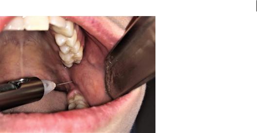
2.5.2.1 Conventional ‘Open-Mouth’ Technique
1)Position the patient in the dental chair, lying flat with head slightly extended and mouth open.
2)With the patient’s mouth open, retract the buccal tissues to place tension on the pterygomandibular soft tissues and obtain sufficient vision and access. The pterygomandibular raphe can usually be visualised. Palpate the anterior border of the ramus to confirm the anatomic location of the pterygomandibular fold.
3)The site of needle penetration is approximately 1 cm above the occlusal plane of the mandibular teeth, into the pterygotemporal depression (Figure 2.5). The angle of insertion of the needle should be such that the barrel of the syringe is aligned with the contralateral premolar teeth. Advance the needle slowly through the soft tissues. As the needle is advanced, it will pierce through buccinator, through to the pterygomandibular space. If the orientation is correct, the needle will contact the medial aspect of the bony ramus near the mandibular canal after having advanced 2–2.5 cm.
4)Retract the needle 1–2 mm so that the needle point is within the pterygomandibular space. Confirm a negative aspirate using the syringe plunger.
5)Deposit the anaesthetic solution slowly; a slow rate of injection significantly reduces discomfort for the patient.
6)Allow the local anaesthetic sufficient time to anaesthetise the tissues, based upon the pharmacokinetic properties of the solution, and monitor the patient for any adverse reaction.
This technique can be difficult when patients have severe trismus, have a prognathic or retrognathic mandible, or are edentulous, or when there is excessive adiposity, making it difficult to locate anatomical landmarks.
If bone is contacted prematurely, then the needle is likely to be orientated too far anteriorly, against the anterior border of the ramus or temporal crest. The needle will need to be directed more posteriorly in order to deposit the anaesthesia in the correct region; that is, adjacent to the mandibular canal.
Care must be taken to not advance the needle too far posteriorly. The posterior wall of the pterygomandibular space is formed by the parotid gland, in which the motor branches of the facial
https://t.me/DentalBooksWorld
2.5 asic Oal Anaesthesia echniiqes 23
Figure 2.5 Conventional‘open-mouth’technique.
nerve course. If local anaesthetic solution is inadvertently injected into the parotid gland, the patient will develop a transient facial nerve palsy for the duration of effect of the local anaesthetic. Local anaesthetic solution should not be deposited until bony contact is noted against the needle point during insertion, confirming the correct location of the needle.
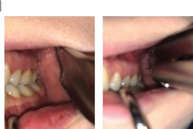
2.5.2.2 Akinosi ‘Closed-Mouth’ Technique
The ‘closed-mouth’ or Akinosi block can be utilised to anaesthetise the inferior alveolar nerve and the lingual nerve. It is useful in cases where there is severe trismus or macroglossia, or if the patient has a prominent gag reflex.
1)Position the patient in the dental chair, lying flat with head slightly extended.
2)Retract the buccal tissues to place tension on the pterygomandibular soft tissues and obtain sufficient vision and access (Figure 2.6).
3)Slowly advance the needle buccal to the maxillary molars, parallel to the occlusal plane at the level of the gingival margin of the maxillary teeth.
4)Advancing the needle in this manner, pierce the mucosa overlying the medial aspect of the mandible. Continue to advance approximately 2 cm. Because of the orientation of the needle, there is no bony anatomic landmark that will indicate the correct location of the needle tip relative to the lingula; as such, it is not recommended to insert the needle further, as the parotid space will be entered.
5)Aspirate the syringe to ensure the needle point has not traversed the intravascular space of a blood vessel.
6)Deposit the anaesthetic solution slowly; a slow rate of injection significantly reduces discomfort for the patient.
7)Allow the local anaesthetic sufficient time to anaesthetise the tissues, based upon the pharmacokinetic properties of the solution, and monitor the patient for any adverse reaction.
2.5.2.3 Gow–Gates Technique
This is a well described but technically challenging method of obtaining mandibular anaesthesia that uses extraoral landmarks to guide the needle path of insertion. It results in deposition of anaesthetic
https://t.me/DentalBooksWorld
24 2 Local Anaesthesia
Figure 2.6 Akinosi‘closed-mouth’technique.SoqOce: Seth Delpachitra.
higher in the pterygomandibular space compared with the conventional and Akinosi techniques. The benefit of a Gow–Gates block is that the inferior alveolar, lingual, and long buccal nerves may all be anaesthetised in a single injection, reducing the need for multiple anaesthetic injections.
1)Position the patient in the dental chair, lying flat with head slightly extended and mouth open.
2)Orientate the syringe and needle along an axis formed between the tragus of the ear of the side of the patient being anaesthetised and the contralateral oral commissure (Figure 2.7).
3)Place the needle tip just distobuccal to the second maxillary molar tooth (where present), and slowly advance it into the buccal mucosa in this area. The needle will advance approximately 2.5 cm before contacting the bone of the pterygoid fovea on the mandibular condyle.
4)Aspirate the syringe to ensure the needle point has not traversed the intravascular space of a blood vessel.
5)Deposit the anaesthetic solution slowly; a slow rate of injection significantly reduces discomfort for the patient.
6)Allow the local anaesthetic sufficient time to anaesthetise the tissues, based upon the pharmacokinetic properties of the solution, and monitor the patient for any adverse reaction.
2.5.2.4Mandibular Long Buccal Block
The conventional and Akinosi block techniques do not anaesthetise the buccal mucosa of mandibular teeth; as such, in order to obtain total anaesthesia for the removal of such teeth, the buccal mucosa will need to be anaesthetised using a separate technique. The long buccal nerve is a branch of the third division of the trigeminal nerve, which travels inferoanteriorly along the condyle and external oblique ridge, descending at the anterior border of the ramus on the lateral aspect of the mandible and supplying the buccal mucosa.
1)Position the patient in the dental chair, lying flat with head slightly extended and mouth open.
2)Retract the buccal mucosa to keep the soft tissues under tension.
3)Palpate the external oblique ridge of the mandibular ramus, just posterior and lateral to the molar teeth.
4)Insert the needle into the mucosa just lateral to the external oblique ridge, to a depth of approximately 2 mm (Figure 2.8).
https://t.me/DentalBooksWorld