

9.5 Managemen tof Lat e Complicatio 117
There is no evidence for routine prescribing of postoperative antibiotics for simple dental extraction. Antibiotics should only be prescribed postoperatively if extraction is being performed as part of the management of an acute odontogenic abscess with facial swelling, or where there is prior evidence of periodontal soft tissue infection with suppuration. Teeth with irreversible pulpitis alone are unlikely to result in acute odontogenic infection following extraction.
9.4 24 -Hour On-Call Service and Tertiary Hospital Referral
It is essential that dental practices which offer a dentoalveolar surgery or extraction service provide a 24-hour on-call service or alternative plan in case complications occur out of hours. Rarely, complications of dental extraction may be life-threatening; this can occur at any time following extraction of teeth.
Upon assessment and identification of complications, patients may require transfer to a local oral and maxillofacial surgery department. Every dentoalveolar outpatient clinic should have planned referral pathways to the local hospital to facilitate this transfer as required. Verbal and written communication with a clinician in the receiving department is an important component of safe and appropriate patient handover.
9.5 Management of Late Complications
Whilst the list of intraoperative complications following dental extraction is extensive, there are fewer late complications that can occur. These are equally important, however, as they are the main reason that patients seek unplanned emergency treatment, which may occur out of hours. Patients may present in an unpredictable or delayed manner, and the surgeon must be well prepared to manage complications that require urgent treatment or hospital transfer.
9.5.1 Alveolar Osteitis
Alveolar osteitis, colloquially referred to as ‘dry socket’, occurs when the fibrin clot in the extraction socket is not maintained, resulting in an empty tooth socket exposed to the oral environment. The typical presentation will be severe pain developing 48 hours after a dental extraction procedure, associated with halitosis and malaise. Alveolar osteitis is not an infection per se, and so findings of fluctuance, vestibular swelling, discharge, or facial abscess are not associated with an isolated dry socket.
This condition is more common in females, usually occurs with extraction of mandibular molar teeth, and has an association with use of the oral contraceptive pill, poor oral hygiene, and smoking.
All patients should be advised of the risk of a dry socket developing after any dental extraction. Overall, the risk is approximately 5%, but patients should be advised that this may be higher when they have one or multiple risk factors present.
Management of alveolar osteitis includes pharmacological treatment with analgesia and chlorhexidine mouthwash and placement of an antiseptic or analgesic dressing such as Alvogyl. Alvogyl combines iodoform, eugenol, and butamben in a fibrous, nonabsorbable paste which has anaesthetic and antiseptic properties and typically provides immediate relief. Patients who have been treated with any dressing with a fibrous or nonresorbable component should be reviewed within 48 hours for its removal; left in situ, such a dressing may become a nidus of infection, requiring evacuation.
https://t.me/DentalBooksWorld

118 9 Postoperative Care and Late Complications
Surgical debridement and washout of the socket under local anaesthesia is another appropriate management strategy for alveolar osteitis, as this allows for removal of any debris and promotes formation of a new clot. However, this may cause additional discomfort to an already disconcerted patient, who will often opt against any further surgical intervention in the short term.
9.5.2 Acute Facial Abscess
Dentoalveolar extractions create an intraoral wound which is susceptible to developing infection. Postoperative infections will present with the cardinal signs of inflammation: swelling, redness, pain, and heat, and associated purulent discharge from the surgical site. Such infections may be small localised collections or a serious life-threatening infection involving deep spaces in the head and neck, leading to airway compromise and trismus.
Patients who have undergone dental extractions should always be provided with a postoperative plan which outlines what to do if a developing infection is suspected. At minimum, this should include urgent review by the treating surgeon.
On review, patients should be assessed for trismus, dysphagia, odynophagia, mouth opening, impending upper airway obstruction, and signs of systemic infection. Their oral intake should be assessed and a clinical examination should be undertaken to determine the extent of the swelling. Important features to note are swelling in the floor of the mouth or that which is firm and crosses the border of the mandible, any parapharyngeal bulging or deviation of the uvula from the midline, and any infection spreading and causing periorbital cellulitis.
Clinical signs and symptoms will dictate the appropriate management for patients who develop a postoperative infection. Infections of the buccal space, with swelling of the cheek and vestibule, generally tend to be minor and to be amenable to intraoral incision and drainage under local anaesthetic. Infections involving the submandibular or sublingual spaces tend to be more severe and much more likely to result in airway compromise – these always require hospital admission and a combination of intraand extraoral drainage. Radiographic investigation will help determine the possible cause. For example, a patient might develop an infection due to a retained root, bone sequestra, or a foreign body (dressings or other materials).
The presence of facial swelling with suspicion of abscess warrants immediate surgical intervention. Antibiotic therapy alone is not appropriate management for acute facial abscesses. The development of an abscess is a late sign of infection, and therefore surgical incision and drainage of the abscess, with washout of the involved fascial spaces, is the mainstay of management of this condition. Antibiotics are a useful adjunct treatment for managing the associated facial cellulitis, but are ineffective against mature, walled-off collections of pus.
Patients who develop severe infections with signs of airway compromise, fever, or trismus preventing oral intake require prompt referral via ambulance to a hospital-based oral and maxillofacial surgery service, where appropriate medical and surgical management can be initiated. These patients may progress to develop infections that involve deeper neck or thoracic spaces, requiring tracheostomy, multiple washout procedures, and a lengthy stay in an intensive care unit.
An acute facial infection may herald the development of osteomyelitis of the jaw: a catastrophic deep infection of the maxillary or mandibular bone. This is typically a disease of immunocompromised patients but can sometimes occur in healthy individuals. Early signs of osteomyelitis resemble those of acute odontogenic or postoperative infections and can be difficult to differentiate in the earlier stage of the disease process. Acute osteomyelitis can result in the accumulation of purulent discharge under the periosteum, with an associated periosteal reaction visible on radiographic imaging. The associated mucosa and skin are usually erythematous and very tender to palpation,
https://t.me/DentalBooksWorld

9.5 Managemen tof Lat e Complicatio 119
with discharge from multiple intraand extraoral fistulae. Osteomyelitis may also cause paraesthesia when the infection is present around the inferior alveolar canal. Radiographic features suggestive of osteomyelitis include a moth-eaten appearance with presence of large sequestra. Immediate referral to a hospital infectious diseases unit is recommended.
9.5.3 Postoperative Haemorrhage
Postoperative haemorrhage may present within the first few days after dentoalveolar extraction. Unlike with intraoperative haemorrhage, patients will already have been discharged, under the presumption that all bleeding has been adequately controlled. A slow postoperative haemorrhage can lead to significant blood loss; as patients are not under observation at home, occult bleeding may be missed, and it may be very late in the picture before they seek medical assistance.
There are two forms of postoperative bleeding in healthy patients: delayed primary bleeding and secondary bleeding. Delayed primary bleeding occurs within the first several hours post-dental extraction, and is caused by the waning of the vasoconstrictive effect of local anaesthetics. Secondary bleeding occurs days after extraction, and is usually related to infection producing an inflammatory and vasodilatatory response in the local tissues.
In the assessment of any patient with postoperative haemorrhage, the primary survey must be undertaken to assess their cardiovascular status and estimated blood loss. Large haemorrhages may cause haemodynamic compromise, which itself requires transfer to a local hospital service for resuscitation. Once the patient has been deemed to be haemodynamically stable, attempts should be made using local methods to control the bleeding (see Chapter 6). Simultaneously, the patient’s history should be revisited in order to determine any systemic medical conditions which might have contributed to the onset of the bleed. The patient should also be examined for any signs of developing soft tissue infection; if this is a potential cause of haemorrhage, antibiotic therapy may be indicated.
Once haemostasis has been achieved, the patient requires close follow-up after 24 hours and then after one week, to ensure that bleeding has subsided and any causes have been identified and managed appropriately.
9.5.4 Temporomandibular Joint Disorder
Temporomandibular joint disorders (TMDs) are common in the general population; a considerable proportion of patients presenting for dentoalveolar extractions will have pre-existing TMD. This should be documented in the patient history, and a comprehensive temporomandibular joint assessment should form part of the preoperative workup, including a record of maximal mouth opening, joint crepitus, hypermobility, locking, and pain on movement. It is uncommon to develop TMD de novo following dental extraction procedures; usually, there will be some evidence suggesting subclinical temporomandibular joint disease.
Patients with TMD may experience an exacerbation of their condition after extractions. This is largely due to the force applied to the joint and masticatory musculature during mandibular extractions, but it can be also related to prolonged mouth opening during maxillary ones. An efficient and safe operator is much less likely to cause TMD; appropriate and effective use of forces, early determination of the need for surgical extraction, and timely use of patient breaks between longer procedures prevent inadvertent stress on the temporomandibular joint.
Patients who develop symptoms of TMD following extraction will be understandably concerned regarding the onset of such syndromes. A full assessment and diagnosis should be undertaken to
https://t.me/DentalBooksWorld
120 9 Postoperative Care and Late Complications
rule out other causes of temporomandibular symptoms. If the patient’s presentation can be explained by intraoperative trauma or a prolonged and forceful procedure, the prognosis is generally good, and there will likely be a return to baseline after three months postoperatively. Advice on conservative management (including jaw rest, physiotherapy, and anti-inflammatory medications) should be provided, with regular follow-up to assess for clinical improvement. Rarely, patients may require advanced management by an oral medicine specialist or oral and maxillofacial surgeon, and a timely referral may be required.
9.5.5Epulis Granulomatosa
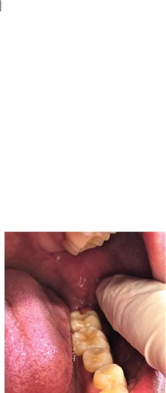
The normal healing of an extraction socket involves the formation of granulation tissue inside it, which is gradually replaced by bone and gingival soft tissue. This normal healing may be complicated by the development of hyperplastic granulation tissue that exudes out of the socket and into the oral cavity, giving the appearance of an epulis (Figure 9.1). This is usually a foreign-body reaction to debris or bony sequestra in the socket, as a result of inadequate debridement following extraction. Occasionally, haemostatic dressings or treatments used for alveolar osteitis may be implicated in this reaction.
Epulis granulomatosa is clinically indistinguishable from intra-alveolar squamous cell carcinoma or other giant cell lesions of the jaws. As such, the presence of this hyperplastic tissue warrants urgent biopsy for formal histopathologic diagnosis. Simultaneous curettage and debridement of the socket is often adequate to treat epulis granulomatosa and encourage normal healing.
Figure 9.1 Epulis granulomatosa following third molar removal.
https://t.me/DentalBooksWorld