

112 8 Management of the Medically Compromised Patient
8.8 The Irradiated Patient
Radiation therapy in conjunction with surgery is an established treatment modality for head and neck cancer patients. The effects of radiation devastate normal oral physiology, resulting in mucositis in the short term and hyposalivation, caries, periodontal disease, and scarring in the long term.
A critical side effect of radiation to the head and neck is the risk of osteoradionecrosis (ORN) of the jaws. Osteoradionecrosis presents as an area of exposed, devitalised bone that has previously been irradiated which fails to heal over a period of greater than three months in the absence of recurrent neoplastic disease. Irradiated bone is said to become hypoxic, hypocellular, and hypovascular, with a resulting diminished ability to withstand trauma and infection. These conditions are what is thought to precipitate the development of osteoradionecrosis. Furthermore, the risk of osteoradionecrosis is lifelong, and there is no amount of time elapsed after which it is considered ‘safe’ to remove teeth in the radiation field.
Osteoradionecrosis may occur spontaneously, but there is often an antecedent dentoalveolar injury such as an extraction which precipitates its development. Osteonecrosis has a wide array of severity, from a small asymptomatic area of exposed bone, to extensive exposure associated with pathologic fracture, extraoral fistulae, or lytic lesions involving the nasal and paranasal sinuses. Advanced osteoradionecrosis may require major and lengthy surgical management, including resection and free flap reconstruction, and can pose significant challenges to the patient and their treating medical and surgical teams.
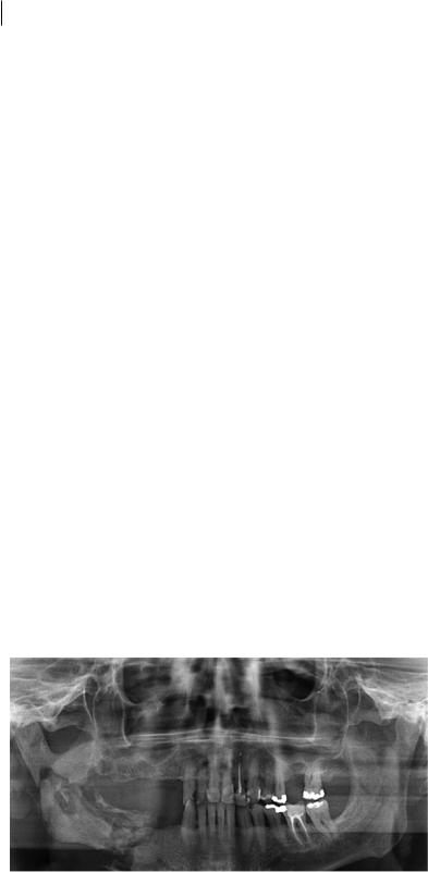
Osteoradionecrosis is predominantly a disease of the mandible, and usually presents in the mandibular body (Figure 8.2). It is most commonly seen in cases where the mandible or maxilla receive a cumulative dose of more than 60 Gray of radiation. A number of other factors contribute to osteoradionecrosis risk, including tumour site and size, malnutrition, poor oral hygiene, and immunosuppression. Surgical extraction of posterior mandible teeth with roots below the mylohyoid line has the highest risk of developing osteoradionecrosis in at-risk groups.
The risk for developing osteoradionecrosis in the past was considerably high, estimated at up to 35%. However, with intensity-modulated radiation therapy and growing awareness about the dental considerations, more recent estimates are lower, with some around 5%.
Patients who have been diagnosed with head and neck cancer are managed by multidisciplinary teams in the hospital setting. Major hospitals may manage all aspects of the patient’s treatment, including dentoalveolar extractions, but in rural settings these may be outsourced to public or private dental clinics.
Figure 8.2 Panoramic radiograph of a patient with end-stage osteoradionecrosis causing mandible fracture.
https://t.me/DentalBooksWorld

8.9 Hepati c Rena l Impairme 113
8.8.1 Management of the Patient with a History of Head and Neck Radiotherapy
Hyperbaric oxygen (HBO) therapy has been considered for patients requiring dental extractions and for those with established osteoradionecrosis. The role of HBO is controversial, as there are conflicting results from the various studies conducted on it to date. Therefore, patients who are likely candidates for HBO should be referred to a specialist oral and maxillofacial surgery unit.
Antibiotic prophylaxis may be useful in reducing the risk of development of osteoradionecrosis following dental extractions, but the risk reduction might be as small as 1%. Generally, antibiotic prophylaxis used in osteoradionecrosis is prescribed similarly to that used in infective endocarditis prophylaxis: a single, high-dose antibiotic providing an appropriate spectrum for oral bacteria is prescribed preoperatively. Local guidelines should be employed where a decision to use antibiotic prophylaxis is made.
8.9 Hepatic or Renal Impairment
The liver and kidneys have synergistic functions in the human body, involved in fluid and electrolyte balance, secretion of wastes, hormone production, and overall metabolism. Hepatic or renal impairment can be found in isolation, but in many patients there will be a combination of liver and kidney dysfunctions due to the intimate physiologic relationship between these two organs. There is a litany of medical aetiologies which culminate in the development of hepatorenal impairment, leading to a wide range of clinical presentations of patients in this disease category.
The sequelae of hepatic or renal impairment on patient physiology can be complex and may influence both the decision to proceed with dentoalveolar surgery and the expected postoperative outcomes. Using the framework described at the start of this chapter, outlining the effects of the disease on planned dentoalveolar surgery, outlining the effect of planned surgery on the disease, and recognising the effects of prescribed medication on the surgery is beneficial in such complex conditions (see Table 8.8).
Table 8.8 Considerations in patients with hepatorenal disease undergoing surgery.
Hepatic |
Renal |
Effect of disease on surgery
Patient’s liver failure may be related to hepatitis B and C, which are communicable diseases
Postoperative medications require hepatic-adjusted dosing
Antibiotic prophylaxis may be indicated
Patients with advanced liver disease may have an associated coagulopathy, increasing bleeding risk
Anaemia of chronic disease may result in a low preoperative haemoglobin level, necessitating specialist management
Postoperative medications require renally adjusted dosing
Antibiotic prophylaxis may be indicated
Hypertension is common, and is usually more severe in patients with a history of renal impairment
Effect of surgery on disease
Postoperative paracetamol and opiates should be avoided, as these may result in acute liver failure
Postoperative NSAIDs should be avoided, as these may result in acute renal failure
Effect of medications on surgery
Patients with end-stage hepatic failure may be using a number of medications that can affect surgery
Patients requiring haemodialysis are anticoagulated during and after dialysis, increasing bleeding risk
https://t.me/DentalBooksWorld

114 8 Management of the Medically Compromised Patient
8.10 Pregnancy and Lactation
When caring for women who are pregnant or lactating, the surgeon should always consider the benefits and risk associated with carrying out a procedure. As a rule, any elective dental treatment, including dentoalveolar surgery, should be postponed until after delivery. Surgical and psychologic stress, postoperative medications, and radiation associated with dental x-rays are best avoided whilst a patient is pregnant.
Dentoalveolar extractions should only be carried out in the pregnant patient if absolutely necessary (e.g. in the case of acute odontogenic abscess). Alternative dental treatments (including endodontic therapy) should be considered, where appropriate. In situations where dental extraction is the only option, referral to a multidisciplinary service with obstetric medicine input is recommended.
https://t.me/DentalBooksWorld

115
9
Postoperative Care and Late Complications
Postoperative care is a critical component of the patient’s journey from treatment planning through to completion of extraction therapy. Adequate provision of early postoperative management and sufficient home care instructions, both written and verbal, can significantly reduce the risk of shortand medium-term complications.
9.1 Immediate Postoperative Period
Immediately following completion of a dental extraction, the following procedures must be performed:
●●Adequate Haemostasis of the Surgical Site. One of the most common presentations following dental extraction is the presence of ongoing bleeding from the surgical site. It is simple to diagnose the cause of post-extraction bleeding in patients with medical comorbidities; in otherwise healthy patients, a more strident approach should be taken. In all patients, regardless of other haemostatic measures utilised during the procedure, simple prevention of surgical-site bleeding can be obtained by immediately placing moist gauze at the site and asking the patient to bite firmly for approximately 10 minutes post-procedure. At the end of the 10-minute period, the gauze should be removed, and the site checked for adequate haemostasis. If bleeding continues, this requires management as outlined in Chapter 6 prior to patient discharge.
●●Temporary Avoidance of Oral Intake or Expectoration. Following extractions, patients will commonly want to rinse their mouth or expectorate. This can disrupt formation of a stable clot in the fresh extraction socket. It is recommended that patients avoid oral intake, rinsing, or spitting for 30 minutes post-extraction. This needs to be explained to them clearly immediately after removal of haemostatic gauze.
●●Assessment of Patient Wellbeing. Dental extraction can sometimes be an overwhelming experience for patients. This, combined with their remaining in the supine position for prolonged periods, can lead to postural hypotension or vasovagal syncope in the moments following extraction. A period of rest in the dental chair is recommended to mitigate the risk of fainting. Postural hypotension is much more likely in elderly patients with a history of antihypertensive medication use. Patient wellbeing should be assessed in an informal manner using open-ended questions. Whilst consumption of fluids is a relative contraindication in the first half hour following an extraction, it may be necessary to provide cool water or a glucose drink if the patient feels faint.
Principles of Dentoalveolar Extractions, First Edition. Seth Delpachitra, Anton Sklavos and Ricky Kumar. © 2021 John Wiley & Sons Ltd. Published 2021 by John Wiley & Sons Ltd.
Companion website: www.wiley.com/go/delpachitradentoalveolarextractions
https://t.me/DentalBooksWorld

116 9 Postoperative Care and Late Complications
9.2 Postoperative Instructions
Postoperative instructions should be provided in verbal and written format, and should generally cover the following information:
●●Summary of Procedure Performed. This should include a list of which teeth were removed and information on whether any changes to the plan of dental extractions were made during the procedure.
●●Open Disclosure Regarding Intraoperative Complications. Any complications, including damage to other teeth, restorations, or vital structures, should be relayed to the patient. If further surgery or referral is required (e.g. in the case of retained roots or oroantral communication), the patient should be informed of the likely need for repeat surgery with referral to an oral and maxillofacial surgeon.
●●Expected Postoperative Course. Depending on the number and type of teeth extracted, the normal postoperative course can vary significantly between patients. The patient must be made aware of the expected nature and intensity of any pain, swelling, bruising, or minor bleeding that might occur. Information on home management of these symptoms should also be provided, regarding use of analgesia, ice packs, and jaw rest.
●●Red Flags for Common and Rare Complications. The patient must be educated on the early warning signs that suggest impending postoperative infection, haematoma, or long-term nerve injury. Early reassessment and appropriate transfer is the key to reducing postoperative morbidity from complications.
●●Planned Reviews and Monitoring. The patient should be informed of all postoperative follow-up appointments. Generally, in low-risk patients with no medical comorbidities, a oneweek planned review is sufficient. In patients with comorbidities or light bleeding risk, a 24-hour review followed by a one-week review ensures early identification of complications.
●●Postoperative Medications and Instructions for Use. Any postoperative medications prescribed should have clear, written instructions for use.
●●Oral Hygiene and Nutrition Instructions. Patients may be unsure about how to care for their oral cavity following dental extraction. The pain and nausea associated with surgery may limit oral intake, resulting in dehydration or malnutrition.
●●Self-Care Advice. Instructions on the avoidance of alcohol and smoking, the need to obtain adequate rest, and the avoidance of exercise can be useful in guiding patients through a smooth recovery period.
9.3 Postoperative Medications
Simple analgesia is often sufficient to manage the minor discomfort following dental extraction. Nonsteroidal anti-inflammatory drugs (NSAIDs) tend to work most effectively and are readily available without prescription. Paracetamol (acetaminophen) can be used in conjunction with NSAIDs if additional analgesia is required.
In lieu of saline mouthwashes, an antiseptic mouthwash such as aqueous chlorhexidine gluconate 0.2% solution has been shown to reduce the incidence of alveolar osteitis and should be considered in high-risk patients with poor oral hygiene or a history of smoking. Use of chlorhexidine mouthwash should be limited to a maximum duration of two weeks, as it can cause significant tooth discoloration.
https://t.me/DentalBooksWorld