


2 1 Principles of Surgery
undergoing tooth extraction as part of a wider treatment plan where multiple other medical comorbidities require interdisciplinary management. In such situations, good communication minimises delays to receiving time-critical treatment, such as in the case of dental extractions prior to head and neck radiotherapy or bisphosphonate treatment.
●●An Individualistic Approach. Patients will have a wide variety of backgrounds, demands, and prior medical knowledge. A tailored and individualised approach is required in order to ensure they understand the proposed procedure, its risks, and its expected outcomes, and are able to compare options in order to make an informed decision.
●●Leadership and Management. A surgeon will often find themselves the leader of a multifaceted treatment team, including nursing staff, dental assistants, anaesthetic staff, and sterilisation technicians. This leadership comes with great responsibility: the expert surgeon must guide others in the team, provide feedback and education, and thus help maintain a standard of excellence. The surgeon must ensure that all staff are orientated towards the goal of achieving the best outcome for the patient. When the highest standard of care is compromised, the responsibility is on the surgeon to make sure the team gets back on track.
●●Decision Making. The word ‘decision’ shares a common root with another word often associated with surgery: incision. Both are derivatives from the Latin word caedis, meaning ‘to cut’. Incision means to cut into something, such as the operative site; decision literally means ‘to cut away’. A decision thus precludes other options, and sets one upon a particular course of action. A skilled surgeon will be able to make treatment planning decisions that are in the best interests of their patients.
1.1 Wound Healing
Good outcomes following surgery depend on satisfactory wound healing. This involves a range of inflammatory, biochemical, and physiologic changes at the operative site, which will ultimately lead to resolution, healing, and bone remodelling. Wound healing does not always follow a predictable course, and therefore an understanding of its key aspects will serve as a foundation for interpreting clinical signs and determining when it is compromised.
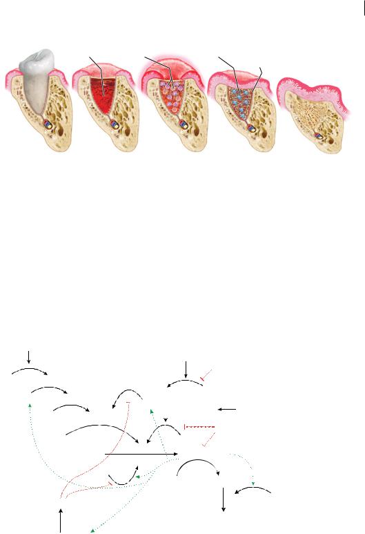
There are four key stages in wound healing (Figure 1.1):
1)Haemostasis.
2)Inflammatory phase.
3)Proliferative phase.
4)Remodelling and resolution.
An interruption at any one of these stages will lead to a protracted recovery period.
1.1.1 Haemostasis
Any tissue trauma will result in bleeding from the local vasculature supplying the tissues. The immediate physiologic reaction is haemostasis, which involves reactive vasospasm, formation of a platelet plug, and activation of the coagulation cascade.
Reactive vasospasm occurs in the seconds to minutes following damage to the blood vessels. This is mediated through neurologic mechanisms, as well as the local release of endothelin. It rapidly reduces blood loss from trauma. In surgery, exogenous vasoactive medications such as adrenaline utilise this response to improve visual access to the surgical field by reducing blood flow.
https://t.me/DentalBooksWorld
1.1 ounnd eeling 3
Swelling, redness, heat and pain
Epithelial cell overgrowth
Fibrin clot |
Phagocytes |
Osteoblasts |
Angiogenesis
Pre-Extraction |
Haemostasis |
Inflammatory Stage |
Proliferative Stage |
Remodeling Stage |
|
(1 second–minutes) |
(1–3 days) |
(3 days-–3 weeks) |
(3 weeks–8 weeks) |
Figure 1.1 Phases of wound healing.
Damaged endothelial cells result in a conformational change in von Willebrand factor expressed on the cell surface. Von Willebrand factor interacts with glycoprotein Ib on circulating platelets, resulting in activation and aggregation of the platelets, forming links to fibrinogen via the GpIIb/ IIIa receptor. This leads to the formation of the platelet plug. Antiplatelet medications inhibit aspects of platelet plug formation and increase the risk of bleeding during surgical procedures.
The coagulation cascade is a series of successive reactions that occur in order to activate thrombin and form a stable fibrin clot (Figure 1.2). There are two pathways in this cascade: intrinsic and extrinsic. The intrinsic pathway is activated within the vascular system through exposure
Contact activation |
|
|
|
Tissue factor |
||
(intrinsic) pathway |
|
|
|
(extrinsic) pathway |
||
Damaged surface |
|
|
|
|
|
|
|
|
|
|
Trauma |
|
|
|
|
|
|
|
TFPI |
|
XII |
XIIa |
|
|
|
|
|
XI |
XIa |
|
|
VIIa |
VII |
|
|
VIII |
|
|
|
||
|
|
|
Tissue factor |
Trauma |
||
|
IX |
IXa VIIIa |
|
|||
|
|
|
|
|
||
|
|
|
|
|
Antithrombin |
|
|
X |
|
|
|
X |
|
|
Prothrombin (II) |
Xa |
|
Thrombin (IIa) |
Common |
|
|
Va |
|
||||
|
|
V |
|
|
pathway |
|
|
|
|
|
|
|
|
|
|
|
|
Fibrinogen (I) Fibrin (Ia) |
|
|
|
Active Protein C |
|
|
XIIIa |
XIII |
|
|
|
|
|
|
||
Protein S |
Cross-linked |
|
fibrin clot |
||
|
||
Protein C + Thrombomodulin |
|
Figure 1.2 The coagulation cascade.
https://t.me/DentalBooksWorld

4 1 Principles of Surgery
to endothelial collagen, whilst the extrinsic pathway is activated by tissue trauma and release of intracellular tissue factor. Anticoagulant medications and coagulopathies increase the tendency to bleed by inhibiting aspects of the coagulation cascade, and awareness of these effects may be clinically relevant in surgical planning.
Coagulation studies used clinically can assess the function of either the intrinsic, the extrinsic, or the shared common pathway. Prothrombin time screens for factors II, V, VII, and X and fibrinogen; these are all part of the extrinsic pathway, which is used to guide treatment for patients treated with warfarin. Warfarin inhibits vitamin K-dependent factors common to both pathways, but because factor VII has the shortest half-life, the extrinsic pathway is used to determine coagulability. The partial thromboplastin time will screen for factors in the intrinsic pathway affected by medications such as heparin and low-molecular-weight heparin.
1.1.2 Inflammatory Phase
This will commence on day one after the procedure and will continue for approximately three days. Important aspects of the inflammatory response include the release of pro-inflammatory mediators and vasoactive factors such as the prostaglandins, leukotrienes, interleukins, and histamine, and recruitment of phagocytes to remove dead tissue and foreign debris. The inflammatory mediators lead to the swelling, redness, heat, pain, and loss of function associated with inflammation. Anti-inflammatory medications are commonly prescribed after dentoalveolar procedures in order to mitigate the postoperative pain and swelling.
1.1.3 Proliferative Phase
This typically starts around day three and lasts for up to three weeks. The proliferative phase relies on the formation of granulation tissue and type III collagen, mediated by fibroblasts; wound contraction starts due to the action of myofibroblasts. Angiogenesis takes place as new capillaries are formed to provide blood and nutrients in order to help the wound heal. A number of growth factors, including vascular endothelial growth factor (VEGF), are also involved. At the wound edges, epithelial cells proliferate and begin to grow over the granulation tissue scaffold that has formed. Bone healing starts to take place as osteoprogenitor cells arrive, differentiating into osteoblasts, which begin depositing an osteoid matrix. Note that any systemic conditions or medications which prevent or suppress components of angiogenesis or inflammation may delay or prolong healing.
1.1.4 Remodelling and Resolution
At the completion of three weeks of healing, granulation tissue and immature bone will fill the extraction site, and the socket should be completely covered by a layer of epithelium. Bone remodelling will continue to take place with active resorption and deposition mediated by osteoblasts and osteoclasts. This important step can be impeded by medications that inhibit osteoclast function, such as bisphosphonates or denosumab. Radiographic evidence of bone remodelling will not become evident until after six to eight weeks.
1.2 Patient Assessment
Any medical or dental intervention requires a comprehensive patient history. This includes: a detailed medical history, including current and past medical treatments; documentation of known drug allergies and reactions; a social history comprising occupation and use of alcohol, cigarettes,
https://t.me/DentalBooksWorld
1.3 endiogreppic ssesssenn 5
and illicit substances; prior surgeries, dental treatments, and adverse outcomes; and, finally, the patient’s chief complaint or main concerns.
Secondary to this is the clinical assessment of the patient’s orofacial region, including both extraoral and intraoral examination. This should include an assessment of the temporomandibular joint, soft and hard tissue pathologies, and the presence of any dental pathology. Simultaneously, a difficulty and risk assessment for dentoalveolar surgery can be undertaken, paying particular attention to mouth opening, gingival biotype, gag reflex, patient anxiety, and previous heavily restored dentition.
Diagnostic tests should be carried out as necessary, including pulp testing, palpation for mobility, and percussion testing. A periodontal probe can be used to examine partially erupted or unerupted teeth, to assess soft tissue opercula, or to explore other communications with the oral cavity.
The patient’s psychological state and level of anxiety can be assessed by asking how well they have tolerated dental treatment in the past. This aspect of the assessment is important; by virtue of temperament, some patients will require a more detailed discussion about their treatment, and some may request or require treatment with sedation or general anaesthesia.
1.3RadiographicAssessment
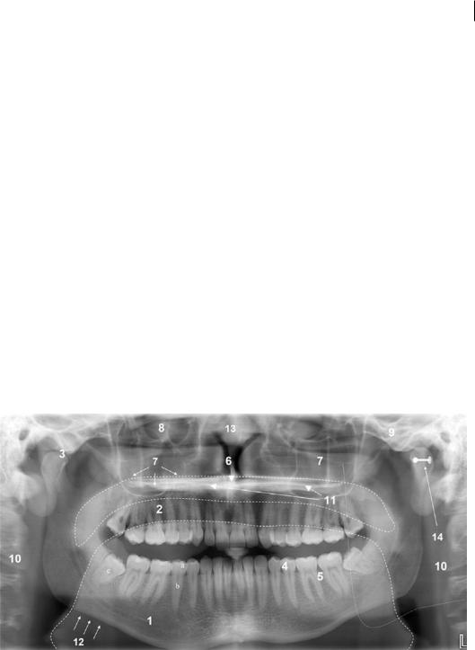
Plain-film orthopantomogram (OPG), periapical (PA) radiograph, and the three-dimensional (3D) cone-beam computed tomogram (CBCT) are the primary imaging modalities used in the assessment of patients prior to dental extractions. As part of the radiographic workup for dental extraction, the minimum required imaging of the tooth for extraction is an intraoral PA radiograph. Where multiple teeth are indicated for extraction, or where third molars are being assessed, a panoramic radiograph is the minimum requirement instead (Figure 1.3).
Figure 1.3 Panoramic radiograph: 1. mandible; 2. maxilla; 3. temporomandibular joint; 4. dentition; 5. alveolar process and periodontium; 6. anterior nasal spine; 7. maxillary antrum; 8. orbit; 9. zygoma;
10.cervical spine (double image); 11.hard palate (double image); 12.lower border of mandible; 13.nasal septum; 14. earring causing artifact. Outlines show oroand nasopharyngeal airspaces and double image of mandibular ramus (left side).Source: Reproduced from The panoramic dental radiograph for emergency physicians by Anton Sklavos, Daniel Beteramia, Seth Navinda Delpachitra, Ricky Kumar, BMJ 36: 565–571. doi:10.1136/emermed-2018-208332. Copyright © 2019 with permission from BMJ Publishing Group Ltd.
https://t.me/DentalBooksWorld
6 1 Principles of Surgery
A procedure should not be undertaken without a diagnostic radiograph, displaying the condition of the tooth, the relevant adjacent structures (inferior alveolar nerve canal, mental foramen, maxillary antrum), and the condition of the adjacent teeth. Generally, a PA radiograph has a limited application and should only be used for emergency single-tooth extractions or procedures limited to one part of the mouth performed under local anaesthesia. A PA radiograph is not considered an appropriate assessment for proximity of the inferior alveolar canal to the lower third molar teeth; errors in patient or film positioning may alter the radiographic relationship between the canal and associated tooth roots, producing a nondiagnostic image.
The OPG gives a better indication of the overall state of the patient’s dentition and of other pathological conditions that may affect the maxillofacial region compared to a PA radiograph. Because the OPG is taken in a standardised manner, it has formed the basis for a number of evi- dence-based risk-assessment tools in the current literature, including assessments of the risk of oroantral communication and inferior alveolar nerve injury.
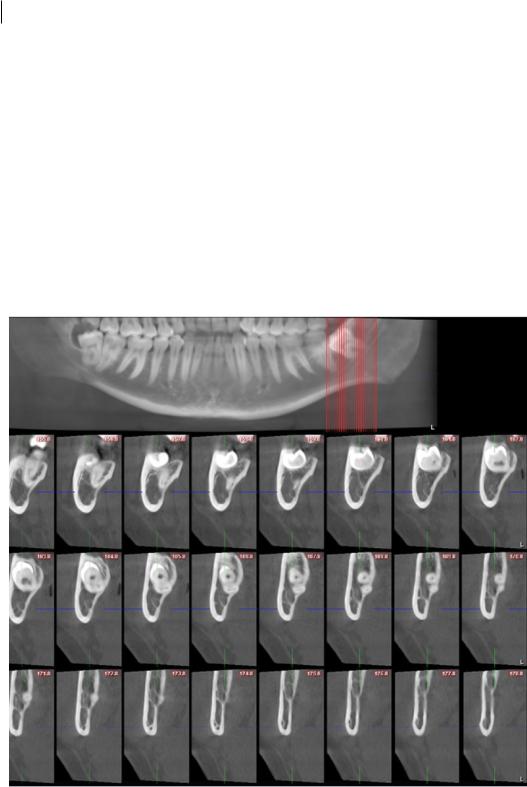
CBCT is a relatively new and inexpensive method of producing 3D images of maxillomandibular structures (Figure 1.4). It is indicated when conventional 2D imaging does not produce enough diagnostic information for treatment planning. Most commonly, this is to investigate jaw
Figure 1.4 Slices of a serial transaxial CBCT.
https://t.me/DentalBooksWorld
1.5 Anaesthesi 7
pathology or the relationship of the inferior alveolar nerve canal to third molar teeth. Whilst CBCT images come in a variety of formats, the serial-transaxial reformat is useful for the determination of the pathway of the inferior alveolar nerve and its relationship to the mandibular teeth.
CBCT images provide a great deal more information than plain-film imaging, and therefore their interpretation can be challenging. For the surgeon who is not experienced with CBCT images, formal reporting by a specialist radiologist is essential; if they are not interpreted carefully, radiographic signs of nerve risk or bone pathology may be easily missed, likely resulting in a poor outcome for the patient and a litigious scenario for the surgeon.
1.4 Informed Consent
In order to carry out any medical or dental procedure, consent must be obtained from either the patient or their legal guardian. A valid consent may only be given when the patient possesses decision-making capacity; that is, when they have the ability to weigh up the pros and cons of treatment and come to a sound decision to either undergo or forgo a proposed procedure. This consent must be given voluntarily, without duress or coercion. The decision must be informed, which is to say that the patient must understand the procedure, the expected outcomes, the risks involved, the anticipated recovery time, and the costs of treatment.
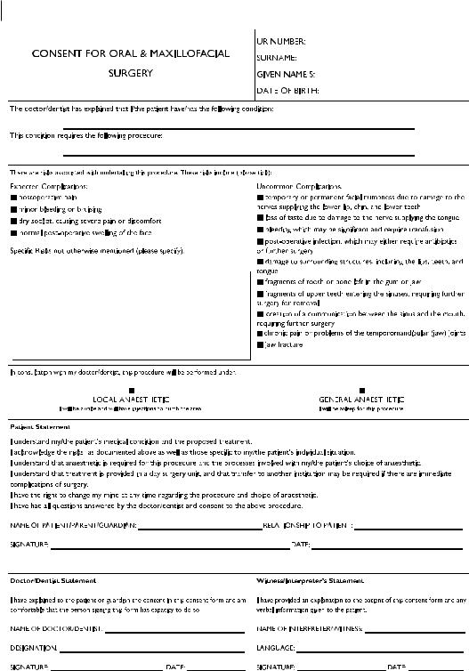
For any planned surgical procedure, consent should be both written and verbal, and patients should be provided by the surgeon with a formal document detailing the procedure and its indications, risks, and expected outcomes (Figure 1.5). When seeking consent from a patient, the surgeon must always provide them sufficient time to ask questions. The patient may request more detailed information about particular risks or outcomes of surgery, and these should be elaborated on and explained in detail.
In addition, the consent should be specific for the particular patient and their individual circumstances. Alternatives to dentoalveolar extraction should be explained, and the reasons for and against their adoption detailed. In cases where extractions will render the patient without functional teeth, future treatment options for replacement should be discussed; this should include an approximate timeline, who will provide the treatment, and a rough cost estimate.
1.5 Anaesthesia
There are a number of different methods of anaesthesia available for dentoalveolar extractions. These include:
●●Local anaesthesia only.
●●Local anaesthesia with relative analgesia.
●●Local anaesthesia with minor oral sedation.
●●Local anaesthesia with intravenous (IV) sedation.
●●Local anaesthesia with general anaesthesia.
Local anaesthesia alone can be administered for in-chair treatment, and is generally considered the safest option. It is appropriate for most simple dentoalveolar procedures and extractions. The main limitation of local anaesthesia is that it may be unsuitable for complex procedures, when a duration of more than 40 minutes is anticipated, or when the patient is anxious or otherwise uncooperative.
https://t.me/DentalBooksWorld
8 1 Principles of Surgery
Figure 1.5 Example consent form for dentoalveolar surgery.
https://t.me/DentalBooksWorld

1.6 Preparatio n o Equipmen 9
Table 1.1 Comparisons between commonly used oral benzodiazepine sedatives.
Drug name |
Dose |
Onset |
|
|
|
Temazepam |
10–20 mg |
30–120 minutes |
Diazepam |
10–15 mg |
30–90 minutes |
Oxazepam |
15–30 mg |
2–3 hours |
|
|
|
Relative analgesia involves the use of inhaled agents, such as nitrous oxide, to produce conscious sedation. It can be used as an adjunct with local anaesthesia, to improve patient comfort without significant airway risk. Nitrous oxide is usually administered through a nasal hood at concentrations of 50–70%. The advantage of this method is that it can be titrated and rapidly adjusted. However, there is a variable dose–response relationship between individuals, and patients may experience a number of unpleasant side effects if too high a dose is administered. Use of relative analgesia requires additional training, and it is recommended that at least two trained personnel are present in the clinic room when employing this technique.
Oral sedation, when employed effectively, can provide a greater level of sedation than nitrous oxide. Common drug classes used for oral sedation include benzodiazepine and barbiturate medications (Table 1.1). These drugs have a significant depressant effect on the central nervous system, and hence carry the serious risk of respiratory depression and loss of airway reflexes. Therefore, the use of oral sedation should only be considered when the surgeon and clinic personnel are sufficiently trained in anaesthesia, resuscitation, and airway management.
IV sedation typically involves more powerful sedative agents, such as midazolam, propofol, or fentanyl. It can only be administered in a setting which is fully prepared for airway management, such as a hospital theatre. As with all methods of anaesthesia where the airway is not secure, there is a risk that the patient will aspirate on any foreign object present in their mouth. This can be particularly dangerous when their reflexes are not protecting the airway. The administration of IV sedation may only be undertaken by a suitably qualified medical specialist or dental specialist with advanced training.
General anaesthesia will render the patient completely unconscious, and involves securing the airway using a laryngeal mask or endotracheal tube. It should be provided by a specialist anaesthetist or a suitably qualified medical professional. Dentoalveolar extractions carried out under general anaesthesia involve a shared airway, and communication with the anaesthetist throughout the procedure is essential.
1.6 Preparation of Equipment
Preparation for dentoalveolar extractions must follow the principles of asepsis, with strict maintenance of an aseptic operative field (Figure 1.6). The fundamental reason for this is to prevent transmission of microorganisms, which may cause surgical-site infections, transmit bloodborne diseases, and prolong postoperative healing. Inadequate or insufficient adherence to appropriate aseptic techniques or inadequate sterilisation of surgical instruments can result in harm to patients.
In hospital settings, sterilisation of instruments is usually performed through a hospital-wide central sterile services department. In the clinic setting, surgeons and their staff are responsible for the setup and proper maintenance of appropriate sterilisation facilities.
https://t.me/DentalBooksWorld