

705 Surgical Extraction Techniques
5)Avoid Incisions that Transect the Crestal Gingiva on the Buccal Convexities of the Teeth. The gingiva is thinnest and under most tension at the midpoint of the buccal gingiva. Incisions in this area that are not carefully repaired can lead to long-term periodontal dehiscence, which may require future periodontal surgery to normalise the clinical crown length. If a relieving incision is to be used that transects the gingiva, it should be closer to the interdental papillae, as these areas are much less likely to dehisce due to increased tissue bulk.
6)Avoid Incising Near Neurovascular Structures. The mental foramen, inferior orbital foramen, lingual nerve, incisive nerve, and greater palatine artery are all susceptible to iatrogenic injury if their position is not factored into the design of a soft tissue flap. Transection of these structures will result in irreversible anaesthesia, paraesthesia, or dysaesthesia for the patient.
7)Use Sharp, Clean, and Precise Incisions to Enable Apposition of the Soft Tissues During Closure. Jagged or poorly defined edges result in a less than ideal closure, which can lead to wound breakdown and future localised periodontal disease.
8)Follow the Cleavage Planes of the Soft Tissues. A mucoperiosteal flap is most robust when all layers (mucosa, submucosa, muscle, and periosteum) are raised together. Failure to maintain a subperiosteal plane predisposes the flap to avascularity or tearing.
5.3 Common Soft Tissue Flaps for Dental Extraction
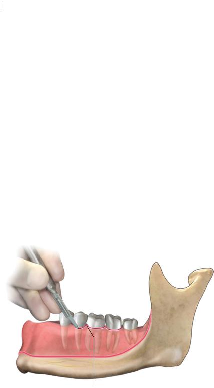
A crestal or envelope approach involves an incision made in the sulcus of the gingiva surrounding teeth and elevation of a mucoperiosteal flap from the bony alveolar crest away from the tooth (Figure 5.1). The gingival papillae are incised between the teeth at the interproximal col to provide as long and broad a pedicle as possible, and to maintain the blood supply to both the buccal and the lingual sides of the transected papillary tip. The envelope approach provides excellent access to the crestal alveolar bone and periodontal structures, but access is limited to the stretch of the
Gingival sulcular incison
Gingival papillae
Figure 5.1 Crestal-only flap with no relieving incision.
https://t.me/DentalBooksWorld
5.3 oo o oc tAASp alrsA o Pf pocral EcPract o 71
keratinised gingival tissue. If deeper apical areas of the tooth or bone require access, use of a crestal incision alone can lead to unfavourable tearing of the flap, which will require careful apposition.
Raising of a crestal flap commences with the sharp gingival sulcular incision along the region of keratinised gingiva that is to be elevated from the bone. A fine scalpel, such as an #11 or #15 blade, is best suited for this task.
Once the sulcus is incised, the pointed end of a periosteal elevator should first be used to free the gingival papillae away from the interdental areas. This area is usually the most tethered, and freeing of the papillae early helps to prevent flap tearing along the remainder of the gingival margin. Once the papillae have been freed, the remainder of the flap can be elevated from the bone, taking care to remain in the subperiosteal plane at all times.
After the surgical procedure is completed, apposition of the flap should be undertaken with interdental mattress sutures to correctly apposition the gingival papillae.
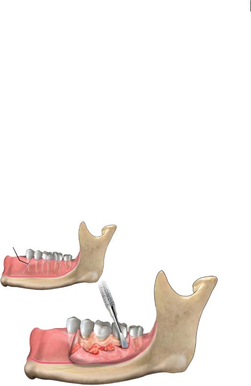
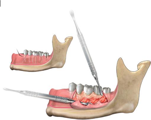
Relieving incisions are apical extensions of a crestal flap that can be used to improve bony exposure through transection of the keratinised gingival margin. Relieving incisions may be placed at the mesial end of the incision (Figure 5.2), at the distal end, or at both the mesial and the distal ends (Figure 5.3). The decision as to where a relieving incision should be made depends upon the area and amount of access required and the surrounding neurovascular structures, with an emphasis on reducing damage to the periodontal tissues as much as possible. Relieving incisions cannot be used alone, as they do not provide access to the required underlying tissues without an associated crestal component.
Mesial relieving incision
Flap elevated with broad end of periosteal elevator
Figure 5.2 Two-sided flap,consisting of a crestal flap with mesial relieving incision.
https://t.me/DentalBooksWorld
72 5 Surgical Extraction Techniques
Distal
Mesial relieving incision relieving incision
Free gingival papilla with pointed
end of periosteal elevator
Figure 5.3 Three-sided flap,consisting of a crestal flap with mesial and distal relieving incisions.
The decision to use a relieving incision may be made after a crestal flap has already been raised, but ideally this should be decided upon beforehand. In either situation, the technique is the same, although it is more difficult if the crestal flap has already been raised, as the tissues are harder to put under tension. Once an appropriate retractor has been used to place tension on the area to be incised, a sharp blade should be used to create a relieving incision through the full thickness of the mucoperiosteal flap. The relieving incision should start at the crestal interdental gingiva, including the adjacent gingival papilla, and end once the free mucosa is reached. Care should be taken to avoid neurovascular structures, such as the mental or inferior orbital nerves, when the relieving incision is in the vicinity of these anatomic areas. Once the flaps have been appropriately incised, they can be gently elevated using the broad end of the periosteal elevator.
When the surgical procedure is completed, large relieving incisions should be reattached using a mattress suture through the included papilla of the flap. It is not essential to obtain primary closure of the incision through the free mucosa; in fact, this opening can be used as a port for surgical drainage.
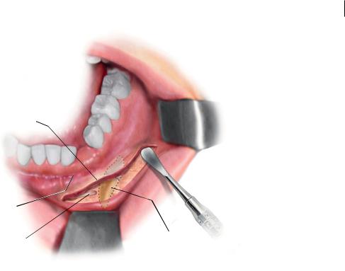
In situations where a tooth is impacted within the soft and hard tissue, away from the crowns of the erupted dentition, a vestibular incision can be used to spare iatrogenic damage to the periodontium and gingiva of that dentition (Figure 5.4). A common example is the buccally erupted upper canine tooth, which can lie as high as the piriform rim. Vestibular incisions are confined to the free mucosa only, and as such, any flaps raised are extremely versatile and offer excellent access to underlying structures.
Vestibular incisions should only be used if the tooth marked for extraction is sufficiently distant from the keratinised gingiva, as the incision requires an adequate ‘cuff’ of mucosa on either side to allow for closure, without putting tension on the gingiva, which can cause recession.
https://t.me/DentalBooksWorld
5.4 one eeooal 73
Vestibular incision
Mucogingival
junction
Mental nerve |
Impacted canine |
Figure 5.4 Vestibular incision used to approach an impacted canine.
To perform a vestibular incision, first the mucosa must be placed under tension. The incision should be at least 3 mm away from the mucogingival junction, and may be parallel to this line or slightly convex from it. The first incision should be made through and perpendicular to the mucosa. Once the mucosa is incised, a second pass should be made with the scalpel down through submucosa and periosteum to bone. The flap can then be gently elevated with a broad periosteal elevator to expose the underlying bone and impacted tooth.
When the procedure is completed, the vestibular incision can be opposed using simple interrupted sutures or a continuous-suture technique.
5.4Bone Removal
Once a tooth and the overlying alveolar bone is exposed, bone removal may be required in order to:
1)Eliminate any bony impaction of the crown of an impacted tooth.
2)Remove bone around bulbous roots, where the diameter of the root is too great and impedes delivery of a tooth root from the socket.
3)Create an application point whereby an elevator can be used to wedge between tooth root and bone to facilitate elevation of the root.
Precision is the key factor in performing successful bone removal. Appropriately planned bone removal ensures that the periodontal space between tooth and bone is maintained, to provide a precise application point for a dental elevator. Careful removal of bone around adjacent tooth roots and nerves prevents iatrogenic damage to these structures, which helps to prevent postsurgical complications.
https://t.me/DentalBooksWorld