


34 Biomolecules
Overview
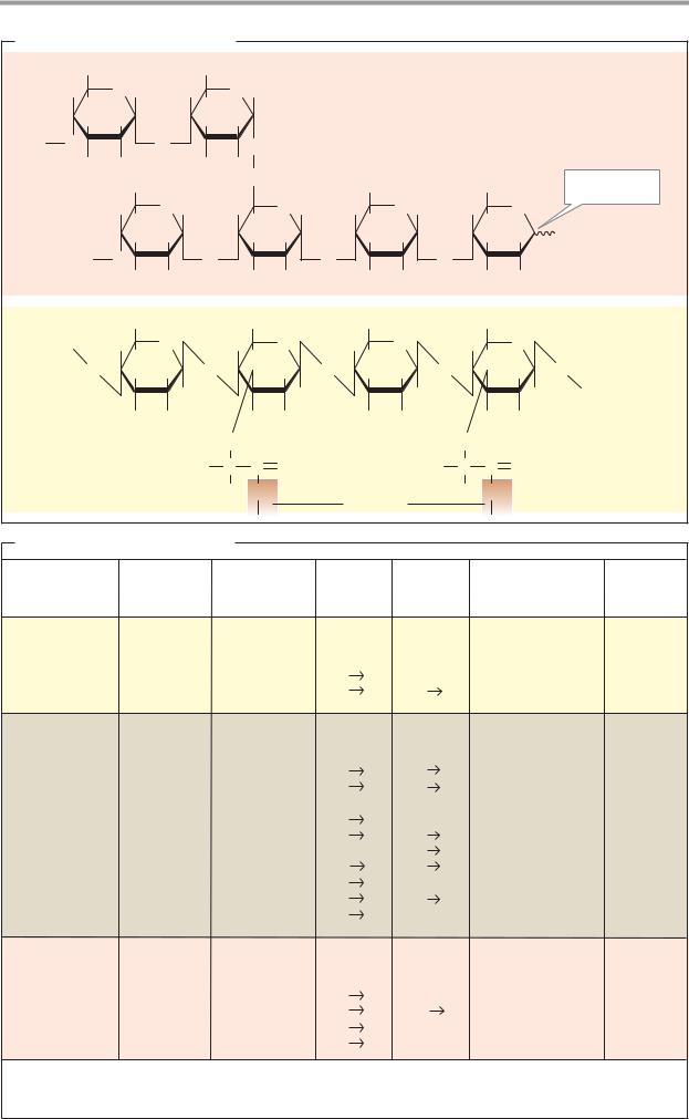
The carbohydrates are a group of naturally occurring carbonyl compounds (aldehydes or ketones) that also contain several hydroxyl groups. The carbohydrates include single sugars (monosaccharides) and their polymers, the oligosaccharides and polysaccharides.
A. Carbohydrates: overview
Polymeric carbohydrates–above all starch, as well as some disaccharides–are important (but not essential) components of food (see p.360). In the gut, they are broken down into monosaccharides and resorbed in this form (see p.272). The form in which carbohydrates are distributed by the blood of vertebrates is glucose (“blood sugar”). This is taken up by the cells and either broken down to obtain energy (glycolysis) or converted into other metabolites (see pp.150–159). Several organs (particularly the liver and muscles) store glycogen as a polymeric reserve carbohydrate (right; see p.156). The glycogen molecules are covalently bound to a protein, glycogenin. Polysaccharides are used by many organisms as building materials. For example, the cell walls of bacteria contain murein as a stabilizing component (see p.40), while in plants cellulose and other polysaccharides fulfill this role (see p.42). Oligomeric or polymeric carbohydrates are often covalently bound to lipids or proteins. The glycolipids and glycoproteins formed in this way are found, for example, in cell membranes (center). Glycoproteins also occur in the blood in solute form (plasma proteins; see p.276) and, as components of proteoglycans, form important constituents of the intercellular substance (see p.346).
B. Monosaccharides: structure
The most important natural monosaccharide, D-glucose, is an aliphatic aldehyde with six C atoms, five of which carry a hydroxyl group (1). Since C atoms 2 to 5 represent chiral centers (see p.8), there are 15 further isomeric aldohexoses in addition to D-glucose, although only a few of these are important in nature (see p.38). Most natural monosaccharides have the same configuration at C-5 as D-glyceraldehyde–they belong to the D series.
The open-chained form of glucose shown in (1) is found in neutral solution in less than 0.1% of the molecules. The reason for this is an intramolecular reaction in which one of the OH groups of the sugar is added to the aldehyde group of the same molecule (2). This gives rise to a cyclic hemiacetal (see p.10). In aldohexoses, the hydroxy group at C-5 reacts preferentially, and a six-membered pyran ring is formed. Sugars that contain this ring are called pyranoses. By contrast, if the OH group at C-4 reacts, a five-part furan ring is formed. In solution, pyranose forms and furanose forms are present in equilibrium with each other and with the open-chained form, while in glucose polymers only the pyranose form occurs.
The Haworth projection (2) is usually used to depict sugars in the cyclic form, with the ring being shown in perspective as viewed from above. Depending on the configuration, the substituents of the chiral C atoms are then found above or below the ring. OH groups that lie on the right in the Fischer projection (1) appear under the ring level in the Haworth projection, while those on the left appear above it.
As a result of hemiacetal formation, an additional chiral center arises at C-1, which can be present in both possible configurations (anomers) (see p.8). To emphasize this, the corresponding bonds are shown here using wavy lines.
The Haworth formula does not take account of the fact that the pyran ring is not plain, but usually has a chair conformation. In B3, two frequent conformations of D-glucopy- ranose are shown as ball-and-stick models. In the 1C4 conformation (bottom), most of the OH groups appear vertical to the ring level, as in the Haworth projection (axial or a position). In the slightly more stable 4C1 conformation (top), the OH groups take the equatorial or e position. At room temperature, each form can change into the other, as well as into other conformations.

|
|
|
|
|
Carbohydrates |
35 |
A. Carbohydrates: overview |
|
|
|
|||
|
|
|
Transporter |
Other monosaccharides |
|
|
CH2OH |
|
|
Glycogenin |
|
||
|
|
|
|
|
||
H H |
O OH |
Glucose |
|
|
||
HO OH |
H |
H |
|
|
||
Mono- |
|
|
|
|||
H |
OH |
|
Gluconeo- |
|
||
saccharide |
Glycolysis |
|
||||
|
|
|
|
|||
|
|
|
|
|
genesis |
|
|
|
|
|
Pyruvate |
Amino |
|
|
|
|
|
|
|
|
|
|
|
Glycoproteins |
|
acids |
|
|
|
|
|
ATP |
|
|
|
|
|
|
|
|
|
|
|
|
|
|
CO2+H2O |
|
|
|
|
Glycolipids |
|
Glycogen |
|
|
|
|
|
|
|
|
|
|
|
Peptidoglycan |
|
|
|
|
|
|
(Murein) |
Proteoglycans |
|
|
|
|
|
Periplasm |
|
|
|
Bacterium |
|
|
|
|
|
|
B. Monosaccharides: structure
H |
1C |
O |
|
|
|
H |
2 C |
OH |
HO |
3 C |
H |
H |
4 C |
OH |
H |
C |
OH |
|
5 |
|
6 CH2OH
Open-chained form of glucose
Chiral center
1. Fischer projection
|
|
|
|
|
|
|
|
|
|
|
|
HOCH2 |
|
|
|
|
|
|||||||||
Open-chained |
H |
|
6 |
OH |
O |
|
|
|
|
|
||||||||||||||||
|
5 |
|
|
|
|
|
||||||||||||||||||||
form (< 0.1%) |
|
|
|
|
|
|
|
|||||||||||||||||||
|
|
|
|
|
|
|
||||||||||||||||||||
|
|
|
|
|
|
|
|
|
|
|
|
|||||||||||||||
|
|
|
|
|
|
|
|
|
|
|
|
|
H |
|
|
|
|
|
|
|
|
H |
|
|||
|
|
|
|
|
|
|
|
|
|
|
|
4 |
OH H C |
|
|
|
|
|||||||||
|
|
|
|
|
|
|
|
|
|
|
|
|
|
|
|
|||||||||||
|
|
|
|
|
|
|
|
|
|
|
|
|
|
|
|
|
|
1 |
|
|
|
|
|
|
|
|
|
|
|
|
|
|
|
|
|
|
|
|
|
|
|
|
|
|
|
|
|
|
|||||
|
|
|
|
|
|
|
|
|
|
|
|
HO |
|
3 |
|
|
2 |
|
|
|
|
|
|
|
|
|
|
|
|
|
|
|
|
|
|
|
|
|
|
H |
OH |
|
|
|
|
|
|||||||
|
|
|
|
|
|
|
|
|
|
|
|
|
|
|
|
|
|
|
|
|
|
|
||||
|
|
|
|
CH2OH |
Hemiacetal formation |
|
|
|||||||||||||||||||
|
|
|
|
|
|
|
|
|
|
|
|
|
|
|
|
|
|
|
|
|
||||||
|
|
|
|
|
|
|
|
HOCH2 |
|
|||||||||||||||||
|
|
|
|
|
|
|
|
|
|
|||||||||||||||||
|
|
|
|
|
|
|
|
|
|
|
|
|
|
|
|
|
|
|
||||||||
HO |
|
C |
|
|
|
H |
OH |
|
|
|
||||||||||||||||
|
|
|
|
|
|
|
H |
|
|
O |
|
|||||||||||||||
|
|
|
|
|
|
|
|
|
||||||||||||||||||
|
|
|
|
|
|
|
|
O |
|
|
|
|
|
OH |
||||||||||||
|
|
|
|
|
|
|
|
|
|
|
|
|
|
|
|
|
|
|||||||||
|
|
|
|
|
|
|
|
|
|
|
|
|
|
|
|
|
|
H |
||||||||
|
|
|
|
|
|
OH H |
|
|
|
|
|
|
|
|
|
|
|
|
||||||||
|
|
|
|
|
|
|
H |
|
|
|
|
|
|
OH H |
H |
|||||||||||
|
|
|
|
|
|
|
|
|
|
|
|
|
|
|
|
|
|
|
|
|
|
|||||
|
|
H |
|
|
|
|
|
|
|
|
|
|
|
|
|
|
|
|
||||||||
|
|
|
|
|
|
|
|
|
|
|
|
|
|
HO |
|
|
|
|
||||||||
|
|
|
|
|
|
|
|
|
|
|
|
|
|
|
|
|
|
|
|
|
|
|
||||
|
|
|
|
|
H |
OH |
|
|
|
|
|
|
|
|
|
|
|
H OH |
|
|||||||
|
D-Gluco- |
|
|
|
|
|
|
|
D-Gluco- |
|
||||||||||||||||
|
furanose (<1%) |
|
|
pyranose (99%) |
||||||||||||||||||||||
2. Ring forms (Haworth projection)
4
1
4C1-conformation
1
4
1C4-conformation
3. Conformations
36 |
Biomolecules |
|
|
Chemistry of sugars |
5. Esterification. The hydroxyl groups of |
||
|
|
monosaccharides can form esters with acids. |
|
A. Reactions of the monosaccharides |
In metabolism, phosphoric acid esters such as |
||
glucose 6-phosphate and glucose 1-phosphate |
|||
|
|
||
The sugars (monosaccharides) occur in the |
(6) are particularly important. |
||
metabolism in many forms (derivatives). |
|
||
Only a few important conversion reactions |
B. Polarimetry, mutarotation |
||
are discussed here, using D-glucose as an ex- |
|
||
ample. |
Sugar solutions can be analyzed by polarim- |
||
1. Mutarotation. In the cyclic form, as op- |
etry, a method based on the interaction be- |
||
posed to the open-chain form, aldoses have a |
tween chiral centers and linearly polarized |
||
chiral center at C-1 (see p.34). The corre- |
light—i.e., light that oscillates in only one |
||
sponding isomeric forms are called anomers. |
plane. It can be produced by passing normal |
||
In the β-anomer (center left), the OH group at |
light through a special filter (a polarizer). A |
||
C-1 (the anomeric OH group) and the CH2OH |
second polarizing filter of the same type (the |
||
group lie on the same side of the ring. In the α- |
analyzer), placed behind the first, only lets the |
||
anomer (right), they are on different sides. |
polarized light pass through when the polar- |
||
The reaction that interconverts anomers into |
izer and the analyzer are in alignment. In this |
||
each other is known as mutarotation (B). |
case, the field of view appears bright when |
||
2. Glycoside formation. When the anome- |
one looks through the analyzer (1). Solutions |
||
ric OH group of a sugar reacts with an alcohol, |
of chiral substances rotate the plane of polar- |
||
with elimination of water, it yields an |
ized light by an angle α either to the left or to |
||
O–glycoside (in the case shown, α –methylglu- |
the right. When a solution of this type is |
||
coside). The glycosidic bond is not a normal |
placed between the polarizer and the ana- |
||
ether bond, because the OH group at C-1 has a |
lyzer, the field of view appears darker (2). |
||
hemiacetal quality. Oligosaccharides and pol- |
The angle of rotation, α, is determined by |
||
ysaccharides also contain O-glycosidic bonds. |
turning the analyzer until the field of view |
||
Reaction of the anomeric OH group with an |
becomes bright again (3). A solution’s optical |
||
NH2 or NH group yields an N-glycoside (not |
rotation depends on the type of chiral com- |
||
shown). N-glycosidic bonds occur in nucleo- |
pound, its concentration, and the thickness of |
||
tides |
(see p.80) and in glycoproteins (see |
the layer of the solution. This method makes it |
|
p.44), for example. |
possible to determine the sugar content of |
||
3. Reduction and oxidation. Reduction of |
wines, for example. |
||
the anomeric center at C-1 of glucose (2) pro- |
Certain procedures make it possible to ob- |
||
duces the sugar alcohol sorbitol. Oxidation of |
tain the α and β anomers of glucose in pure |
||
the aldehyde group at C-1 gives the intramo- |
form. A 1-molar solution of α-D-glucose has a |
||
lecular ester (lactone) of gluconic acid (a gly- |
rotation value [α]D of +112°, while a corre- |
||
conic acid). Phosphorylated gluconolactone is |
sponding solution of β -D-glucose has a value |
||
an intermediate of the pentose phosphate |
of +19°. These values change spontaneously, |
||
pathway (see p.152). When glucose is oxi- |
however, and after a certain time reach the |
||
dized at C-6, glucuronic acid (a glycuronic |
same end point of +52°. The reason for this is |
||
acid) is formed. The strongly polar glucuronic |
that, in solution, mutarotation leads to an |
||
acid plays an important role in biotransforma- |
equilibrium between the α and β forms in |
||
tions in the liver (see pp.194, 316). |
which, independently of the starting condi- |
||
4. Epimerization. In weakly alkaline solu- |
tions, 62% of the molecules are present in the |
||
tions, glucose is in equilibrium with the |
β form and 38% in the α form. |
||
ketohexose D-fructose and the aldohexose D- |
|
||
mannose, via an enediol intermediate (not shown). The only difference between glucose and mannose is the configuration at C-2. Pairs of sugars of this type are referred to as epimers, and their interconversion is called epimerization.

|
|
|
|
|
|
|
|
|
|
|
Carbohydrates |
37 |
||
A. Reactions of the monosaccharides |
|
|
|
|
|
|
|
|
|
|||||
|
|
Glucuronate |
|
Gluconolactone |
|
|
Sorbitol |
|
|
|||||
|
|
6 COO |
|
HOCH2 |
|
|
|
|
HOCH2 |
|
|
|||
|
|
H |
O OH |
|
|
H |
O |
|
|
|
H |
OH |
1 |
|
|
|
H |
|
|
|
H |
|
|
|
|
H |
|
|
|
|
|
OH H |
|
|
OH H |
O |
|
|
OH H CH2OH |
|
||||
|
|
HO |
H |
|
HO |
1 |
|
|
|
HO |
|
|
|
|
|
|
|
|
|
|
|
|
|
|
|||||
|
|
H |
OH |
|
|
H |
OH |
|
|
|
H |
OH |
|
|
|
|
Oxidation |
|
|
Oxidation |
|
|
Reduction |
|
|
||||
|
|
|
HOCH2 |
|
|
|
|
|
|
HOCH2 |
|
|
|
|
|
|
|
H |
O OH β |
|
|
|
|
H |
O H |
|
|
|
|
|
|
|
H |
|
|
|
|
|
|
H |
|
|
|
|
|
|
|
OH H 1 |
|
Mutarotation |
|
OH H 1 |
|
|
|
||||
|
|
|
HO |
H |
|
|
|
|
|
HO |
OH α |
|
|
|
|
|
|
H |
OH |
|
|
|
|
|
H |
OH |
|
|
|
|
|
|
β-D-Glucose |
|
|
|
|
|
α-D-Glucose |
|
|
|
||
|
|
Esterification |
|
|
Epimerization |
|
Glycoside formation |
|
||||||
|
O |
|
|
|
|
|
|
|
|
|
|
|
|
|
O |
P |
6 |
|
|
H |
|
|
HOCH2 |
HOCH2 |
|
|
|||
O CH2 |
|
1 |
|
|
|
|||||||||
|
|
|
|
|
|
|
|
|
|
|
|
|
|
|
|
O |
H |
O OH |
H |
O CH2OH |
H |
H |
O H |
H |
H |
O H |
|
||
|
|
H |
|
|
H |
2 |
|
|
|
|
|
|
||
|
|
OH H |
|
H HO |
|
|
OHHO |
|
OH H 1 |
|
||||
|
|
HO |
H |
HO |
3 |
OH |
|
HO |
|
2 OH |
HO |
|
α O |
CH3 |
|
|
H |
OH |
|
OH H |
|
|
|
H |
H |
|
H |
OH |
|
|
Glucose 6- |
|
D-Fructose |
|
α-D-Mannose |
|
α-Methyl- |
|
||||||
|
phosphate |
|
|
|
|
|
|
|
|
|
glucoside |
|
||
B. Polarimetry, mutarotation
|
Polarizer |
Analyzer |
|
|
|
|
|
|
|
Water |
|
α (˚) |
α-D-Glucose: [ α ]D = +112° |
|
|||
1. |
|
|
|
|
|
|
||
|
100 |
|
|
|
|
|
||
|
|
|
|
|
|
|
|
|
|
Sugar |
a |
80 |
|
|
|
+ 52° |
62% β |
2. |
|
60 |
|
|
|
|||
|
|
|
|
|
|
|||
|
|
|
|
|
|
|
||
|
|
a |
40 |
|
|
|
|
38% α |
|
|
20 |
|
|
|
|
|
|
|
Sugar |
|
|
|
|
|
|
|
3. |
|
0 |
20 |
30 |
40 |
50 |
Time (min) |
|
|
|
|
10 |
|||||
|
|
|
|
β-D-Glucose: [α]D = +19° |
|
|||

38 Biomolecules
Monosaccharides and disaccharides
A. Important monosaccharides
Only the most important of the large number of naturally occurring monosaccharides are mentioned here. They are classified according to the number of C atoms (into pentoses, hexoses, etc.) and according to the chemical nature of the carbonyl function into aldoses and ketoses.
The best-known aldopentose (1), D-ribose, is a component of RNA and of nucleotide coenzymes and is widely distributed. In these compounds, ribose always exists in the furanose form (see p.34). Like ribose, D-xylose and L-arabinose are rarely found in free form. However, large amounts of both sugars are found as constituents of polysaccharides in the walls of plant cells (see p.42).
The most important of the aldohexoses (1) is D-glucose. A substantial proportion of the biomass is accounted for by glucose polymers, above all cellulose and starch. Free D-glucose is found in plant juices (“grape sugar”) and as “blood sugar” in the blood of higher animals. As a constituent of lactose (milk sugar), D- galactose is part of the human diet. Together with D-mannose, galactose is also found in glycolipids and glycoproteins (see p.44).
Phosphoric acid esters of the ketopentose D-ribulose (2) are intermediates in the pentose phosphate pathway (see p.152) and in photosynthesis (see p.128). The most widely distributed of the ketohexoses is D-fructose. In free form, it is present in fruit juices and in honey. Bound fructose is found in sucrose (B) and plant polysaccharides (e.g., inulin).
In the deoxyaldoses (3), an OH group is replaced by a hydrogen atom. In addition to 2-deoxy-D-ribose, a component of DNA (see p.84) that is reduced at C-2, L-fucose is shown as another example of these. Fucose, a sugar in the λ series (see p.34) is reduced at C-6.
The acetylated amino sugars N-acetyl-D- glucosamine and N-acetyl-D-Galactosamine
(4) are often encountered as components of glycoproteins.
N-acetylneuraminic acid (sialic acid, 5), is a characteristic component of glycoproteins. Other acidic monosaccharides such as D-glu- curonic acid, D-galacturonic acid, and liduronic acid, are typical constituents of the glycosaminoglycans found in connective tissue.
Sugar alcohols (6) such as sorbitol and mannitol do not play an important role in animal metabolism.
B. Disaccharides
When the anomeric hydroxyl group of one monosaccharide is bound glycosidically with one of the OH groups of another, a disaccharide is formed. As in all glycosides, the glycosidic bond does not allow mutarotation. Since this type of bond is formed stereospecifically by enzymes in natural disaccharides, they are only found in one of the possible configurations (α or β).
Maltose (1) occurs as a breakdown product of the starches contained in malt (“malt sugar”; see p.148) and as an intermediate in intestinal digestion. In maltose, the anomeric OH group of one glucose molecule has an α- glycosidic bond with C-4 in a second glucose residue.
Lactose (“milk sugar,” 2) is the most important carbohydrate in the milk of mammals. Cow’s milk contains 4.5% lactose, while human milk contains up to 7.5%. In lactose, the anomeric OH group of galactose forms a β - glycosidic bond with C-4 of a glucose. The lactose molecule is consequently elongated, and both of its pyran rings lie in the same plane.
Sucrose (3) serves in plants as the form in which carbohydrates are transported, and as a soluble carbohydrate reserve. Humans value it because of its intensely sweet taste. Sources used for sucrose are plants that contain particularly high amounts of it, such as sugar cane and sugar beet (cane sugar, beet sugar). Enzymatic hydrolysis of sucrose-containing flower nectar in the digestive tract of bees— catalyzed by the enzyme invertase—produces honey, a mixture of glucose and fructose. In sucrose, the two anomeric OH groups of glucose and fructose have a glycosidic bond; sucrose is therefore one of the non-reducing sugars.

|
|
|
|
|
|
|
|
|
|
|
|
|
|
|
|
|
|
Carbohydrates |
|
39 |
||||||||
A. Important monosaccharides |
|
|
|
|
|
|
|
|
|
|
|
|
|
|
|
|
|
|
|
|
|
|||||||
|
|
|
|
|
|
|
|
|
|
|
|
|
|
|
|
|
|
|
|
|
||||||||
|
|
|
|
|
|
|
|
|
|
|
|
|
|
|
|
|
|
|
|
|
|
|
|
|
|
|
|
|
1 Aldoses |
|
|
|
|
|
|
|
|
|
2 |
Ketoses |
|
|
|
|
|
|
|
|
|
||||||||
D-Ribose (Rib) |
D-Xylose (Xyl) |
L-Arabinose (Ara) |
|
CH2OH |
D-Ribulose (Rub) |
|
|
|
||||||||||||||||||||
HOCH2 |
HOCH2 |
H |
|
|
|
H |
|
|
|
|
|
|
||||||||||||||||
|
|
|
|
|
|
|
O OH |
|
|
|
||||||||||||||||||
|
|
O |
|
|
O |
|
|
O |
|
|
|
|
|
|
|
|
|
|
|
|
|
|||||||
|
|
|
|
|
|
OH |
|
C |
|
O |
|
|
|
|
|
|
||||||||||||
|
H H OH |
|
OH H OH |
|
OH H |
|
|
|
|
|
|
H |
H |
|
|
|
|
|||||||||||
|
|
|
|
|
|
|
|
|
|
|
|
|
||||||||||||||||
|
|
|
|
|
|
|
|
|
|
|
|
|
|
H |
|
C |
|
OH |
|
|
|
|
|
CH2OH |
|
|
|
|
H |
|
|
H |
|
|
|
HOH2C |
|
|
|
|
|
|
|
|
H |
|
|
|
|
|
|
|
|||||
|
|
|
|
|
|
|
|
H |
|
C |
|
OH |
|
|
|
|
|
|
|
|||||||||
|
OH OH |
|
H OH |
|
H OH |
|
|
|
|
OH OH |
|
|
|
|
||||||||||||||
|
|
|
|
|
|
|
|
|
|
|
|
|
|
|
||||||||||||||
Pentoses |
|
|
|
|
|
|
|
|
|
|
|
|
|
CH2OH |
|
|
|
|
|
|
|
|
|
|||||
D-Glucose (Glc) |
D-Mannose (Man) |
D-Galactose (Gal) |
|
|
|
|
|
D-Fructose (Fru) |
|
|
|
|||||||||||||||||
|
HOCH2 |
|
|
|
|
HOCH2 |
|
|
|
HOCH2 |
|
|
|
|
|
CH2OH |
|
|
|
|
|
|||||
|
|
|
|
|
|
|
|
|
|
|
|
|
C |
O |
|
|
|
|
|
|
||||||
|
H |
|
O |
|
|
|
H |
|
O |
|
HO |
O |
|
|
|
|
|
|
|
|
|
|
|
|||
|
|
|
|
|
|
|
|
|
|
|
HO |
C |
H |
|
|
|
|
|
|
|||||||
|
|
H |
|
OH |
|
|
H |
|
OH |
|
H |
|
|
OH |
|
|
HOCH2 |
|
OH |
|||||||
|
|
OH H |
|
|
OHHO |
|
OH H |
|
|
|
|
|
|
|
||||||||||||
|
HO |
|
|
|
|
|
HO |
|
|
|
|
H |
|
|
|
|
|
H |
C |
OH |
|
O |
|
|
||
|
|
|
|
|
|
|
|
|
|
|
|
|
|
|
H HO |
|
|
|||||||||
|
|
|
|
|
|
|
|
|
|
|
|
|
|
|
H |
C |
OH |
|
CH2OH |
|||||||
|
|
H |
OH |
|
|
|
H |
H |
|
|
H |
OH |
|
|
|
H |
|
|
|
|||||||
|
|
|
|
|
|
|
|
|
|
|
|
|
|
|||||||||||||
Hexoses |
|
|
|
|
|
|
|
|
|
|
|
|
|
|
|
CH2OH |
OH H |
|
|
|||||||
|
|
|
|
|
|
|
|
|
|
|
|
|
|
|
|
|
|
|
|
|
||||||
3 Deoxyaldoses |
|
|
|
|
|
|
|
|
4 |
|
Acetylated amino sugars |
|
|
|
|
|
||||||||||
|
2-Deoxy- |
|
|
|
L-Fucose (Fuc) |
|
|
|
N-Acetyl-D-glucos- |
|
N -Acetyl-D-galac- |
|||||||||||||||
|
D-ribose (dRib) |
|
|
|
|
|
|
|
amine (GlcNAc) |
|
|
tosamine (GalNAc) |
||||||||||||||
|
|
HOCH2 |
|
|
|
H |
|
|
|
|
|
|
HOCH2 |
|
|
|
|
|
HOCH2 |
|
|
|
||||
|
|
|
|
O |
|
OH |
|
H CH6 O |
|
|
|
|
|
H H |
O |
|
|
|
|
HO H |
|
O |
|
|
||
|
|
|
H H |
|
|
OH |
|
|
|
|
|
OH |
|
|
|
|
OH |
|||||||||
|
|
H |
2 |
|
|
|
H |
3HO |
|
|
|
|
OH H |
|
|
|
|
OH H |
|
|||||||
|
|
|
|
|
|
|
|
|
|
|
|
HO |
|
2 |
|
|
|
H |
|
|
2 |
|
|
|||
|
|
|
|
|
|
HO |
|
|
|
|
|
|
|
|
|
|
|
|
|
|
||||||
|
|
|
OH H |
|
|
|
|
|
|
|
|
|
|
|
|
|
|
|
|
|||||||
|
|
|
|
|
|
OH H |
|
|
|
|
|
H HN |
C |
CH3 |
|
|
H HN |
C |
CH3 |
|||||||
|
|
|
|
|
|
|
|
|
|
|
|
|
|
|
||||||||||||
|
|
|
|
|
|
|
|
|
|
|
|
|
|
|
|
|
O |
|
|
|
|
|
|
O |
|
|
5 Acidic monosaccharides |
|
|
N-Acetylneuraminic acid |
|
|
6 |
Sugar alcohols (alditoles) |
|||||||||||||||||||
D-Glucuronic acid |
|
|
|
|
(NeuAc) |
|
|
|
|
|
|
D-Sorbitol |
|
D-Mannitol |
||||||||||||
|
L-Iduronic acid |
|
9 CH2OH |
|
|
|
|
|||||||||||||||||||
(GlcUA) |
|
|
|
|
(IduUA) |
|
|
|
HO |
C |
H |
|
|
|
|
CH2OH |
|
|
|
CH2OH |
||||||
|
6 COO |
|
|
|
|
|
|
|
|
|
|
|
|
|
|
|
|
|||||||||
|
|
|
|
|
H |
|
|
|
|
H |
|
|
|
|
|
|
H |
C |
OH |
|
HO |
C |
H |
|||
|
|
|
|
|
|
|
|
|
|
|
2 HO |
C |
H |
|
|
|
|
|||||||||
|
H |
O |
|
|
|
|
H |
O |
|
|
|
|
|
|
|
|
|
|
|
|
|
|||||
|
|
|
|
|
|
|
H |
O |
7 |
|
|
|
|
HO |
C |
H |
|
HO |
C |
H |
||||||
|
H |
|
|
OH |
|
|
COO |
OH |
3 |
COO |
|
|
|
|
|
|
||||||||||
|
OH H |
|
|
|
OH H |
OH H |
6 |
|
|
|
|
|
H |
C |
OH |
|
|
H |
C |
OH |
||||||
HO |
|
|
|
|
HO |
|
|
|
|
5 |
H |
|
|
|
|
|
|
|
||||||||
|
|
|
|
|
|
|
H4 |
|
|
|
|
|
H |
C |
OH |
|
|
H |
C |
OH |
||||||
|
H |
OH |
|
|
|
H |
OH |
|
|
H HN |
C |
|
CH3 |
|
|
|
|
|
||||||||
|
|
|
|
|
|
|
|
|
|
|
|
|
|
|
|
|
|
|||||||||
|
|
|
|
|
|
|
|
|
|
|
|
|
O |
|
|
|
|
|
|
CH2OH |
|
|
|
CH2OH |
||
B. Disaccharides |
|
|
|
|
|
|
|
|
|
|
|
|
|
|
|
|
|
|
|
|
|
|||||
|
CH2OH |
|
|
CH2OH |
|
|
CH2OH |
CH2OH |
|
|
|
|
CH2OH |
|
|
|
|
|
||||||||
H |
|
O H |
|
H |
|
O |
|
|
|
HO |
O β |
H |
|
O |
|
|
|
H |
|
O H |
|
|
CH2OH H |
|||
|
H |
|
|
|
H |
|
|
|
|
H |
|
|
H |
|
|
|
|
|
H |
|
|
|
|
O |
|
|
|
OH H 1 |
4 |
OH H |
OH |
|
OH H 1 O 4 OH H |
OH |
|
|
OH H 1 |
|
2 |
H HO |
|||||||||||||
HO |
|
α |
|
O |
|
|
|
|
|
H |
|
H |
|
|
|
|
|
|
HO |
|
α |
O |
β |
|
CH2OH |
|
|
H |
OH |
|
|
H |
OH |
|
|
H |
OH |
H |
OH |
|
|
|
|
H |
|
|
|
||||||
|
|
|
|
|
|
|
|
|
OH |
|
|
OH H |
||||||||||||||
|
|
|
|
|
|
|
|
|
|
|
|
|
|
|
|
|
|
|
|
|
|
|
|
|||
1. Maltose |
|
|
|
|
|
|
2. Lactose |
|
|
|
|
|
|
3. Sucrose |
|
|
|
|
||||||||
α-D-Glucopyranosyl- |
|
|
|
β-D-Galactopyranosyl- |
|
|
|
α-D-Glucopyranosyl- |
|
|||||||||||||||||
(1 |
4)-D-glucopyranose |
|
|
|
(1 |
4)-D-glucopyranose |
|
|
|
(1 |
2)-β-D-fructofuranoside |
|||||||||||||||

40 Biomolecules
Polysaccharides: overview
Polysaccharides are ubiquitous in nature. They can be classified into three separate groups, based on their different functions.
Structural polysaccharides provide mechanical stability to cells, organs, and organisms.
Waterbinding polysaccharides are strongly hydrated and prevent cells and tissues from drying out. Finally, reserve polysaccharides serve as carbohydrate stores that release monosaccharides as required. Due to their polymeric nature, reserve carbohydrates are osmotically less active, and they can therefore be stored in large quantities within the cell.
A. Polysaccharides: structure
Polysaccharides that are formed from only one type of monosaccharide are called homoglycans, while those formed from different sugar constituents are called heteroglycans. Both forms can exist as either linear or branched chains.
A section of a glycogen molecule is shown here as an example of a branched homoglycan. Amylopectin, the branched component of vegetable starch (see p.42), has a very similar structure. Both molecules mainly consist of α1 4-linked glucose residues. In glycogen, on average every 8th to 10th residue carries —via an α1 6 bond—another 1,4-linked chain of glucose residues. This gives rise to branched, tree-like structures, which in animal glycogen are covalently bound to a protein, glycogenin (see p.156).
The linear heteroglycan murein, a structural polysaccharide that stabilizes the cell walls of bacteria, has a more complex structure. Only a short segment of this thread-like molecule is shown here. In murein, two different components, both β1 4-linked, alternate: N-acetylglucosamine (GlcNAc) and N-acetylmuraminic acid (MurNAc), a lactic acid ether of N-acetylglucosamine. Peptides are bound to the carboxyl group of the lactyl groups, and attach the individual strands of murein to each other to form a three-dimen- sional network (not shown). Synthesis of the network-forming peptides in murein is inhibited by penicillin (see p.254).
B. Important polysaccharides
The table gives an overview of the composition and make-up both of the glycans mentioned above and of several more.
In addition to murein, bacterial polysaccharides include dextrans—glucose polymers that are mostly α1 6-linked and α1 3- branched. In water, dextrans form viscous slimes or gels that are used for chromatographic separation of macromolecules after chemical treatment (see p.78). Dextrans are also used as components of blood plasma substitutes (plasma expanders) and foodstuffs.
Carbohydrates from algae (e.g., agarose and carrageenan) can also be used to produce gels. Agarose has been used in microbiology for more than 100 years to reinforce culture media (“agar-agar”). Algal polysaccharides are also added to cosmetics and ready-made foods to modify the consistencyof these products.
The starches, the most important vegetable reserve carbohydrate and polysaccharides from plant cell walls, are discussed in greater detail on the following page. Inulin, a fructose polymer, is used as a starch substitute in diabetics’ dietary products (see p.160). In addition, it serves as a test substance for measuring renal clearance (see p.322).
Chitin, a homopolymer from β 1 4-linked N-acetylglucosamine, is the most important structural substance in insect and crustacean shells, and is thus the most common animal polysaccharide. It also occurs in the cell wall of fungi.
Glycogen, the reserve carbohydrate of higher animals, is stored in the liver and musculature in particular (A, see pp.156, 336). The formation and breakdown of glycogen are subject to complex regulation by hormones and other factors (see p.120).
|
|
|
|
|
|
|
|
|
|
|
|
|
|
Carbohydrates |
41 |
||
A. Polysaccharides: structure |
|
|
|
|
|
|
|
|
|
|
|
|
|
|
|||
HOCH2 |
HOCH2 |
|
|
|
|
|
|
|
|
|
|
|
|
Glycogen – |
|
||
H |
O H |
H |
O H |
|
|
|
|
|
|
|
|
|
|
|
|
||
|
|
|
|
|
|
|
|
|
|
|
branched |
|
|||||
H |
|
4 |
H |
1 |
|
|
|
|
|
|
|
|
|
|
|
|
|
|
|
|
|
|
|
|
|
|
|
|
|
homopolymer |
|
||||
OH H 1 |
OH H |
|
|
|
|
|
|
|
|
|
|
|
|
||||
O |
α |
O |
|
Oα |
|
|
|
|
|
|
|
|
|
|
|
|
|
H |
OH |
|
H HO |
|
|
|
|
|
|
|
|
|
|
|
|
|
|
|
HOCH2 |
6CH2 |
|
|
HOCH2 |
|
|
|
HOCH2 |
|
Reducing end |
|
|||||
|
H |
O H |
H |
|
O H |
|
H |
|
O H |
|
H |
|
|
O |
|
|
|
|
H |
|
4 |
H |
|
|
4 |
H |
|
1 |
|
4 |
H |
|
|
OH |
|
|
OH H 1 |
OH H 1 |
|
OH H |
|
OH H |
|
||||||||||
|
O |
α |
O |
H |
α |
O |
|
H |
|
α |
O |
|
H |
|
OH |
|
|
|
H |
OH |
|
OH |
|
|
OH |
|
|
|
|
|
|||||
|
HOCH2 |
HOCH2 |
β |
|
HOCH2 |
|
β |
|
HOCH2 |
|
|
|
|||||
|
H |
O β |
H |
|
|
H |
|
O |
|
H |
|
|
O |
|
|
||
|
|
O |
|
|
|
|
|
|
|
|
|||||||
|
H |
|
O 4 |
H |
H 1 O |
4 |
H |
|
1 O |
4 |
H |
|
H O |
|
|||
O OH H 1 |
|
OH H |
3 |
|
|
||||||||||||
|
|
H |
|
|
|
|
|
|
|
H |
|
|
2 |
|
|
||
|
|
|
|
H |
|
|
|
|
|
|
|
|
H |
|
|
||
|
H |
NHCOCH3 |
H |
NHCOCH3 |
H |
NHCOCH3 |
H |
|
NHCOCH3 |
|
|||||||
|
|
|
O |
|
|
|
|
|
|
|
|
O |
|
|
|
|
|
|
|
H3C C C O |
|
|
|
|
H3C |
C C O |
Murein – linear |
|
|||||||
|
|
|
|
|
|
heteropolymer |
|
||||||||||
|
|
|
H |
NH |
|
|
|
|
|
|
|
H |
NH |
|
|
||
|
|
|
|
|
Peptide |
|
|
|
|
|
|||||||
|
|
|
|
|
|
|
|
|
|
|
|
|
|
|
|||
B. Important polysaccharides
Poly- |
Mono- |
Mono- |
Linkage |
|||
saccharide |
saccharide1 |
saccharide 2 |
|
|
||
Bacteria |
|
|
|
|
|
|
Murein |
D-GlcNAc |
D-MurNAc1) |
β 1 |
4 |
||
Dextran |
D-Glc |
|
|
|
α 1 |
6 |
|
|
|
||||
Plants |
|
|
|
|
|
|
Agarose |
D-Gal |
L-aGal2) |
β 1 |
4 |
||
Carrageenan |
D-Gal |
|
|
|
β 1 |
3 |
|
|
|
||||
Cellulose |
D-Glc |
|
|
|
β 1 |
4 |
|
|
|
||||
Xyloglucan |
D-Glc |
D-Xyl (D-Gal, |
β 1 |
4 |
||
Arabinan |
L-Ara |
L-Fuc) |
α 1 |
5 |
||
|
|
|
||||
|
|
|
||||
Amylose |
D-Glc |
|
|
|
α 1 |
4 |
|
|
|
||||
Amylopectin |
D-Glc |
|
|
|
α 1 |
4 |
|
|
|
||||
Inulin |
D-Fru |
|
|
|
β 2 |
1 |
Animals |
|
|
|
|
|
|
Chitin |
D-GlcNAc |
|
|
|
β 1 |
4 |
|
|
|
||||
Glycogen |
D-Glc |
|
|
|
α 1 |
4 |
|
|
|
||||
Hyaluronic |
D-GlcUA |
D-GlcNAc |
β 1 |
4 |
||
acid |
|
|
|
|
β 1 |
3 |
Branch- Occurrence |
Function |
ing |
|
|
|
Cell wall |
SC |
|
|
||
α 1 3 Slime |
WB |
||
β 1 |
3 |
Red algae (agar) |
WB |
|
α 1 |
4 |
Red algae |
WB |
|
|
|
|
Cell wall |
SC |
|
|
|
||
β 1 |
6 |
Cell wall |
SC |
|
(β 1 |
2) |
(Hemicellulose) |
SC |
|
α 1 |
3 |
Cell wall (pectin) |
|
|
|
|
|
Amyloplasts |
RC |
|
|
|
||
α 1 |
6 |
Amyloplasts |
RC |
|
|
|
|
Storage cells |
RC |
|
|
|
||
|
|
Insects, crabs |
SK |
|
|
||
α 1 6 Liver, muscle |
RK |
||
|
|
Connective tissue |
SK,WB |
|
|
||
SC= structural carbohydrate, RC= reserve carbohydrate,
WB = water-binding carbohydrate; 1) N-acetylmuramic acid, 2) 3,6-anhydrogalactose
42 Biomolecules
Plant polysaccharides
Two glucose polymers of plant origin are of special importance among the polysaccharides: β1 4-linked polymer cellulose and starch, which is mostly α1 4-linked.
A. Cellulose
Cellulose, a linear homoglycan of β1 4- linked glucose residues, is the most abundant organic substance in nature. Almost half of the total biomass consists of cellulose. Some 40–50% of plant cell walls are formed by cellulose. The proportion of cellulose in cotton fibers, an important raw material, is 98%. Cellulose molecules can contain more than 104 glucose residues (mass 1–2 106 Da) and can reach lengths of 6–8 µm.
Naturally occurring cellulose is extremely mechanically stable and is highly resistant to chemical and enzymatic hydrolysis. These properties are due to the conformation of the molecules and their supramolecular organization. The unbranched β 1 4 linkage results in linear chains that are stabilized by hydrogen bonds within the chain and between neighboring chains (1). Already during biosynthesis, 50–100 cellulose molecules associate to form an elementary fibril with a diameter of 4 nm. About 20 such elementary fibrils then form a microfibril (2), which is readily visible with the electron microscope.
Cellulose microfibrils make up the basic framework of the primary wall of young plant cells (3), where they form a complex network with other polysaccharides. The linking polysaccharides include hemicellulose, which is a mixture of predominantly neutral heteroglycans (xylans, xyloglucans, arabinogalactans, etc.). Hemicellulose associates with the cellulosefibrilsvianoncovalent interactions. These complexes are connected by neutral and acidic pectins, which typically contain galacturonic acid. Finally, a collagen-related protein, extensin, is also involved in the formation of primary walls.
In the higher animals, including humans, cellulose is indigestible, but important as roughage (see p.273). Many herbivores (e.g., the ruminants) have symbiotic unicellular organisms in their digestive tracts that break down cellulose and make it digestible by the host.
B. Starch
Starch, a reserve polysaccharide widely distributed in plants, is the most important carbohydrate in the human diet. In plants, starch is present in the chloroplastsin leaves, as well as in fruits, seeds, and tubers. The starch content is especially high in cereal grains (up to 75% of the dry weight), potato tubers (approximately 65%), and in other plant storage organs.
In these plant organs, starch is present in the form of microscopically small granules in special organelles known as amyloplasts. Starch granules are virtually insoluble in cold water, but swell dramatically when the water is heated. Some 15–25% of the starch goes into solution in colloidal form when the mixture is subjected to prolonged boiling. This proportion is called amylose (“soluble starch”).
Amylose consists of unbranched α1 4- linked chains of 200–300 glucose residues. Due the α configuration at C-1, these chains form a helix with 6–8 residues per turn (1). The blue coloring that soluble starch takes on when iodine is added (the “iodine–starch reaction”) is caused by the presence of these helices—the iodine atoms form chains inside the amylose helix, and in this largely nonaqueous environment take on a deep blue color. Highly branched polysaccharides turn brown or reddishbrown in the presence of iodine.
Unlike amylose, amylopectin, which is practically insoluble, is branched. On average, one in 20–25 glucose residues is linked to another chain via an α1 6 bond. This leads to an extended tree-like structure, which— like amylose—contains only one anomeric OH group (a “reducing end”). Amylopectin molecules can contain hundreds of thousands of glucose residues; their mass can be more than 108 Da.
|
|
|
|
|
|
|
|
|
|
Carbohydrates |
43 |
||
A. Cellulose |
|
|
|
|
|
|
|
|
|
|
|
|
|
|
|
Microfibril |
|
|
|
|
|
|
|
|
|
|
|
Elementary |
|
|
|
|
|
|
|
|
Ca2 |
Ca2 |
Pectin |
|
|
|
|
|
|
|
|
|
|
|
|
||||
fibril |
|
|
|
|
|
|
|
|
|
|
|
||
2. |
|
|
|
|
|
|
|
|
|
|
|
Hemi- |
|
|
|
|
|
|
|
|
|
|
|
|
|
|
|
|
|
|
|
|
|
|
|
|
|
|
|
cellulose |
|
1. |
|
|
|
|
|
|
|
|
|
|
|
|
|
|
|
|
β |
4 |
6 |
|
|
|
Extensin |
|
Cellulose |
|
|
|
3 |
|
|
3. |
|
|
|
microfibril |
|
||||
|
2 |
1 |
|
5 |
|
|
|
|
|
|
|
||
|
|
|
|
|
|
|
|
|
|
||||
4 |
|
|
|
|
|
|
|
|
|
|
|||
|
5 |
|
3 |
2 |
1 |
|
|
|
|
|
|
|
|
|
6 |
|
|
|
|
|
|
|
|
|
|
|
|
|
|
|
|
|
|
|
|
|
|
|
|
|
|
|
Golgi apparatus |
|
|
|
|
|
Mitochondria |
|
|
||||
|
|
|
|
|
|
|
Nucleus |
|
|
||||
|
Endoplasmic |
|
|
|
|
|
|
|
|
||||
|
|
|
|
|
|
|
|
|
|
|
|||
|
reticulum |
|
|
|
|
|
|
|
|
|
|
|
|
|
|
|
|
|
|
|
|
|
|
Plasma membrane |
|
||
B. Starch |
|
|
|
|
|
|
|
|
|
|
|
|
|
|
Chloroplasts |
|
|
|
|
|
|
Vacuole |
|
||||
|
|
|
|
|
|
|
|
|
|
|
|||
Plant cell |
|
|
Starch |
|
|
|
Starch |
|
|
|
|
||
|
|
|
|
|
|
|
|
|
|
|
|||
|
1. Amylose 20% |
|
|
2. Amylopectin 80% |
|
|
|||||||
|
|
|
|
|
|
OH |
|
|
|
|
OH |
|
|
|
|
|
|
|
|
O |
|
|
|
|
|
||
|
|
|
|
|
|
|
|
|
|
O |
|
|
|
|
|
|
|
|
|
|
|
|
|
|
|
|
|
|
|
|
|
|
|
|
O |
OH |
|
O |
|
O |
|
|
1 |
|
|
|
|
OH |
|
|
|
|
|||
|
|
|
|
|
|
O |
|
|
|
HO |
|
||
|
|
α |
|
|
|
O |
|
|
|
OH |
|
||
|
|
|
|
|
|
|
|
|
|
|
|
||
|
|
|
|
|
|
6 |
1 |
α |
|
O |
O |
|
|
|
|
|
|
|
|
|
|
|
|||||
|
4 |
|
|
|
|
|
O |
O |
|
|
|
O |
|
|
|
|
|
|
|
|
O |
|
O |
HO |
|
||
|
|
|
|
|
|
|
|
|
|
||||
|
|
|
|
|
|
|
|
1α |
|
|
|
|
|
|
|
|
|
|
|
|
|
O |
4 |
O |
O |
|
|
|
|
|
|
|
|
|
|
|
O |
O |
|
|
|
|
|
|
|
|
|
|
|
|
|
|
|
|
|
|
|
|
|
|
|
|
|
|
O |
|
|
|
|
|
|
|
|
|
|
|
|
HO |
Reducing end |
|
|
||

44 Biomolecules
Glycosaminoglycans and glycoproteins
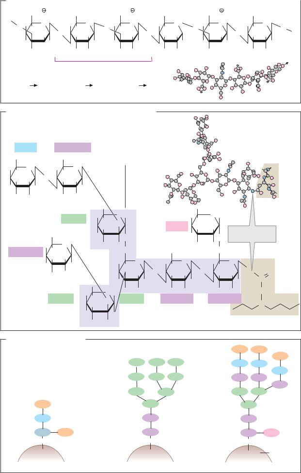
A. Hyaluronic acid
As constituents of proteoglycans (see p.346), the glycosaminoglycans—a group of acidic heteropolysaccharides—are important structural elements of the extracellular matrix.
Glycosaminoglycans contain amino sugars as well as glucuronic acid and iduronic acid as characteristic components (see p.38). In addition, most polysaccharides in this group are esterified to varying extents by sulfuric acid, increasing their acidic quality. Glycosaminoglycans can be found in free form, or as components of proteoglycans throughout the organism.
Hyaluronic acid, an unesterified glycosaminoglycan with a relatively simple structure, consists of disaccharide units in which N- acetylglucosamine and glucuronic acid are alternately β1 4-linked and β 1 3-linked. Due to the unusual β 1 3 linkage, hyaluronic acid molecules–which may contain several thousand monosaccharide residues—are coiled like a helix. Three disaccharide units form each turn of the helix. The outwardfacing hydrophilic carboxylate groups of the glucuronic acid residues are able to bind Ca2+ ions. The strong hydration of these groups enables hyaluronic acid and other glycosaminoglycans to bind water up to 10 000 times their own volume in gel form. This is the function which hyaluronic acid has in the vitreous body of the eye, which contains approximately 1% hyaluronic acid and 98% water.
B. Oligosaccharide in immunoglobulin G (IgG)
Many proteins on the surface of the plasma membrane, and the majority of secreted proteins, contain oligosaccharide residues that are post-translationally added to the endoplasmic reticulum and in the Golgi apparatus (see p.230). By contrast, cytoplasmic proteins are rarely glycosylated. Glycoproteins can contain more than 50% carbohydrate; however, the proportion of protein is generally much greater.
As an example of the carbohydrate component of a glycoprotein, the structure of one of the oligosaccharide chains of immunoglobulin G (IgG; see p.300) is shown here. The oligosaccharide has an N-glycosidic link to the amide group of an asparagine residue in the Fc part of the protein. Its function is not known.
Like all N-linked carbohydrates, the oligosaccharide in IgG contains a T-shaped core structure consisting of two N-acetylglucos- amines and three mannose residues (shown in violet). In addition, in this case the structure contains two further N-acetylglucos- amine residues, as well as a fucose residue and a galactose residue. Glycoproteins show many different types of branching. In this case, we not only have β1 4 linkage, but also β1 2, α1 3, and α1 6 bonds.
C. Glycoproteins: forms
On the cell surface of certain glycoproteins, O-glycosidic links are found between the carbohydrate part and a serine or threonine residue, instead of N-glycosidic links to asparagine residues. This type of link is less common than the N-glycosidic one.
There are two types of oligosaccharide structure with N-glycosidic links, which arise through two different biosynthetic pathways. During glycosylation in the ER, the protein is initially linked to an oligosaccharide, which in addition to the core structure contains six further mannose residues and three terminal glucose residues (see p.230). The simpler from of oligosaccharide (the mannose-rich type) is produced when only the glucose residues are cleaved from the primary product, and no additional residues are added. In other cases, the mannose residues that are located outside the core structure are also removed and replaced by other sugars. This produces oligosaccharides such as those shown on the right (the complex type). At the external end of the structure, glycoproteins of the complex type often contain N-acetylneuraminic acid residues, which give the oligosaccharide components negative charges.
|
|
|
|
|
|
|
|
|
|
|
|
|
|
Carbohydrates |
45 |
||||
A. Hyaluronic acid |
|
|
|
|
|
|
|
|
|
|
|
|
|
|
|
|
|
||
|
|
|
|
|
|
|
|
|
|
|
|
|
|
|
|
|
|||
COO |
HOCH2 |
β |
COO |
β |
HOCH2 |
|
|
|
COO |
HOCH2 |
|
|
|
||||||
O H H |
O |
|
H H |
O H H |
O |
H H |
O |
|
|
O H H |
O |
H H |
|
|
|
||||
|
O |
|
|
|
O |
O |
|||||||||||||
|
|
|
|||||||||||||||||
|
H O |
|
OH H 1 |
3 |
H 1 O |
OH H |
|
|
|
H |
O OH H |
|
|
||||||
|
|
|
|
|
|||||||||||||||
HO |
|
H |
|
|
H |
HO |
|
H |
4 H |
H |
HO |
|
H |
|
H |
|
|
||
|
|
|
|
|
|
|
|
||||||||||||
H |
OH |
|
H |
NHCOCH3 H |
OH |
NHCOCH3 H |
OH |
H |
NHCOCH3 |
||||||||||
Disaccharide unit
|
|
|
|
|
|
|
|
|
|
|
|
|
II |
|
|
|
|
V |
VI |
|
|
|
|
|
|
|
|
|
|
|
|
|
|
|
|
|
|
||
|
[ |
3)-β-D-GlcNAc-(1 |
4)-β-D-GlcUA-(1 |
|
4]n |
|
I |
|
|
III |
IV |
|
|
|
|||||
|
|
|
|
|
|
|
|
|
|||||||||||
|
|
|
|
|
|
|
|
|
|
|
|||||||||
B. Oligosaccharide in immunoglobulin G (IgG) |
|
|
|
|
|
|
|
|
|
||||||||||
D-Gal |
|
|
D-GlcNAc |
|
|
|
|
|
|
|
|
|
|
|
|
|
|
||
HOCH2 |
|
|
HOCH2 |
|
|
Core structure |
|
|
|
|
|
|
|
|
|
||||
HO |
O |
|
|
H |
O |
|
|
|
|
|
|
|
|
|
|
|
|
|
|
H |
|
O |
|
H |
|
|
|
|
|
|
|
|
|
|
|
|
|
|
|
OH H |
|
OH H |
|
|
|
|
|
|
|
|
|
|
|
|
|
|
|||
H |
H |
|
|
|
H |
|
|
|
|
|
|
|
|
|
|
|
|
|
|
H |
OH |
|
|
H |
NHCOCH3 |
|
|
|
|
|
|
|
|
|
|
|
|
|
|
|
|
|
|
|
HOH2C |
|
|
|
|
|
|
H |
|
|
|
|
|
|
|
|
|
|
|
D-Man |
H |
|
O H |
|
|
|
H |
|
O H |
|
|
|
|
||
|
|
|
|
H |
|
O |
|
|
L-Fuc |
CH3 |
|
|
|
|
|
|
|||
|
|
|
|
|
|
|
|
|
|
|
|
|
|
|
|||||
|
|
|
|
|
|
OH |
|
|
H |
HO |
|
N-glycosidic |
|
||||||
|
|
HOCH2 |
|
|
|
|
|
|
|||||||||||
|
|
|
HO |
|
|
|
|
|
HO |
|
|
|
|
||||||
|
|
|
|
O |
|
|
|
|
|
O |
bond |
|
|
||||||
|
|
H |
|
O |
|
|
|
|
|
|
|
|
|
||||||
|
|
H |
|
H |
|
H CH2 |
|
HOCH2 |
|
OH |
H CH2 |
|
|
|
|
||||
D-GlcNAc |
|
|
|
|
|
|
|
|
|
||||||||||
|
|
|
OH H |
|
|
|
H |
O |
H |
|
O |
|
H |
|
O |
|
|
|
|
|
|
HO |
|
H |
|
|
|
|
|
|
|
|
|
||||||
|
|
|
|
|
|
H |
|
O |
H |
|
O |
|
H |
|
HN |
|
|
||
|
|
|
H |
NHCOCH3 |
|
HO |
OH H |
|
OH H |
O |
|
||||||||
|
|
|
|
|
|
|
|||||||||||||
|
|
|
|
|
HOH2C |
HO |
H |
|
|
H |
|
|
|
H |
|
C |
|
||
|
|
|
|
|
|
|
|
|
|
|
|
|
|||||||
|
|
|
D-Man |
H |
|
O H |
H |
H |
|
H |
NHCOCH3 |
H |
NHCOCH3CH2 |
|
|||||
|
|
|
|
H |
O |
D-Man |
D-GlcNAc |
|
D-GlcNAc |
|
C |
|
|||||||
|
|
|
|
|
|
OH |
|
|
|
|
|
|
|
|
|
|
|
||
|
|
|
|
|
|
|
|
|
|
|
|
|
|
|
|
|
|
||
|
|
|
|
|
HO |
|
O |
|
|
|
|
|
|
|
|
|
Asn-297 |
|
|
|
|
|
|
|
|
H |
H |
|
|
|
|
|
|
|
|
|
|
||
|
|
|
|
|
|
|
|
|
|
|
|
|
|
|
|
|
|
||
C. Glycoproteins: forms |
|
|
|
|
|
|
|
O-linked |
|
|
|
N-linked |
NeuAc |
NeuAc |
|
|
|
|
|
|
NeuAc |
||
|
|
|
|
|
|
|
|
|
Man |
Man |
Man |
|
Gal |
|
Gal |
|
|
|
|
|
|
||
|
|
|
|
|
|
|
Gal |
|
Man |
Man |
Man |
|
GlcNAc |
GlcNAc |
|
|
|
|
|
|
|
|
GlcNAc |
|
Man |
|
Man |
|
Man |
|
Man |
NeuAc |
|
Man |
|
|
Man |
|
|
Gal |
|
GlcNAc |
|
|
GlcNAc |
|
|
GalNAc |
NeuAc |
GlcNAc |
|
|
GlcNAc |
Fuc |
|
O |
|
NH |
|
|
|
NH |
|
Ser |
|
Asn |
|
|
Asn |
Protein |
|
|
Mannose-rich type |
|
Complex type |
||||