

- •About the Authors
- •Preface
- •Contents
- •Basics
- •Chemistry
- •Physical Chemistry
- •Biomolecules
- •Carbohydrates
- •Lipids
- •Amino Acids
- •Peptides and Proteins
- •Nucleotides and Nucleic Acids
- •Metabolism
- •Enzymes
- •Metabolic Regulation
- •Energy Metabolism
- •Carbohydrate Metabolism
- •Lipid Metabolism
- •Protein Metabolism
- •Nucleotide Metabolism
- •Porphyrin Metabolism
- •Organelles
- •Basics
- •Cytoskeleton
- •Nucleus
- •Mitochondria
- •Biological Membranes
- •Endoplasmic Reticulum and Golgi Apparatus
- •Lysosomes
- •Molecular Genetics
- •Genetic engineering
- •Tissues and organs
- •Digestion
- •Blood
- •Immune system
- •Liver
- •Kidney
- •Muscle
- •Connective tissue
- •Brain and Sensory Organs
- •Nutrition
- •Nutrients
- •Vitamins
- •Hormones
- •Hormonal system
- •Lipophilic hormones
- •Hydrophilic hormones
- •Other signaling substances
- •Growth and development
- •Cell proliferation
- •Viruses
- •Metabolic charts
- •Annotated enzyme list
- •Abbreviations
- •Quantities and units
- •Further reading
- •Source credits
- •Index
- •Introduction
- •Basics
- •Biomolecules
- •Carbohydrates
- •Lipids
- •Metabolism
- •Organelles
- •Cytoskeleton
- •Nucleus
- •Lysosomes
- •Molecular Genetics
- •Genetic engineering
- •Tissues and organs
- •Digestion
- •Immune system
- •Nutrition
- •Vitamins
- •Hormones
- •Lipophilic hormones
- •Hydrophilic hormones
- •Growth and development
- •Viruses
- •Metabolic charts
- •Annotated enzyme list
- •Abbreviations
- •Quantities and units
- •Further reading
- •Source credits
- •Index

404 Growth and development
Viruses
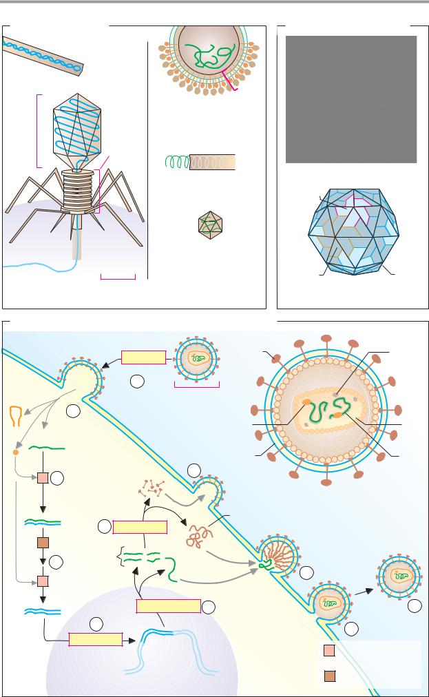
Viruses are parasitic nucleoprotein complexes.
They often consist of only a single nucleic acid molecule (DNA or RNA, never both) and a protein coat. Viruses have no metabolism of their own, and can therefore only replicate themselves with the help of host cells. They are therefore not regarded as independent organisms. Viruses that damage the host cell when they replicate are pathogens. Diseases caused by viruses include AIDS, rabies, poliomyelitis, measles, German measles, smallpox, influenza, and the common cold.
A. Viruses: examples
Only a few examples from the large number of knownviruses are illustrated here. They are all shown on the same scale.
Viruses that only replicate in bacteria are known as bacteriophages (or “phages” for short). An example of a phage with a simple structure is M13. It consists of a singlestranded DNA molecule (ssDNA) of about 7000 bp with a coat made up of 2700 helically arranged protein subunits. The coat of a virus is referred to as a capsid, and the complete structure as a nucleocapsid. In genetic engineering, M13 is important as a vector for foreign DNA (see p. 258).
The phage T4 (bottom left), one of the largest viruses known, has a much more complex structure with around 170 000 base pairs (bp) of double-stranded DNA (dsDNA) contained within its “head.”
The tobacco mosaic virus (center right), a plant pathogen, has a structure similar to that of M13, but contains ssRNA instead of DNA. The poliovirus, which causes poliomyelitis, is also an RNA virus. In the influenza virus, the pathogen that causes viral flu, the nucleocapsid is additionally surrounded by a coat derived from the plasma membrane of the host cell (C). The coat carries viral proteins that are involved in the infection process.
B. Capsid of the rhinovirus
Rhinoviruses cause the common cold. In these viruses, the capsid is shaped like an icosahe- dron—i.e., an object made up of 20 equilateral triangles. Its surface is formed from three different proteins, which associate with one an-
other to form pentamers and hexamers. In all, 60 protein molecules are involved in the structure of the capsid.
C. Life cycle of HIV
The human immunodeficiency virus (HIV) causes the immunodeficiency disease known as AIDS (acquired immune deficiency syndrome). The structure of this virus is similar to that of the influenza virus (A).
The HIV genome consists of two molecules of ssRNA (each 9.2 kb). It is enclosed by a double-layered capsid and a protein-contain- ing coating membrane. HIV mainly infects T helper cells (see p. 294) and can thereby lead to failure of the immune system in the longer term.
During infection (1), the virus’s coating membrane fuses with the target cell’s plasma membrane, and the core of the nucleocapsid enters the cytoplasm (2). In the cytoplasm, the viral RNA is initially transcribed into an RNA/DNA hybrid (3) and then into dsDNA (4). Both of these reactions are catalyzed by reverse transcriptase, an enzyme deriving from the virus. The dsDNA formed is integrated into the host cell genome (5), where it can remain in an inactive state for a long time.
When viral replication occurs, the DNA segment corresponding to the viral genome is first transcribed by host cell enzymes (6). This gives rise not only to viral ssRNA, but also to transcription of mRNAs for precursors of the viral proteins (7). These precursors are integrated into the plasma membrane (8, 9) before undergoing proteolytic modification (10). The cycle is completed by the release of new virus particles (11).
The group of RNA viruses to which HIV belongs are called retroviruses, because DNA is produced from RNA in their replication cy- cle—the reverse of the usual direction of transcription (DNA RNA).
Viruses 405
A. Viruses: examples
|
Phage M13 |
|
ssDNA 7 kb |
|
Helical |
|
T4-Phage |
|
ds DNA |
Eicosa- |
170 kbp |
complex |
|
hedral |
structure |
head |
|
|
Tail |
Phage DNA
Bacterial cell 30 nm
1. Bacteriophages
Coat
Influenza virus
ssRNA (8 molecules) 3.6 kb Nucleocapsid with coat
Tobacco mosaic virus ssRNA6.4 kb
Helical
Poliovirus ssRNA 7 kb
Eicosahedral capsid
2. Plant and animal pathogenic viruses
B. Capsid of the rhinovirus
1. Structure
Pentamer
Hexamer |
Capsid |
|
Eicosahedron of |
2. Diagram |
180 monomers |
C. Life cycle of the human immunodeficiency virus (HIV)
|
|
|
|
Glycoprotein |
|
|
Other |
|
|
|
|
GP120 |
|
|
enzymes |
|
|
|
|
Infection |
|
|
|
|
|
|
|
1 |
|
|
|
|
|
|
|
100 nm |
|
|
|
|
|
2 |
|
Reverse |
|
|
|
|
|
|
|
transcriptase |
|
|
Core |
|
|
Viral |
|
|
|
|
|
|
|
RNA |
|
Membrane |
|
|
Viral |
|
|
|
|
|
|
||
|
|
|
|
8 |
|
|
RNA |
1 |
3 |
|
|
|
|
|
|
|
|
GP120 |
|
|
|
||
|
|
|
|
|
|
|
|
|
|
|
|
Precursors |
|
|
|
|
|
|
|
of core proteins |
|
|
|
|
|
RNA/ |
|
and enzymes |
|
|
|
|
|
DNA |
7 |
Translation |
|
|
|
|
|
hybrid |
|
|
|
||
|
|
|
|
|
|
|
|
2 |
|
|
|
|
|
|
|
|
4 |
|
mRNA |
|
|
|
|
|
|
|
Viral |
9 |
|
|
|
1 |
|
|
|
RNA |
|
|
|
|
|
|
|
|
|
|
|
|
|
ds |
|
Transcription 6 |
|
|
Mature 11 |
|
|
DNA |
5 |
|
|
|
virus |
|
|
|
|
|
10 |
particle |
|
|
|
Integration |
|
|
|||
|
|
|
Reverse transcriptase |
||||
|
|
|
|
|
1 |
||
|
|
|
|
|
|
2.7.7.49 |
|
|
|
Nucleus |
Cytoplasm |
2 |
Ribonuclease H |
||
|
|
Host DNA |
|||||
|
|
|
|
|
|
3.1.26.4 |
|