

- •About the Authors
- •Preface
- •Contents
- •Basics
- •Chemistry
- •Physical Chemistry
- •Biomolecules
- •Carbohydrates
- •Lipids
- •Amino Acids
- •Peptides and Proteins
- •Nucleotides and Nucleic Acids
- •Metabolism
- •Enzymes
- •Metabolic Regulation
- •Energy Metabolism
- •Carbohydrate Metabolism
- •Lipid Metabolism
- •Protein Metabolism
- •Nucleotide Metabolism
- •Porphyrin Metabolism
- •Organelles
- •Basics
- •Cytoskeleton
- •Nucleus
- •Mitochondria
- •Biological Membranes
- •Endoplasmic Reticulum and Golgi Apparatus
- •Lysosomes
- •Molecular Genetics
- •Genetic engineering
- •Tissues and organs
- •Digestion
- •Blood
- •Immune system
- •Liver
- •Kidney
- •Muscle
- •Connective tissue
- •Brain and Sensory Organs
- •Nutrition
- •Nutrients
- •Vitamins
- •Hormones
- •Hormonal system
- •Lipophilic hormones
- •Hydrophilic hormones
- •Other signaling substances
- •Growth and development
- •Cell proliferation
- •Viruses
- •Metabolic charts
- •Annotated enzyme list
- •Abbreviations
- •Quantities and units
- •Further reading
- •Source credits
- •Index
- •Introduction
- •Basics
- •Biomolecules
- •Carbohydrates
- •Lipids
- •Metabolism
- •Organelles
- •Cytoskeleton
- •Nucleus
- •Lysosomes
- •Molecular Genetics
- •Genetic engineering
- •Tissues and organs
- •Digestion
- •Immune system
- •Nutrition
- •Vitamins
- •Hormones
- •Lipophilic hormones
- •Hydrophilic hormones
- •Growth and development
- •Viruses
- •Metabolic charts
- •Annotated enzyme list
- •Abbreviations
- •Quantities and units
- •Further reading
- •Source credits
- •Index

204 Organelles
Cytoskeleton: components
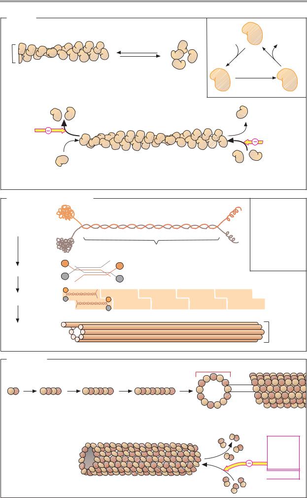
The cytoplasm of eukaryotic cells is traversed by three–dimensional scaffolding structures consisting of filaments (long protein fibers), which together form the cytoskeleton. These filaments are divided into three groups, based on their diameters: microfilaments (6–8 nm), intermediate filaments (ca. 10 nm), and microtubules (ca. 25 nm). All of these filaments are polymers assembled from protein components.
A. Actin
Actin, the most abundant protein in eukaryotic cells, is the protein component of the microfilaments (actin filaments). Actin occurs in two forms—a monomolecular form (G actin, globular actin) and a polymer (F actin, filamentous actin). G actin is an asymmetrical molecule with a mass of 42 kDa, consisting of two domains. As the ionic strength increases, G actin aggregates reversibly to form F actin, a helical homopolymer. G actin carries a firmly bound ATP molecule that is slowly hydrolyzed in F actin to form ADP. Actin therefore also has enzyme properties (ATPase activity).
As individual G actin molecules are always oriented in the same direction relative to one another, F actin consequently has polarity. It has two different ends, at which polymerization takes place at different rates. If the ends are not stabilized by special proteins (as in muscle cells), then at a critical concentration of G actin the (+) end of F actin will constantly grow, while the (–) end simultaneously decays. These partial processes can be blocked by fungal toxins experimentally. Phalloidin, a toxin contained in the Amanita phalloides mushroom, inhibits decay by binding to the
(–) end. By contrast, cytochalasins, mold toxins with cytostatic effects, block polymerization by binding to the (+) end.
Actin–associated proteins. The cytoplasm contains more than 50 different proteins that bind specifically to G actin and F actin. Their actin uptake has various different functions. This type of bonding can serve to regulate the G actin pool (example: profilin), influence the polymerization rate of G actin (villin), stabilize the chain ends of F actin (fragin, β–actinin), attach filaments to one another or
to other cell components (villin, –actinin, spectrin), or disrupt the helical structure of F actin (gelsolin). The activity of these proteins is regulated by protein kinases via Ca2+ and other second messengers (see p. 386).
B. Intermediate filaments
The components of the intermediate filaments belong to five related protein families. They are specific for particular cell types.Typical representatives include the cytokeratins, desmin, vimentin, glial fibrillary acidic protein
(GFAP), and neurofilament. These proteins all have a rod–shaped basic structure in the center, which is known as a superhelix (“coiled coil”; see keratin, p. 70). The dimers are arranged in an antiparallel fashion to form tetramers. A staggered head-to–head arrangement produces protofilaments. Eight protofilaments ultimately form an intermediary filament.
Free protein monomers of intermediate filaments rarely occur in the cytoplasm, in contrast to microfilaments and microtubules. Their polymerization leads to stable polymers that have no polarity.
C. Tubulins
The basic components of the tube-shaped microtubules are α– and β–tubulin (53 and 55 kDa). These form α,β-heterodimers, which in turn polymerize to form linear protofilaments. Thirteen protofilaments form a ringshaped complex, which then grows into a long tube as a result of further polymerization.
Like microfilaments, microtubules are dynamic structures with (+) and (–) ends. The
(–) end is usually stabilized by bonding to the centrosome. The (+) end shows dynamic instability. It can either grow slowly or shorten rapidly. GTP, which is bound by the microtubules and gradually hydrolyzed into GDP, plays a role in this. Various proteins can also be associated with microtubules.
|
|
|
Cytoskeleton |
205 |
|
A. Actin |
|
|
|
|
|
|
|
|
Associates |
Dissociates |
|
|
|
|
more easily |
more easily |
|
|
Polymerization |
|
ATP |
|
ADP |
8 nm |
|
|
|
Pi |
|
|
|
|
|
||
Depolymerization |
|
|
|
||
|
|
|
|
||
F-actin |
|
G-Actin |
ATP |
|
ADP |
helical polymer |
|
|
|||
|
Slow |
|
Pi |
||
microfilament (detail) |
monomers |
|
|||
hydrolysis |
|
||||
|
|
42 kDa |
|
|
|
Phalloidin |
|
|
|
|
|
|
|
|
|
Cytochalasin |
|
|
(–) End: dissociation |
(+) End: polymerization |
|
|
|
|
favored |
|
favored |
|
|
B. Intermediate filaments |
|
|
|
IF Proteins: |
|
|
Cytokeratins |
|
Dimer |
Desmin |
|
|
Vimentin |
|
Superhelical structure |
Glial fibrillary |
|
acidic protein |
||
|
Neurofilaments |
|
Tetramer |
Lamins |
|
|
||
Protofilament |
|
|
Intermediate |
nm |
|
filament |
||
10 |
||
|
C. Tubulins
binds GTP and |
25 nm |
slowly hydrolyzes it |
|
α β |
Protofilament |
Tubulin |
|
53 and 55 kDa |
|
heterodimer |
|
|
|
|
|
|
|
|
|
|
|
|
|
|
|
|
|
|
|
|
|
(–) End: stabilized by binding |
(+) End: grows |
to the centromere |
or shortens |
Microtubule (cylindrical polymer)
Plant alkaloids: Vinblastine, vincristine, colchicine
 Taxol
Taxol

206 Organelles
Structure and functions
The cytoskeleton carries out three major tasks:
•It represents the cell’s mechanical scaffolding, which gives it its typical shape and connects membranes and organelles to each other. This scaffolding has dynamic properties; it is constantly being synthesized and broken down to meet the cell’s requirements and changing conditions.
•It acts as the motor for movement of animal cells. Not only muscle cells (see p. 332), but also cells of noncontractile tissues contain many different motor proteins, which they use to achieve coordinated and directed movement. Cell movement, shape changes during growth, cytoplasmic streaming, and cell division are all made possible by components of the cytoskeleton.
•It serves as a transport track within the cell. Organelles and other large protein complexes can move along the filaments with the help of the motor proteins.
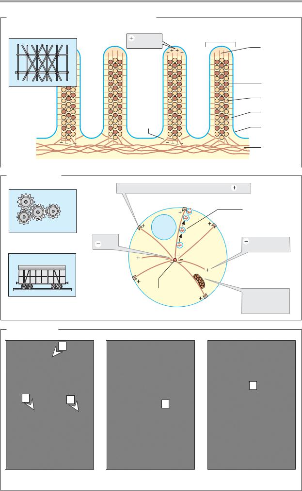
A. Microfilaments and intermediate filaments
The illustration schematically shows a detail of the microvilli of an intestinal epithelial cell as an example of the structure and function of the components of the cytoskeleton (see also
C1).
Microfilaments of F actin traverse the microvilli in ordered bundles. The microfilaments are attached to each other by actin–as- sociated proteins, particularly fimbrin and villin. Calmodulin and a myosin–like ATPase connect the microfilaments laterally to the plasma membrane. Fodrin, another microfila- ment–associated protein, anchors the actin fibers to each other at the base, as well as attaching them to the cytoplasmic membrane and to a network of intermediate filaments. In this example, the microfilaments have a mainly static function. In other cases, actin is also involved in dynamic processes. These include muscle contraction (see p. 332), cell movement, phagocytosis by immune cells, the formation of microspikes and lamellipodia (cellular extensions), and the acrosomal process during the fusion of sperm with the egg cell.
B. Microtubules
Only the cell’s microtubules are shown here. They radiate out in all directions from a center near the nucleus, the centrosome. The tubeshaped microtubules are constantly being synthesized and broken down at their (+) ends. In the centriole, the (–) end is blocked by associated proteins (see p. 204). The (+) end can also be stabilized by associated pro- teins—e.g., when the microtubules have reached the cytoplasmic membrane.
The microtubules are involved in defining the shape of the cell and also serve as guiding tracks for the transport of organelles. Together with associated proteins (dynein, kinesin), microtubules are able to carry out mechanical work—e.g., during the transport of mitochondria, the movement of cilia (hairlike cell protrusions in the lungs, intestinal epithelium, and oviduct) and the beating of the flagella of sperm. Microtubules also play a special role in the mitotic period of cell division (see p. 394).
C. Architecture
The complex structure and net-like density of the cytoskeleton is illustrated here using three examples in which the cytoskeletal components are visualized with the help of antibodies.
1.The border of an intestinal epithelial cell is seen here (see also B). There are microfilaments (a) passing from the interior of the cell out into the microvilli. The filaments are firmly held together by spectrin (b), an associated protein, and they are anchored to intermediate filaments (c).
2.Only microtubules are seen in this fibroblast cell. They originate from the microtubule organizing center (centrosome) and radiate out as far as the plasma membrane.
3.Keratin filaments are visible here in an epithelial cell. Keratin fibers belong to the group of intermediate filaments (see pp. 70, 204; d = nucleus).
Cytoskeleton 207
A. Microfilaments and intermediate filaments
End of |
Microvillus |
F-Actin |
Actin |
|
filament |
|
(micro- |
|
filament) |
|
|
Villin |
Mechanical |
|
Fimbrin |
framework |
|
Calmodulin |
|
|
|
|
Fodrin |
Plasma |
|
membrane |
|
|
|
Intermediate |
|
|
filament |
B. Microtubules |
|
|
|
2. Mikrotubuli |
end |
|
Microtubule – associated protein stabilizes |
|
|
|
Secretory |
|
|
vesicle |
|
Nucleus |
|
Motor for movement |
Stable |
End grows or |
|
ends |
|
|
becomes shorter |
|
|
|
|
|
Nucleus |
|
|
Centrosome |
A mitochondrion |
Rail for transport |
|
migrates along |
|
a microtubule |
C. Architecture |
|
|
|
a |
|
|
|
d |
b |
c |
d |
|
|
|
1. Microfilaments |
2. Microtubules |
3. Intermediate filaments |