

John Wiley & Sons - 2004 - Analysis of Genes and Genomes
.pdf388 ENGINEERING ANIMALS 31
conditional knock-outs may be produced (see below). Knock-outs that fall into the last category (no observable phenotype) may arise as a result of genes acting in parallel pathways compensating for each others’ functions. It is also possible that the techniques are simply too crude to detect any subtle differences between the wild-type and the knock-out animals. The complexity of animal genomes also means that a knock-out may have a profound effect in one strain of mouse, but quite a different effect in another. For example, the deletion of the gene encoding epidermal growth factor in one mouse strain (CF-1) results in embryos that die around the time of implantation into the uterus. If, however, the same knockout is introduced into a different mouse strain (CD-1), then the animals can survive for up to three weeks after birth (Threadgill et al., 1995). Ideally, knockout experiments should be performed in a variety of strain backgrounds, but the length of time required to do that, and the costs involved, often preclude this analysis.
One problem with this type of approach for producing transgenic animals, which we have seem previously when looking at engineering in plants (Chapter 11), is that the selectable maker gene is transferred to the transgenic animal. The high-level expression of an antibiotic-resistance gene within a transgenic animal is generally undesirable. The expression of the marker may induce the abnormal expression of other neighbouring genes, and the potential for transfer of the marker gene to non-transgenic animals should be avoided. The marker gene can effectively be removed after the transgene has been established within the ES cell if its sequences are flanked by loxP sites – the recognition sequences for the Cre recombinase (Kilby, Snaith and Murray, 1993). Transfection of the transgenic cell line with a plasmid expressing Cre recombinase catalyses the excision of the DNA between the two loxP sites to remove the marker gene and leave a single loxP site in its place.
There are many instances where the expression of an inserted transgene is required only in a specific tissue or set of cells. This can readily be achieved by constructing the foreign gene such that it is under the control of a tissuespecific promoter. For example, the promoter of the calcium –calmodulin dependent kinase II (CaMKIIα) gene drives expression only in the neurons of the hippocampus (Mayford et al., 1996). Such an approach works well, provided that a suitable tissue-specific promoter is available (Table 13.1).
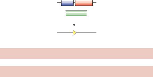
Conditional knock-outs can also be produced, again using the loxP-Cre site-specific recombination system (Gossen and Bujard, 2002). If, for example, the knock-out of a gene results in an embryonic-lethal phenotype, then it may be necessary to delete the gene from the genome after the animal has been born. A method by which this can be achieved is shown in Figure 13.5 (Kuhn¨ et al., 1995). The normal copy of the gene to be deleted is replaced in the
13.2 EMBRYONIC STEM CELLS |
389 |
|
|
Mx1 Cre
 Target gene
loxP |
loxP |
|
+ inducer |
|
|
loxP
Deletion of target gene
Figure 13.5. Tissue-specific gene knock-outs. See the text for details
Table 13.1. Some tissue-specific promoters in mice. Adapted from Lewandoski (2001)
Promoter |
Gene normally |
Tissue or cells |
Reference |
|
controlled |
of expression |
|
|
|
|
|
Alb |
Albumin |
Liver |
(Postic et al., 1999) |
Camk2α Ca2+/calmodulin- |
Forebrain |
(Mayford et al., 1995) |
|
|
dependent protein |
|
|
|
kinase II, α |
|
|
Cryαa |
Crystallin αA |
Eye lens |
(Lakso et al., 1992) |
En2 |
Engrailed |
Mid/hindbrain |
(Logan et al., 1993) |
Gcg |
Glucagon |
Pancreatic α-cells |
(Herrera, 2000) |
Ins2 |
Insulin II |
Pancreatic β-cells |
(Rommel et al., 1994) |
KRT5 |
Keratin 5 |
Epidermis |
(Ramirez et al., 1994) |
Lck |
Lymphocyte-specific |
T cells |
(Chaffin et al., 1990) |
|
tyrosine kinese |
|
|
Msx2 |
Msh-like homeobox |
Apical ectodermal |
(Liu et al., 1994) |
|
gene 2 |
ridge of limb bud |
|
Myog |
Myogenin |
Skeletal muscle |
(Yee and Rigby, 1993) |
Nes |
Nestin |
Neuronal cells |
(Zimmerman et al., |
|
|
|
1994) |
Pax6 |
Paired-box gene 6 |
Retina |
(Gruss and Walther, |
|
|
|
1992) |
Wnt1 |
Wingless related |
Neural crest |
(Echelard, Vassileva and |
|
MMTV integration |
|
McMahon, 1994) |
|
site 1 |
|
|
|
|
|
|

390 ENGINEERING ANIMALS 31
genome by a version that is flanked by loxP sites (often referred to as a floxed gene – flanked by loxP). In addition, the transgenic animal is also modified to carry a copy of the gene encoding the Cre recombinase under the control of an inducible promoter, e.g. Mx1. Mx1 is part of the mouse viral defence system and is transcriptionally inert in healthy mice (Hug et al., 1988). The promoter can, however, be activated by high levels of interferon or by adding synthetic double-stranded RNA to cells (which induces interferon expression). Transgenic animals produced in this way retain a functional copy of the gene to be deleted until they are injected with double-stranded RNA. The effect of the lost gene may then be investigated.
Rather than constructing a transgenic mouse containing both the tissuespecific promoter expressing the Cre recombinase and the target gene surrounded by loxP sites, a series of transgenic mice have been constructed that each contain a different tissue-specific promoter controlling the expressing of Cre. These can then be used as a ‘bank’ of mice strains to which transgenic mice containing a particular floxed gene can be crossed. Mating these strains will result in the formation of progeny in which the gene in inactivated only in those tissues that express Cre (Gu et al., 1994). This means that a single transgenic floxed gene can be deleted in a variety of tissues without having to resort to further in vitro manipulation.
The tetracycline-inducible expression system (see Chapter 8) may be used to drive Cre expression to regulate knock-out function. In this system, a transactivator fusion protein composed of the tetracycline repressor (tetR) and the acidic activation domain of the herpes simplex virus 16 (VP16) protein regulate the expression of the Cre gene from a promoter containing tet-operator (tetO) sequences. In the absence of tetracycline, the Cre gene is expressed and will induce site-specific recombination between two loxP sites. In the presence of tetracycline, the Cre gene will not be expressed and recombination will not occur (St-Onge, Furth and Gruss, 1996).
13.3Nuclear Transfer
Although animal cells become increasingly committed as differentiation and development proceeds, the DNA contained within each differentiated cell still retains all the information necessary to form the whole animal. If the nucleus of a differentiated cell is introduced into an enucleated egg then, under appropriate conditions, the nucleus can become ‘reprogrammed’ such that development of the animal reoccurs. The production of cloned animals – all of which have originated from a single, possibly recombinant, cell line – has several potential uses.
13.3 NUCLEAR TRANSFER |
391 |
|
|
•Recombinant protein production. We have discussed previously that the expression level of recombinant protein production is not strictly inherited (Chapter 12). Therefore, the ability to create large number of animals each expressing identical levels of, say, a therapeutic protein can only be achieved using cloned animals.
•The conservation of endangered species. Rare animals could be cloned to repopulate dwindling natural levels.
The idea of transferring a nucleus from one cell to another is not new. Over 50 years ago it was discovered that the nuclei of blastocyst frog cells could be implanted into eggs that lacked a nucleus to created a series of cloned frogs that were identical to the donor cells (Briggs and King, 1952). It was found, however, that as the donor cells became more differentiated, it became increasing difficult to reprogramme them to produce new animals. The few embryos cloned from differentiated cells that survived to become tadpoles grew abnormally. This led to the speculation that genetic potential diminished as a cell differentiated and that it was impossible to clone an organism from adult differentiated cells. In 1975, however, John Gurdon developed a method of nuclear transfer using fully differentiated cells and Xenopus eggs (Gurdon, Laskey and Reeves, 1975). This is a two-step process.
•Production of enucleated eggs. Delicate needles and a powerful microscope were used to suck the nucleus from a frog oocyte to produce an enucleated oocyte. With the genetic material removed the enucleated oocyte would not divide or differentiate even when fertilized.
•Introduction of a new nucleus. Using the same equipment, the nuclei of keratinized skin cells of adult Xenopus foot-webs were transfered into the enucleated oocytes. Many of these new cells behaved like normal fertilized eggs and were capable of producing tadpoles. Since the tadpoles arose from the cells of the same adult, they all contained the same genetic material and were clones of each other produced from apparently fully differentiated cells. This indicates that DNA is not discarded or permanently inactivated even in highly specialized cells.
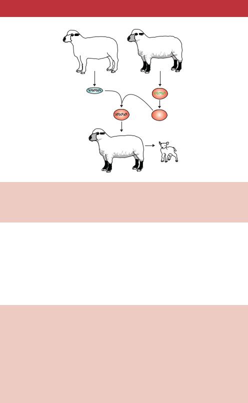
A somewhat modified procedure has been used recently to produce cloned mammals (Figure 13.6). This was first achieved by taking cells from the blastocyst stage of a sheep embryo and fusing them with enucleated eggs (Smith and Wilmut, 1989). The reconstituted cells were subjected to a brief electrical pulse to stimulate embryonic development prior to implantation into a surrogate ewe. Live sheep have subsequently been produced from the
392 ENGINEERING ANIMALS 31
Sheep 1 |
Sheep 2 |
Sheep 1 |
Sheep 2 |
udder cell |
oocyte |
Fuse |
|
cells |
Enucleate |
Culture cells to blastocyst
Implant into surrogate ewe
Clone of sheep 1
Figure 13.6. Nuclear transfer. The cells of an adult sheep (sheep 1) are fused with the enucleated eggs of a sheep of a different breed (sheep 2). The fusion between the two is grown in culture to the blastocyst stage prior to implantation into a surrogate ewe. The resulting lamb contains the nuclear genome of sheep 1
nuclei of cultured embryonic cells (Campbell et al., 1996), and from cultured adult breast epithelial cells (Wilmut et al., 1997). This last example produced probably the most famous sheep in the world – Dolly (Box 13.1). The success of these experiments appears to be dependent on the synchronization of the cell cycles of the donor and recipient cells that are to be fused. In the case of Dolly, quiescence of the donor cell was induced prior to the cell fusion process. Unsynchronized cells appear to be less successful in forming fruitful fusions.
Box 13.1. The life and death of Dolly.
Dolly was the first mammal clone to be produced from an adult cell. She was produced following the procedures described below (Wilmut et al., 1997).
•Donor cells. Mammary gland tissue of a 6-year-old Finn Dorset ewe was used to prepare a primary cell culture. This culture contained a mixture of mammary epithelial cells (>90 per cent), myoepithelial cells and fibroblasts. An important step in the success of the cloning process was to induce these donor cells to exit their growth cycle and enter the G0 phase of the cell cycle before nuclear transfer. This was accomplished

394 ENGINEERING ANIMALS 31
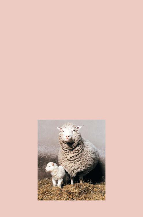
The precise cell type from which Dolly was derived remains unclear. Further analysis indicated that she was indeed derived from the cells of the mammary gland of the donor sheep, rather than from a contaminating cell (Ashworth et al., 1998). She is not, however, an exact clone of the sheep whose cells were used to create her. The DNA of her mitochondria are derived exclusively from recipient enucleated oocytes (Evans et al., 1999). Therefore she is a chimera, containing somatic cell derived nuclear DNA but oocyte derived mitochondrial DNA. It also is interesting to note that the scientific paper in which Dolly was introduced to the world (Wilmut et al., 1997) does not include the words ‘clone’ or ‘cloning’ anywhere within its text. Perhaps the authors realized the potential impact of their findings and chose less inflammatory language to describe their results. Dolly subsequently grew into an adult sheep have bore her own offspring (Box Figure 13.1). Finn Dorset sheep have an average life expectancy of about 12 years, but in January 2002 Dolly was reported to be suffering from arthritis, which is highly unusual for a sheep of her age. On 14 February 2003, aged only six, Dolly was put to sleep following a diagnosis that she was suffering from a progressive lung disease.
The method of nuclear transfer to produce viable offspring from differentiated adult cells is not without its problems (Wilmut et al., 2002). It is likely that not all of the difficulties described below are due to the nuclear transfer process itself, as some similar abnormalities have been reported after embryo culture.
•The process is extremely inefficient. In the case of Dolly, only one of the 277 cell fusions produced was capable of developing into a lamb. Similar efficiency levels have also been reported for other whole animal cloning experiments.
•Many of the embryos produced by nuclear transfer suffer gross abnormalities. In addition to embryonic loss, nuclear transfer is also associated with very high rates of foetal, perinatal and neonatal loss, and production of abnormal offspring.
•Although Dolly was born following a normal gestation period and was a normal weight, many offspring produced by nuclear transfer suffer from large offspring syndrome (LOS) in which gestation period and birth-weight are greatly increased (Lazzari et al., 2002). The frequency and severity of the symptoms of LOS appear to vary widely even under similar experimental conditions. Early deviations from the normal developmental pattern,
13.3 NUCLEAR TRANSFER |
395 |
|
|
particularly with regard to embryonic gene expression, may be involved in this phenomenon.
•Since Dolly was created from a cell that was potentially 6 years old, what genetic age was she when she was born? This has been addressed by looking at the length of the telomeres at the ends of chromosomes. Telomeres generally shorten as aging progresses, although the precise effects of this phenomenon are not well understood. Dolly has been found to have short telomeres when compared with other sheep of the same age (Shiels et al., 1999). It was recently reported that Dolly developed arthritis, which is highly unusual in a sheep of her age (Williams, 2002). It remains to be seen whether this and other potential age-related effects, including Dolly’s death, are a result of the nuclear transfer process.
•Widespread disruptions in the DNA methylation patterns have been described in cloned embryos of a number of cloned animals (Fairburn, Young and Hendrich, 2002). The effects of these changes remain unclear.
•The technique of nuclear transfer is still in its infancy. This means that the effects of aging and genetic inheritance have not been fully assessed. In two independent studies, animals cloned from one cell type became obese in adult life (Tamashiro et al., 2002) whereas those from another cell type died at an unusually early age (Ogonuki et al., 2002). Further work in this area is required.
The technique of nuclear transfer by which Dolly was produced has been replicated or modified to produced clones from adult cells using a variety of other farm animals, e.g. cows, goats and pigs (Cibelli et al., 1998; Baguisi et al., 1999; Polejaeva et al., 2000), and in more experimentally amenable laboratory animals such as mice (Wakayama et al., 1998). In addition, cloned domestic pets such as cats (Shin et al., 2002) and rabbits (Chesne´ et al., 2002) have also been reported. In early 2003, news reports suggested that the first cloned human child had been born. Although such claims have not been scrutinized scientifically, it seems inevitable that a cloned human will be produced at some stage. The difficulties encountered with cloned animals described above should serve as a warning to anyone considering the procedure. The temptation to replace a dead or dying child with an ‘exact copy’ may be more than some parents can bear, but the potentially disastrous consequences should not be underestimated.
Aside from the very negative impact of nuclear transfer technology described above, the process has proved useful for the creation of animals with specific traits. The ability to recreate a whole animal from cells that have been

396 ENGINEERING ANIMALS 31
extensively manipulated in vitro could have a profound positive impact on medicine. For example, there is great potential for the replacement of damaged human organs (e.g. liver, heart) with their equivalents from animals. This process, termed xenotransplantation, is often unsuccessful because some of the cell surface carbohydrates are different between humans and animals. With the exception of catarrhines (Old World monkeys, apes and humans), all animals possess the enzyme α(1,3)-galactosyl transferase, which catalyses the formation of the disaccharide galactose-α(1,3)-galactose that is found on the cell surface. The presence of the disaccharide causes hyperactue rejection of the organ in humans. This problem can only be partially overcome by temporarily removing antibodies to galactose-α(1,3)-galactose from the recipient through affinity adsorption. However, returning antibodies can damage the transplanted organ and severely limit its survival even in the presence of high levels of immunosuppressive drugs. Sheep have been produced that lack the GGTA1 gene encoding the α(1,3)-galactosyl transferase enzyme (Denning et al., 2001). GGTA1 was replaced in tissue culture cells by a copy of the neomycin-resistance gene, and nuclear transfer was used to generate sheep embryos. Unfortunately, the foetuses died before birth, so it remains to be seen whether organs from animals produced in this way may be suitable for human transplantation. More recently, pigs knocked out for either one (Lai et al., 2002; Dai et al., 2002) or both (Phelps et al., 2002) alleles of GGTA1 have been produced. Some of the knock-out pigs are apparently healthy and further work will assess the suitability of their organs for human transplantation.
13.4Gene Therapy
Gene therapy is an approach to treat, cure or ultimately prevent disease by changing the expression of genes within an individual. The idea seems simple – a healthy copy of a mutated gene is introduced into an affected individual such that the normal protein can be made, and the disease symptoms thereby alleviated (Morgan and Anderson, 1993). Although the idea of gene therapy has been around for some time, actual treatments are still in their infancy. Most human clinical trials are only in the research stages. Gene therapy is most applicable to the correction of single gene disorders, especially recessive diseases where a functional copy of the defective gene will restore the activity of the mis-functional protein (Table 13.2). The insertion of the transgene to bring about the desired change can be targeted to either germ (egg and sperm) or somatic (body) cells.
•Germ-line gene therapy. The egg or sperm cells are changed with the goal of passing on the changes to their offspring. Human germ-line gene therapy is

13.4 GENE THERAPY |
397 |
|
|
Table 13.2. Examples of some single-gene human genetic disorders
Disorder |
Symptoms |
|
|
Autosomal recessive: |
|
Cystic fibrosis |
Recurrent lung infection, increased mucus |
|
production |
α1-antitrypsin deficiency |
Liver failure, emphysema |
Phenylketonuria |
Mental retardation |
Tay-Sachs disease |
Neurological degeneration, blindness, paralysis |
Sickle cell anaemia |
Anaemia |
Thalassemia |
Anaemia |
Autosomal dominant: |
|
Neurofibromatosis type 1 |
Tumours of peripheral nerves |
Huntington’s disease |
Involuntary dance-like movements, dementia |
Mytonic dystrophy |
Heart defects and cataracts |
Familial retinoblastoma |
Tumours of the eye |
|
X-linked: |
Haemophilia |
Deficient blood clotting |
Duschenne muscular dystrophy |
Progressive muscle wasting |
Fragile-X syndrome |
Mental retardation |
|
|
prohibited in most countries since the consequences of producing a human with artificially altered genetic traits are far from clear.
•Somatic gene therapy. The genome of the recipient is altered, but this change is not passed to the next generation. Somatic gene therapy can be classed as being performed either in vivo or ex vivo (Figure 13.7). In vivo therapy involves the addition of a gene directly to a patient. Ex vivo therapy involves the removal of cells from the patient and their culturing and genetic manipulation in vitro before the return of the modified cells to the patient.
The type of therapy used depends on the sorts of cell that need to be modified. If the cells in which the gene defect is apparent can be easily cultured, then the ex vivo route offers tremendous advantages. For example, all blood cells are derived from multipotent stem cells in the bone marrow. These differ from ES cells that we have previously discussed in that they can only differentiate into a limited number of different cell types. Multipotent stem cells can, however, be