

Laser-Tissue Interactions Fundamentals and Applications - Markolf H. Niemz
.pdf48 3. Interaction Mechanisms
idea of photochemical treatment is to use a chromophore receptor acting as a catalyst. Its excited states are able to store energy transferred from resonant absorption, and their deactivation leads to toxic compounds leaving the photosensitizer in its original state. Therefore, this type of interaction is also called photosensitized oxidation.
Most photosensitizers belong to the group of organic dyes. Their electronic states are characterized by singlet states (total electron spin momentum s=0) and triplet states (s=1). Furthermore, each electronic state is subdivided into a band of vibrational states. Intersystem crossing is permitted but is associated with an increased lifetime.
Potential reaction kinetics of the photosensitizer are listed in Table 3.1. The reaction types can be characterized by either excitation, decays, Type I or Type II reactions, and carotenoid protection.
Table 3.1. Kinetics of photosensitization (S: photosensitizer, RH: substrate with H-bond, CAR: carotenoid). Modified from Boulnois (1986)
Excitation |
|
• Singlet state absorption |
1S + hν = 1S |
|
|
Decays |
|
• Radiative singlet decay |
1S = 1S + hν (fluorescence) |
• Nonradiative singlet decay |
1S = 1S |
• Intersystem crossing |
1S = 3S |
• Radiative triplet decay |
3S = 1S + hν (phosphorescence) |
• Nonradiative triplet decay |
3S = 1S |
|
|
Type I reactions |
|
• Hydrogen transfer |
3S + RH = SH• + R• |
• Electron transfer |
3S + RH = S•− + RH•+ |
• Formation of hydrogen dioxide |
SH• + 3O2 = 1S + HO2• |
• Formation of superoxide anion |
S•− + 3O2 = 1S + O2• |
|
|
Type II reactions |
|
• Intramolecular exchange |
3S + 3O2 = 1S + 1O2 |
• Cellular oxidation |
1O2 + cell = cellox |
|
|
Carotenoid protection |
|
• Singlet oxygen extinction |
1O2 + 1CAR = 3O2 + 3CAR |
• Deactivation |
3CAR = 1CAR + heat |
|
|
After the absorption of laser photons, the photosensitizer is first transferred to an excited singlet state 1S . Then, three potential decay channels are available: nonradiative and radiative singlet decay to the singlet ground state, and intersystem crossing to an excited triplet state. The latter, finally, may also promote to the singlet ground state by either nonradiative
3.1 Photochemical Interaction |
49 |
or radiative triplet decay. The radiative singlet and triplet decays are called fluorescence and phosphorescence, respectively. Typical lifetimes of fluorescence are of the order of nanoseconds, whereas phosphorescence may last up to a few milliseconds or seconds. According to Foote (1968), two alternative reaction mechanisms exist for the decay of the excited triplet state which are called Type I and Type II reactions (see Table 3.1). They are characterized by either the generation of free radicals (Type I) or the transfer of excitation energy to oxygen molecules (Type II).
During Type I reactions, the triplet state next interacts with a target molecule, other than oxygen, resulting in the release of free neutral or ionized radicals. Further reaction with triplet oxygen may lead to the formation of hydrogen dioxide or superoxide anions. In Type II reactions, the triplet state of the photosensitizer directly interacts with molecular triplet oxygen 3O2 which is then transferred to an excited singlet state 1O2. During this reaction, the electronic spins are flipped in the following manner:
↑↑ |
↑↑ |
↑↓ |
↑↓ |
3S |
+ 3O2 |
= 1S |
+ 1O2 . |
Excited singlet oxygen 1O2 is very reactive, thus leading to cellular oxidation and necrosis. For instance, Weishaupt et al. (1986) identified singlet oxygen as the toxic agent in photoactivation of tumor cells. To avoid additional oxidation of healthy cells, carotin is injected after laser exposure which then promotes the toxic singlet oxygen to harmless triplet oxygen. Usually, Type I and Type II reactions take place at the same time. Which mechanism is favored mainly depends on the concentration of available triplet oxygen and appropriate target molecules.
3.1.1 Photodynamic Therapy (PDT)
Since the beginning of the 20th century, certain dyes have been known to induce photosensitizing e ects as reported by von Tappeiner (1900). The first intended therapeutic application of dyes in combination with light was proposed by von Tappeiner and Jesionek (1903). Later, it was observed by Auler and Banzer (1942) that certain porphyrins have a long clearance period in tumor cells. If these dyes could somehow be transferred to a toxic state, e.g. by laser light, tumor cells could be preferentially treated. Kelly and Snell (1976) have reported on the first endoscopic application of a photosensitizer in the case of human bladder carcinoma. Today, the idea of photodynamic therapy has become one of the major pillars in the modern treatment of cancer.
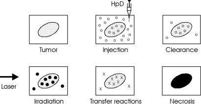
Photodynamic therapy is performed as follows: first, a photosensitizer, e.g. hematoporphyrin derivative (HpD), is injected into a vein of the patient. In the case of HpD, 2.5–5mg per kg body weight are applied. Within the next few hours, HpD is distributed among all soft tissues except the brain. The basic characteristic of a photosensitizer is that it remains inactive until irradiated. After 48–72 hours, most of it is cleared from healthy tissue. However,
50 3. Interaction Mechanisms
its concentration in tumor cells has not decreased much even after a period of 7–10 days. Thus, HpD does not accumulate in tumor cells immediately after injection, but these cells show a longer storage ability (a nity) for HpD. The initial concentration is the same as in healthy cells, but the clearance is faster in the latter cells. After about three days, the concentration of HpD in tumor cells is about thirty times higher than in healthy cells.
Laser irradiation usually takes place after the third day and up to the seventh day after injection if several treatments are necessary. Within this period, tumor cells are still very sensitive and selective necrosis of tumor cells is enabled. However, many healthy tissues may retain certain constituents of HpD and are thus photosensitized, as well. Cellular e ects of HpD were studied in detail by Moan and Christensen (1981) and Berns et al. (1982). The general procedure of photodynamic therapy is illustrated in Fig. 3.2.
Fig. 3.2. Scheme of photodynamic therapy
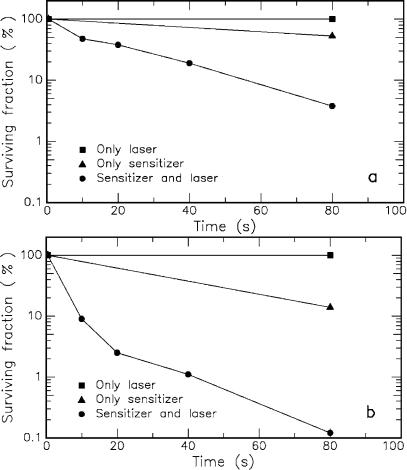
Tumor treatment is the principal but not the only application field of photodynamic therapy. Malik et al. (1990) have observed bactericidal e ects of laser-activated porphyrins. And recently, Wilson et al. (1993) have investigated the e ect of di erent photosensitizers on streptococcus sanguis, a common bacterium of dental plaques. Some of their results are summarized in Figs. 3.3a–b. Obviously, only the combined action of photosensitizer and laser exposure significantly reduces the fraction of surviving bacteria.
One of the most commonly used photosensitizers in photodynamic therapy is a hematoporphyrin derivative called HpD. It is derived from calf blood and is a complex collection of di erent porphyrins, mainly
–dihematoporphyrin,
–hydroxyethylvinyl-deuteroporphyrin,
–protoporphyrin.
3.1 Photochemical Interaction |
51 |
Fig. 3.3. (a) E ect of methylene blue and/or helium–neon laser (power: 7.3mW) on the viability of streptococcus sanguis. (b) E ect of hematoporphyrin ester and/or helium–neon laser (power: 7.3mW) on the viability of streptococcus sanguis. Data according to Wilson et al. (1993)
Among those substances, dihematoporphyrin is the active constituent in providing the photosensitizing e ect. Medical application of HpD was first performed and reported by Lipson and Baldes (1961). Proprietary names for HpD include Photofrin I and Photofrin II. Both of these agents are complex chemical mixtures with the latter being enriched in the tumor-localizing fraction during PDT.
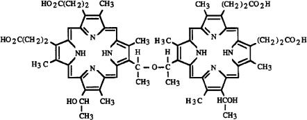
The chemical structure of dihematoporphyrin is shown in Fig. 3.4. It consists of two porphyrin rings connected by a C–O–C chain. According to Dolphin (1979), porphyrins are characterized by a high thermal stability and
52 3. Interaction Mechanisms
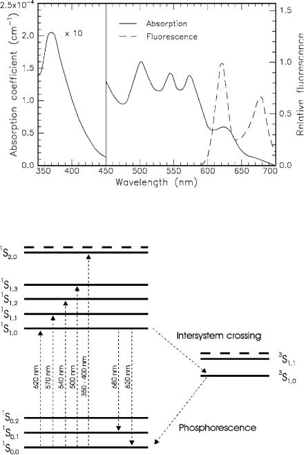
a dark color (from Greek: πoρφυρα = purple). The absorption and fluorescence spectra of HpD are shown in Fig. 3.5, whereas Fig. 3.6 illustrates the corresponding energy level diagram. The strong absorption at 350–400nm originates from the broad excitation band 1S2 of the dye. The relative low absorption at approximately 620–630nm is used for clinical purposes. At this wavelength, deeper structures can be reached compared to using UV light due to the lower absorption coe cient of most tissues in the red spectrum. Detailed studies on porphyrin photosensitizers are found in the book edited by Kessel and Dougherty (1983).
Fig. 3.4. Chemical structure of the active substance dihematoporphyrin which consists of two symmetric porphyrin rings
Primarily, the fluorescence spectrum of HpD is characterized by two peaks at 620nm and 680nm. They stem from the transitions 1S1,0 −→ 1S0,0 and 1S1,0 −→ 1S0,1, respectively. As in all macromolecules, the ground and the excited electronic states are further split into several vibrational states. After excitation, the macromolecule first relaxes to the lowest vibrational state belonging to the same excited electronic state. From there, it reaches the ground state by emitting fluorescence radiation.
The dependence of fluorescence on the concentration level of HpD is of considerable interest. It was found by Kinoshita (1988) that the emission peak at 620nm decreases toward higher concentrations of HpD. This e ect is explained by self-absorption which becomes a dominant process at concentrations higher than about 10−3 mol/l. Therefore, the comparison of relative fluorescence intensities is an indicator for the concentration level of HpD and, thus, for the distinction of tumor cells from healthy cells.
An even more powerful technique is given by time-resolved fluorescence. It was observed by Yamashita (1984) that the duration of the fluorescence decay of HpD also depends on its respective concentration level. The lowest investigated concentration of 8.4 ×10⁻⁶ mol/l yields a fluorescence decay time in the nanosecond range, whereas higher concentrations such as 8.4×10⁻³ mol/l are characterized by decays as short as a few hundred picoseconds. The time-
3.1 Photochemical Interaction |
53 |
Fig. 3.5. Absorption and fluorescence spectra of hematoporphyrin derivative (HpD) dissolved in phosphate-bu ered saline solution (PBS). Data according to Yamashita (1984)
Fig. 3.6. Energy level diagram of HpD. Singlet (1S) and triplet (3S) states are shown. Dashed lines indicate higher excited states
54 3. Interaction Mechanisms
resolved fluoresecence signals are summarized in Fig. 3.7. Tumor diagnosis can thus be established by time-gated detection of the fluorescence. For instance, Fig. 3.8 illustrates the decay in fluorescence intensity of healthy cells with respect to tumor cells as found by Kinoshita (1988). Since clearance of HpD is faster in healthy cells, the corresponding decay duration is significantly longer than in tumor cells. According to Uns¨old et al. (1987), the simultaneous diagnosis and therapy of tumors with photosensitizers is one of the key advantages of PDT. Extensive in vivo studies of time-resolved fluorescence have been performed by Schneckenburger et al. (1993).
Meanwhile, the results of several experiments and clinical applications have been collected. It was found that other photosensitizers might even be more useful than HpD. The major disadvantage of HpD is the fact that the patient needs to remain in a dark room during the first weeks of therapy. This is necessary, because HpD is distributed all over the body, and sun light or artificial light would kill healthy tissue cells, as well. According to Kessel (1987), the primary adverse reaction of photodynamic therapy relates to the photosensitization of skin. Other disadvantages of HpD are:
–since HpD absorbs very poorly in the red and near infrared spectrum, only tumors very close to the surface can be treated,
–its concentration gradient among tumor and healthy cells could be steeper,
–the production of HpD from calf blood is very expensive.
However, the initial isolation in the period between injection of HpD and laser irradiation remains and is one of the biggest problems for patients. This context also explains the injection of carotenoids immediately after laser irradiation. These agents act as a protection system on a molecular basis, because they reverse the production of singlet oxygen by means of a triplet carotenoid state (compare Table 3.1). The protective property of carotenoids has been successfully tested by Mathews-Roth (1982).
Currently, further photosensitive compounds of a so-called second generation are under investigation concerning their applicability for PDT. These are, for instance, certain groups of phthalocyanins, naphthalocyanins and phorbides (reduced porphyrins) as reported by Spikes (1986), Firey and Rodgers (1987), and R¨oder et al. (1990). Hopefully, a dye will soon be found that is more e cient than HpD and overcomes its major disadvantage, the need for carotenoid protection. One potential candidate is meso-tetra-hydroxyphenyl- chlorin (mTHPC), a chemically well-defined substance compared to HpD. In order to achieve a similar extent of tumor necrosis, it was reported by Gossner et al. (1994) that in the case of mTHPC only one fifth of the light energy is needed compared to HpD. Another interesting substance is 5-aminolaevulinic acid (ALA) which itself is not even a photosensitizer but a precursor of the endogeneous synthesis of porphyrins. According to Loh et al. (1993), it can be orally administered rather than being injected like most of the porphyrins.

3.1 Photochemical Interaction |
55 |
Fig. 3.7. Time-resolved fluorescence signals of HpD dissolved in phosphate-bu ered saline solution (PBS). Concentrations of HpD as labeled. The corresponding decay durations of the fluorescence vary between approximately 350ps and 2ns. Data according to Yamashita (1984)
Fig. 3.8. Time-resolved HpD fluorescence of healthy cells versus tumor cells. The approximate decay durations are 2.5ns and 1.0ns, respectively. Data according to Kinoshita (1988)
56 3. Interaction Mechanisms
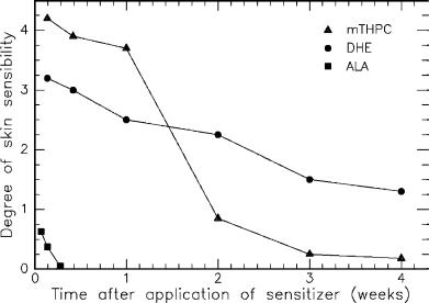
Gossner et al. (1994) have compared the skin sensibility after application of DHE (dihematoporphyrin ester), mTHPC, and ALA. The results are graphically summarized in Fig. 3.9. The sensibility is measured in terms of a commonly used index of skin edema. It is interesting to observe that ALA induces least toxic skin damage. An increased sensibility is detectable only during the first two days after application. Although mTHPC is associated with the highest sensibility during the first week, only a slightly higher sensibility remains after the second week. This negative side e ect during the first week is acceptable, though, since mTHPC is highly e cient in achieving tumor necrosis according to Gossner et al. (1994). The sensibility after application of DHE gradually decreases within the first four weeks and is still significantly enhanced at the end of this period. Thus, patients treated with DHE will have to remain in dark rooms for at least four weeks.
Fig. 3.9. Degree of skin sensibility after application of various photosensitizers as a function of time. Data according to Gossner et al. (1994)
The field of photodynamic tumor therapy has just begun to develop. Although much e ort has already been made concerning treatment with HpD, a lot more research still needs to be done. Alternative photosensitizers such as mTHPC and ALA should be investigated in a large group of patients to further improve this type of treatment. Careful conclusions should then be drawn regarding their e ciency, indications, and potential contraindications. Finally, an approved committee has to compare all results with those obtained with other minimally invasive methods of tumor treatment such as exposure to ionizing radiation.

3.1 Photochemical Interaction |
57 |
3.1.2 Biostimulation
Biostimulation is believed to occur at very low irradiances and to belong to the group of photochemical interactions. Unfortunately, the term biostimulation has not been scientifically very well defined, so far. The potential e ects of extremely low laser powers (1–5mW) on biological tissue have been a subject of controversy, since they were first claimed by the Hungarian surgeon Mester at the end of the 1960s. Wound healing and anti-inflammatory properties by red or near infrared light sources such as helium–neon lasers or diode lasers were reported. Typical energy fluences lie in the range 1–10J/cm2. In several cases, observers have noticed improvements for the patients. But in a few studies only, results could be verified by independent research groups. Moreover, contradictory results were obtained in many experiments.
The most significant studies concerning biostimulation are summarized in Table 3.2. They demonstrate the variety of potential application fields. However, often only very few patients were treated, and no clinical protocols were established. Furthermore, the success is rather doubtful, since in many of these diseases 50% of the patients are spontaneously cured even without treatment. According to Wilder-Smith (1988), the distinction from an ordinary placebo e ect is thus rather di cult to perform.
Table 3.2. Biostimulative e ects investigated by di erent studies
Observation |
Target |
Laser type |
Reference |
|
|
|
|
Hair growth |
Skin |
Ruby |
Mester et al. (1968) |
Wound healing |
Skin |
Ruby |
Mester et al. (1969) |
|
|
|
Mester et al. (1971) |
|
|
He-Ne |
Brunner et al. (1984) |
|
|
|
Lyons et al. (1987) |
No wound healing |
Skin |
He-Ne |
Hunter et al. (1984) |
|
|
|
Strube et al. (1988) |
|
|
Argon ion |
Jongsma et al. (1983) |
|
|
|
McCaughan et al. (1985) |
Stimulated collagen |
Fibroblasts |
Nd:YAG |
Castro et al. (1983) |
synthesis |
|
He-Ne |
Kubasova et al. (1984) |
|
|
|
Boulton et al. (1986) |
Suppressed collagen |
Fibroblasts |
Nd:YAG |
Abergel et al. (1984) |
synthesis |
|
|
|
Increased growth |
Cells |
Diode |
Dyson and Young (1986) |
Suppressed growth |
Cells |
He-Cd |
Lin and Chan (1984) |
|
|
He-Ne |
Quickenden et al. (1993) |
Vascularization |
Oral soft tissue |
Diode |
Kovacs et al. (1974) |
|
|
|
Cho and Cho (1986) |
Pain relief |
Teeth |
He-Ne |
Carrillo et al. (1990) |
|
|
Diode |
Taube et al. (1990) |
No pain relief |
Teeth |
He-Ne |
Lundeberg et al. (1987) |
|
|
Diode |
Roynesdal et al. (1993) |
|
|
|
|