

Laser-Tissue Interactions Fundamentals and Applications - Markolf H. Niemz
.pdf208 4. Medical Applications of Lasers
The most frequent malformations of the external genital are called condylomata acuminata. These benign warts must be treated as early as possible, because they tend to be very infectious and degenerating. After circumcision and application of 4% acetic acid to suspected areas, they are coagulated with either Nd:YAG or CO2 lasers. Occasional recurrences cannot be excluded, especially in the treatment of intraurethral condylomata. With both laser types, however, the rate of recurrence is less than 10% as reported by Baggish (1980) and Rosemberg (1983). Hemangiomas of the external genital should be treated with radiation from Nd:YAG lasers because of its higher penetration depth. Hofstetter and Pensel (1993) stated that additional cooling of the tissue surface may even improve the procedure. Carcinoma of the external genital are best treated with a Nd:YAG laser if they are at an early stage. This significantly reduces the risk of having to perform a partial amputation. According to Eichler and Seiler (1991), powers of 40W and focal spot sizes of 600μm are usually applied. At an advanced stage, the tumor is first mechanically extirpated. Afterwards, the remaining tissue surface should be additionally coagulated.
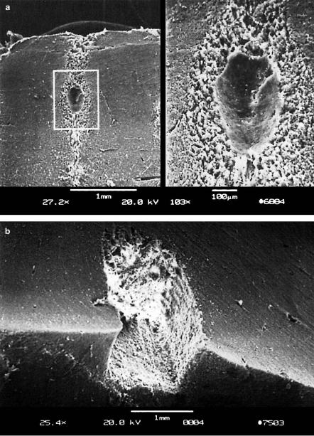
Frequent diseases of the lower urinary tract are stenoses induced by either inflammation, tumor growth, or unknown origins. In these cases, urethrotomy by endoscopic control is usually performed as proposed by Sachse (1974). During this conventional technique, stenotic material is removed with a cold scalpel. Unfortunately, restenoses often occur due to scarring of the tissue. Further urethrotomies are not of great help, since they only enhance additional scarring. The first recanalizations of urethral stenoses with an argon ion laser were performed by Rothauge (1980). However, the results obtained were not as promising as initially expected. Then, no further progress was made until Wieland et al. (1993) recently published first results using a Ho:YAG laser. Meanwhile, follow-up periods of 20 months after Ho:YAG laser treatment were reported by Nicolai et al. (1995). They concluded that this technique is a considerable alternative to mechanical urethrotomy in virgin stenoses as well as restenoses. The probability for the occurrence of laser-induced restenoses is approximately 10% only. In Figs. 4.48a–b, the effects of the Ho:YAG laser on the urethra and ureter are shown, respectively. In both samples, thirty pulses with an energy of 370mJ and an approximate duration of 1ms were applied.
Tumors of the bladder are very di cult to treat, since they tend to recur after therapy. It is yet unknown whether this is due to metastasation induced either prior to or by the treatment. Unfortunately, bladder tumors also easily break through the bladder wall. Thus, a treatment is successful only if it completely removes the tumor, does not perforate the bladder wall, and does not damage the adjacent intestine. Frank et al. (1982) have compared the e ects of CO2, Nd:YAG, and argon ion lasers on bladder tissue. Among these, the Nd:YAG laser has proven to be best suited in coagulating bladder tumors. Argon ion lasers are applicable only in superficial bladder tumors.

4.4 Lasers in Urology |
209 |
Fig. 4.48. (a) E ect of thirty pulses from a Ho:YAG laser (pulse duration: 1ms, pulse energy: 370mJ, bar: 250μm) on the urethra. (b) E ect of thirty pulses from a Ho:YAG laser (pulse duration: 1ms, pulse energy: 370mJ, bar: 250μm) on the ureter. Photographs kindly provided by Dr. Nicolai (Regensburg)
210 4. Medical Applications of Lasers
According to Hofstetter et al. (1980), the rate of recurrence after laser treatment is approximately 1–5%, whereas it ranges from 40–60% if conventional transurethral resection (TUR) is performed. Even advanced tumors can be e ciently removed with Nd:YAG lasers, since the hemostatic treatment guarantees best vision. Pensel (1986) suggests the application of 30–40W of laser power and a working distance between 1mm and 2mm. The tumor should be irradiated until it visibly pales. Afterwards, coagulated necrotic tissue is mechanically removed. For safety reasons, the remaining tissue surface should be coagulated, as well.
It was emphasized by Hofstetter and Pensel (1993) that tumors can still be graded and staged by biopsies after coagulation. Usually, control biopsies should be obtained within the next 3–6 months. The laser treatment itself is extremely safe, since perforations of the bladder wall are very unlikely, and the function of the bladder remains una ected. All transurethral treatments are performed with a rigid cytoscope and a flexible fiber. In most cases, local anesthetization is su cient.
Recently, photodynamic therapy (PDT) has gained increasing significance in the treatment of bladder tumors. First endoscopic applications of HpD have already been investigated by Kelly and Snell (1976). Several clinical reports on PDT are available, e.g. by Benson (1985), Nseyo et al. (1985), and Shumaker and Hetzel (1987). A complete treatment system including in vivo monitoring and dose control was decribed by Marynissen et al. (1989). A list of potential complications arising when using dihematoporphyrin ether was given by Harty et al. (1989). Today, photodynamic therapy is considered as a useful supplement to other techniques, since it enables the resection of tumors which are not visible otherwise. The ability of simultaneous diagnosis
–by means of laser-induced fluorescence – and the treatment of tumors is thus one of the key advantages of photodynamic therapy. So far, red dye lasers at 630nm and energy densities between 10–50J/cm2 are usually applied. In most cases, laser treatment is still restricted to superficial tumors due to the limited penetration depth at the specific wavelength. However, the recent discovery of novel photosensitizers like 5-aminolaevulinic acid (ALA)
–as already discussed in Sect. 3.1 – will certainly improve photodynamic therapy in urology during the next few years.
Lithotripsy of urinary calculi is often based on ultrasound techniques. However, not all calculi are equally indicated for such an external therapy. In particular, those calculi which are stuck inside the ureter are in an extremely inconvenient location. In these cases, laser-induced lithotripsy o ers the advantage of directly applying energy to the vicinity of the calculus by means of a flexible fiber. First experiments regarding laser lithotripsy have already been performed by Mulvaney and Beck (1968) using a ruby laser. From today’s perspective, though, it is quite obvious that these initial studies had to be restricted to basic research, since they were associated with severe thermal side e ects. Watson et al. (1983) first proposed the application of a Q-
4.4 Lasers in Urology |
211 |
switched Nd:YAG laser. Shortly after, pulsed dye lasers were investigated by Watson et al. (1987). With the decrease in pulse durations, additional complications arose concerning induced damage of the fiber. Extensive calculations of the limits of fiber transmission were published by Hering (1987). The advantages of di erent approaches like bare fibers or focusing fiber tips were studied by D¨orschel et al. (1987) and Hofmann and Hartung (1987). Furthermore, a review of 20 years of laser lithotripsy experience was given by Dretler (1988).
Today, dye lasers and Nd:YAG lasers are preferably used for lithotripsy of urinary calculi inside the ureter. A detailed description of the procedure was given by Hofstetter et al. (1986). Typically, pulse energies of 50–200mJ and pulse durations between 10ns and 1μs are applied. The diameter of the optical fiber varies between 200μm and 600μm. With these parameters, optical breakdown is achieved close to the target. As described in Sect. 3.5, plasma formation at high pulse energies is associated with shock waves, cavitations, and jets. This photodisruptive interaction finally leads to the fragmentation of urinary calculi.
Since the 1980s, research in urology has increasingly focused on various treatments of the prostate. This very sensitive organ embraces the urethra. Diseases of the prostate, e.g. benign hyperplasias or carcinoma, thus often tend to handicap the discharge of urine. A profound analysis of the development of benign prostatic hyperplasia (BPH) was given by Berry et al. (1984). Several conventional therapies are available, e.g. the initial application of phytopharmaka or transurethral resection in severe cases. Other techniques such as cryotherapy or photodynamic therapy have also been investigated, e.g. by Bonney et al. (1982) and Camps et al. (1985). A complete list of potential treatment methods was provided by Mebust (1993). During the first few years, research was restricted to the treatment of prostatic carcinoma. B¨owering et al. (1979) were the first to investigate the e ect of Nd:YAG laser radiation on tumors of the prostate. Shortly after, several detailed reports followed, e.g. by Sander et al. (1982) and Beisland and Stranden (1984). The latter study pointed out the extreme importance of temperature monitoring of the adjacent rectum. Extensive clinical results were reported by McNicholas et al. (1988). Usually, indication for laser treatment is given only if the tumor cannot be completely resected otherwise.
At the beginning of the 1990s, the demand for minimally invasive techniques significantly increased. In the treatment of BPH, two milestones were achieved with the development of improved surgical techniques called transurethral ultrasound-guided laser-induced prostatectomy (TULIP) and laser-induced interstitial thermotherapy (LITT). The idea of TULIP was proposed by Roth and Aretz (1991) and Johnson et al. (1992). Detailed clinical results were published by McCullough et al. (1993). The key element of TULIP is to position a 90◦ prism inside the urethra by ultrasound control. Thereby, the precision in aiming at the target is strongly enhanced.
212 4. Medical Applications of Lasers
In other studies, Siegel et al. (1991) have shown that hyperthermia alone, i.e. temperatures up to 45◦C, is not su cient in treating BPH. This has led to the idea of LITT as already described in Sect. 3.2. During LITT, the tissue is completely coagulated, i.e. temperatures above 60◦C are obtained. The technical realization of suitable ITT fibers was discussed by Hessel and Frank (1990). In urology, initial experimental results with LITT were published by McNicholas et al. (1991) and Muschter et al. (1992). With typical laser powers of 1–5W, coagulation volumes with diameters of up to 40mm are achieved. Meanwhile, Muschter et al. (1994) have reported on clinical studies with approximately 200 patients. Roggan et al. (1994) have determined the optical parameters of prostatic tissue for diode lasers at 850nm and Nd:YAG lasers at 1064nm, respectively. Their data are found in Table 2.3. Moreover, they observed that the scattering coe cient of prostatic tissue increases during coagulation by an appoximate factor of two. With these data and appropriate computer simulations, they were able to optimize the parameters for an e cient procedure.
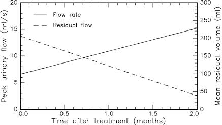
In Fig. 4.49, the most significant postoperative results of LITT in the treatment of BPH are summarized. According to Muschter et al. (1993), the peak urinary flow rate increased from 6.6ml/s to 15.2ml/s two months after treatment, whereas the residual urinary volume decreased from 206ml to 38ml. The mean weight of the prostate dropped from 63g to 44g during the same period. These data are based on mean values obtained from 15 patients. Severe complications were not observed. From these results, it can be concluded that LITT is an excellent therapy for BPH.
Fig. 4.49. Peak urinary flow and mean residual urinary volume after LITT of benign prostatic hyperplasia. The data represent mean values from 15 patients. Data according to Muschter et al. (1993)

4.5 Lasers in Neurosurgery |
213 |
4.5 Lasers in Neurosurgery
Neurosurgery deals with diseases of the central nervous system (CNS), i.e. the brain and the spine. Surgery of brain tumors is very di cult, since extremely localized operations are necessary due to the complicated structure and fragility of the brain. Moreover, the tumor itself is often not easily accessible, and very important vital centers are situated beside it. Therefore, it is not surprising that a considerable amount of research funds is currently being spent in this field, especially since any kind of brain tumor – even benign tumors – are extremely life-threatening. This is because space inside the skull is very limited. Hence, growth of new tissue increases the pressure inside the brain which leads to mechanical damage of other neurons. A schematic cross-section of the brain is shown in Fig. 4.50.
Fig. 4.50. Scheme of a human brain
The major parts of the brain are the cerebrum, diencephalon, cerebellum, and brainstem. The diencephalon can be further divided into the hypothalamus, hypophysis, thalamus, and epiphysis, whereas the brainstem consists of the mesencephalon, pons, and medulla oblongata. Usually, tumors of the brainstem are highly malignant and, unfortunately, they reside in an inaccessible location. In general, brain tissue can be divided into gray matter and white matter which are made up of cell nuclei and axons, respectively. Blood perfusion of gray and white matter di ers remarkably. The corresponding ratio is about five to one.
214 4. Medical Applications of Lasers
The application of lasers in neurosurgery has been extremely slow compared with other medical fields, e.g. ophthalmology. This was mainly due to two reasons. First, studies by Rosomo and Caroll (1966) revealed that the ruby laser was not of great help in neurosurgery. Second, initial experiments with the CO2 laser were performed at too high energy levels, e.g. by Stellar et al. (1970), which was dangerous and completely unnecessary. It then took some time until Ascher (1979), Beck (1980), and Jain (1983) reawakened interest in neurosurgical lasers, especially moderate CO2 lasers and Nd:YAG lasers. The principal advantages of lasers in neurosurgery are evident. Lasers are able to cut, vaporize, and coagulate tissue without mechanical contact. This is of great significance when dealing with very sensitive tissues. Simultaneous coagulation of blood vessels eliminates dangerous hemorrhages which are extremely life-threatening when occurring inside the brain. Moreover, the area of operation is sterilized as lasing takes place, thereby reducing the probability of potential infections.
According to Ascher and Heppner (1984) and Stellar (1984), the main advantage of the CO2 laser is that its radiation at a wavelength of 10.6μm is strongly absorbed by brain tissue. By this means, very precise cuts can be performed. However, CO2 lasers are not appropriate for the coagulation of all blood vessels. In particular, arteries and veins with diameters > 0.5mm tend to bleed after being hit by the laser beam. Nd:YAG lasers, on the other hand, are e ective in coagulating blood vessels as stated by Fasano et al. (1982) and Wharen and Anderson (1984b). Ulrich et al. (1986) even observed very good results on both ablation and coagulation when combining a Nd:YAG laser emitting at 1.319μm and a 200μm fiber. The biological response of brain tissue to radiation from Nd:YAG lasers was extensively studied by Wharen and Anderson (1984a). A preliminary report on the clinical use of a Nd:YAG laser was given by Ascher et al. (1991). Moreover, neurosurgical applications of argon ion lasers had been investigated by Fasano (1981) and Boggan et al. (1982), but they seem to be rather limited, since radiation from these lasers is strongly scattered inside brain tissue.
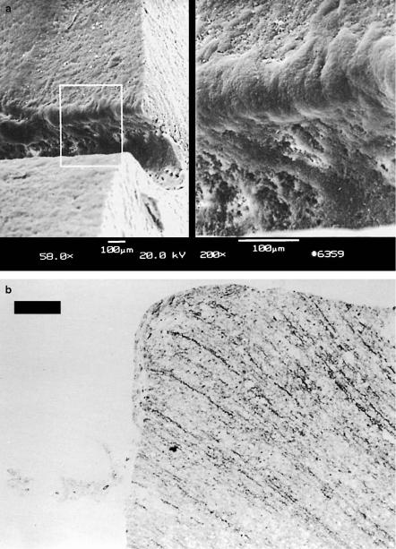
The main problem with CW lasers is that they do not remove brain tumors but only coagulate them. Necrotic tissue remains inside the brain and can thus lead to the occurrence of severe edema. Moreover, adjacent healthy tissue might be damaged due to heat di usion, as well. Recently, two alternative lasers have been investigated concerning their applicability to neurosurgery: Er:YAG and Nd:YLF lasers. Cubeddu et al. (1994) and Fischer et al. (1994) have studied the ablation of brain tissue using free-running and Q-switched Er:YAG lasers. They observed limited thermal alterations of adjacent tissue. However, mechanical damage was very pronounced. Since the Er:YAG laser emits at a wavelength of 2.94μm, its radiation is strongly absorbed in water as already discussed in Sect. 3.2. Thus, soft brain tissue – having a high water content – is suddenly vaporized which leads to vacuoles inside the tissue with diameters ranging up to a few millimeters. In Fig. 4.51a, mechanical damage
4.5 Lasers in Neurosurgery |
215 |
Fig. 4.51. (a) Brain tissue after exposure to an Er:YAG laser (pulse duration: 90μs, pulse energy: 60mJ). Mechanical damage is evident. (b) Voluminous ablation of brain tissue achieved with the same laser. Reproduced from Fischer et al. (1994) by permission. c 1994 Springer-Verlag

216 4. Medical Applications of Lasers
up to a depth of at least 1.5mm is clearly visible. Therefore, it is not very helpful that even large volumes of brain tissue can be ablated with Er:YAG lasers as shown in Fig. 4.51b.
The ablation of brain tissue with a picosecond Nd:YLF laser system was investigated by Fischer et al. (1994). In Fig. 4.52, the ablation depths of white and gray brain matter are given, respectively. Obviously, there is no significant di erence in ablating either substance. It is interesting to observe, though, that there is no saturation in ablation depth even at energy densities as high as 125J/cm2. Thus, higher laser powers will probably enable ablation depths > 200μm. Fischer et al. (1994) state that the corresponding ablation threshold is at approximately 20J/cm2.
Fig. 4.52. Ablation curve of white and gray brain matter obtained with a Nd:YLF laser (pulse duration: 30ps, focal spot size: 30μm). Data according to Fischer et al. (1994)
Two samples of brain tissue which were exposed to the Nd:YLF laser are shown in Figs. 4.53a–b. A rectangular ablation geometry was achieved by scanning the laser beam. The lesion in Fig. 4.53a is characterized by steep walls and is approximately 600μm deep. In Fig. 4.53b, a histologic section of an ablation edge is shown as obtained with the Nd:YLF laser. The tissue was stained with cluever barrera to visualize any thermal e ects. There is no evidence of either thermal or mechanical damage to adjacent tissue. Hence, removal of tissue can be attributed to the process of plasmainduced ablation as described in Sect. 3.4. Nonthermal ablation of tissue is a mandatory requirement for precise functional surgery of the brain.
4.5 Lasers in Neurosurgery |
217 |
Fig. 4.53. (a) Brain tissue after exposure to a picosecond Nd:YLF laser (pulse duration: 30ps, pulse energy: 0.5mJ). (b) Histologic section of brain tissue after exposure to the same laser (bar: 50μm). Reproduced from Fischer et al. (1994) by permission. c 1994 Springer-Verlag