

Laser-Tissue Interactions Fundamentals and Applications - Markolf H. Niemz
.pdf148 3. Interaction Mechanisms
Fig. 3.74. Bubble collapse with jet formation (top). Pictures taken at 20000 frames per second (frame size: 7.3mm×5.6mm). Bubble collapse with counterjet formation (bottom). Reproduced from Vogel et al. (1989) by permission. c 1989 Cambridge University Press
Fig. 3.75. (a) Experimentally obtained pathline portrait of the flow around a collapsing cavitation bubble near a solid wall. Bubble shape 0 represents the bubble at maximum expansion, whereas shapes 1 and 2 correspond to later stages during the collapse. (b) Calculated pathline portrait from Kucera and Blake (1988) for several points on the wall of the collapsing bubble. Reproduced from Vogel and Lauterborn (1988) by permission. c 1988 Optical Society of America

3.6 Questions to Chapter 3 |
149 |
force by the slower collapsing opposite wall – is delayed. This explains why jet formation occurs toward the solid boundary. If the jet is relatively slow, the velocity of the central part of the slower collapsing side might even be higher than the jet itself. This is conceivable, because that side of the bubble is accelerated until the very end of the collapse. In this case, a counterjet is formed pointing in the opposite direction. Pathline portraits of the flow around collapsing bubbles have been experimentally and theoretically determined by Vogel and Lauterborn (1988) and Kucera and Blake (1988), respectively. In Figs. 3.75a–b, two of them are shown which o er a good visualization of the fluid flow during the collapse.
The damaging e ect of jet formation is extremely enhanced if a gas bubble remaining from an earlier laser pulse is hit by acoustic transients generated by subsequent pulses. According to Vogel et al. (1990), the damage range induced by a 4mJ pulse can reach diameters of up to 2–3.5mm if gas bubbles are attached to the corneal tissue. Very small gas bubbles, however, quickly dissolve due to their small volume and strong surface tension. Therefore, these microbubbles should not cause any problem in achieving a certain predictable e ect if the repetition rate of the laser pulses is adequately chosen.
3.5.5 Summary of Photodisruption
• Main idea: |
fragmentation and cutting of tissue by |
|
mechanical forces |
• Observations: |
plasma sparking, generation of shock waves |
|
cavitation, jet formation |
• Typical lasers: |
solid-state lasers, e.g. Nd:YAG, Nd:YLF |
|
Ti:Sapphire |
• Typical pulse durations: |
100fs... 100ns |
•Typical power densities: 1011 ... 1016 W/cm2
•Special applications: lens fragmentation, lithotripsy
3.6 Questions to Chapter 3
Q3.1. Which energy density is typical for a laser–tissue interaction? A: 1J/m2. B: 1mJ/cm2. C: 1J/cm2.
Q3.2. Which is toxic to a biological cell?
A: carotenoid. B: excited singlet oxygen. C: photosensitizer. Q3.3. Coagulation occurs at approximately
A: 60◦C. B: 80◦C. C: 100◦C. Q3.4. UV photons have an energy
A: < 0.3eV. B: < 3eV. C: > 3eV.
Q3.5.√In a plasma with electron density N, the coe cient of absorption is A: N. B: N. C: N2.
150 3. Interaction Mechanisms
Q3.6. Why should there be an appropriate time gap in photodynamic therapy between application of a photosensitizer and laser exposition?
Q3.7. Two di erent laser pulses from a Nd:YAG laser are used to irradiate living liver tissue: a 100mJ pulse at a pulse duration of 1ms and a 100pJ pulse at a pulse duration of 1ps. Both laser pulses have an average power of 100W and are focused to a spot of 1mm in diameter. How will the tissue react in either case?
Q3.8. Why can a frequency-quadrupoled Nd:YAG laser at a wavelength of 266nm induce photoablation, while a frequency-doubled Nd:YAG laser at a wavelength of 532nm cannot?
Q3.9. Which three processes determine the temporal behavior of the free electron density in a plasma?
Q3.10. What is the basic physical mechanism that plasma-induced ablation and photodisruption have in common, and which laser parameter has to be altered to switch from one type of interaction to the other?
4. Medical Applications of Lasers
In this chapter, we will discuss principal applications of lasers in modern medicine. Due to the present boom in developing new laser techniques and due to the limitations given by the dimensions of this book, not all disciplines and procedures can be taken into account. The main intention is thus to focus on the most significant applications and to evoke a basic feeling for using certain techniques. The examples are chosen to emphasize substantial ideas and to assist the reader in grasping some technical solutions. Potential di culties and complications arising from either method are addressed, as well. However, we should always keep in mind that any kind of laser therapy will not be indicated if alternative methods are available which o er a better rate of success, are less dangerous to the patient, and/or easier to perform.
Because of the historic sequence, the first section will be concerned with laser applications in ophthalmology. Even today, the majority of medical lasers sold is applied in this field. Dentistry was the second clinical discipline to which lasers were introduced. However, although considerable research has been done, the results were not quite as promising in most cases, and the discussion on the usefulness of dental lasers still proceeds. Today, the major e ort of clinical laser research is focusing on various kinds of tumor treatments such as photodynamic therapy (PDT) and laser-induced interstitial thermotherapy (LITT). These play a significant role in many other medical disciplines like gynecology, urology, and neurosurgery. Due to recent advancements in instrumentation for minimally invasive surgery (MIS), e.g. the development of miniature catheters and endoscopes, novel techniques are under present investigation in angioplasty and cardiology. Very interesting laser applications were found in dermatology and orthopedics. And, recently, successful laser treatments have been reported in gastroenterology, otorhinolaryngology, and pulmology as discussed at the end of this chapter.
Thus, it can be concluded that – at the present time – laser medicine is a rapidly growing field of both research and application. This is not at all astonishing, since neither the development of novel laser systems nor the design of appropriate application units have yet come to stagnation. Moreover, laser medicine is not restricted to one or a few disciplines. Instead, it has meanwhile been introduced to almost all of them, and it is expected that additional clinical applications will be developed in the near future.
152 4. Medical Applications of Lasers
4.1 Lasers in Ophthalmology
In ophthalmology, various types of lasers are being applied today for either diagnostic or therapeutic purposes. In diagnostics, lasers are advantageous if conventional incoherent light sources fail. One major diagnostic tool is confocal laser microscopy which allows the detection of early stages of retinal alterations. By this means, retinal detachment and also glaucoma1 can be recognized in time to increase the probability of successful treatment. In this book, however, our interest focuses on therapeutic laser applications. The first indications for laser treatment were given by detachments of the retina. Meanwhile, this kind of surgery has turned into a well-established tool and only represents a minor part of today’s ophthalmic laser procedures. Others are, for instance, treatment of glaucoma and cataract. And, recently, refractive corneal surgery has become a major field of research, too.
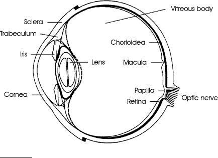
The targets of all therapeutic laser treatments of the eye can be classified into front and rear segments. The front segments consist of the cornea, sclera, trabeculum, iris, and lens. The rear segments are given by the vitreous body and retina. A schematic illustration of a human eye is shown in Fig. 4.1. In the following paragraphs, we will discuss various treatments of these segments according to the historic sequence, i.e. from the rear to the front.
Fig. 4.1. Scheme of a human eye
1Since glaucoma is usually associated with a degeneration of the optical nerve fibers, it can be detected by measuring either the thickness of these fibers or alterations of the optic disc. Further details are given by Bille et al. (1990).
4.1 Lasers in Ophthalmology |
153 |
Retina
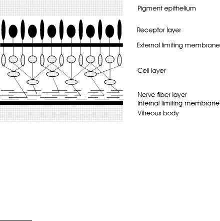
The retina is a part of the central nervous system. Its function is to convert an optical image focused on it into nerve impulses of the optic nerve emerging from it. The retina is a thin and rather transparent membrane which is permeated with blood vessels. According to Le Grand and El Hage (1980), the thickness of the retina varies from 0.5mm near the papilla to 0.1mm at the macula2. Anatomically, the retina is subdivided into several di erent layers, each of them having their own distinct function: pigment epithelium, receptor layer, external limiting membrane, cell layer, nerve fiber layer, and internal limiting membrane. A schematic cross-section of a human retina is shown in Fig. 4.2.
Fig. 4.2. Cross-section of a human retina
The pigment epithelium is strongly attached to the chorioidea. The receptor layer consists of two types of cells – rods and cones. Rods are used in dim light and are primarily located around the macula. Cones are recepting colors in good light and are found especially in the fovea. Obviously, light has to pass through virtually the whole retina beyond the external limiting membrane, before it can stimulate any receptor cells. This structural arrangement is known as the “reversed retina” and can be explained by the fact that the retina is an invagination of the embryonic cerebral wall. The cell layer is made up of horizontal cells, bipolar cells, amacrine cells, and ganglion cells.
2The papilla is a certain location where the optic nerve exits the retina. The macula is the region with the highest density of color receptors. An image formed on the fovea, the central section of the macula, is characterized by best vision. Thus, macula and fovea are the most important segments of the retina.
154 4. Medical Applications of Lasers
The main function of these cells is to serve as a first network with corresponding receptive fields. Finally, the nerve fiber layer contains the axons of the ganglion cells, whereas the internal limiting membrane forms a boundary between the retina and vitreous body.
The ophthalmologist Meyer-Schwickerath (1949) was the first to investigate the coagulation of the retina with sunlight for therapeutic purposes. Because of the inconvenient circumstances of this kind of surgery, e.g. the necessity of sunshine, he continued his studies with his famous xenon photocoagulator as reported in 1956. Shortly after the invention of the laser by Maiman (1960), first experimental studies with the ruby laser were performed by Zaret et al. (1961). The first reports on the treatment of patients were given by Campbell et al. (1963) and Zweng et al. (1964). They discovered that the ruby laser was a very suitable tool when welding detached segments of the retina to the chorioidea located underneath. However, it also became evident that the ruby laser was not able to close open blood vessels or stop bleeding. It was soon found that the argon ion laser is better suited for this aim. Its green and blue wavelengths are strongly absorbed by the hemoglobin of blood – in contrast to the red light from a ruby laser – which finally leads to the coagulation of blood and blood vessels. At typical exposure durations ranging from 0.1s to a few seconds, applied laser powers of 0.1–1W, and spot diameters of approximately 200–1000μm, almost all incident laser energy is converted to heat. Thus, coagulation of retinal tissue is achieved by means of thermal interaction. As discussed in Sect. 3.2, proteins are denaturated and enzymes are inactivated, thereby initiating the process of congealment.
The surgeon conducts the laser coagulation through a slit lamp and a contact glass. He approaches the necessary laser power from below threshold until the focused area just turns greyish. Coagulation of the macula is strictly forbidden, since it would be associated with a severe loss in vision. The temperatures achieved should generally remain below 80◦C to prevent unnecessary vaporization and carbonization. A good localization of blood vessels, i.e. by confocal laser microscopy, and a precise application of the desired energy dose are mandatory when striving for satisfactory results.
At the beginning of the 1970s, the krypton ion laser became very significant for ophthalmic applications. Its red and yellow wavelengths at 647nm and 568nm, respectively, turned out to be very useful when trying to restrict the interaction zone to either the pigment epithelium or the chorioidea. Detailed histologic studies on this phenomenon were conducted by Marshall and Bird (1979). It was found that the red line is preferably absorbed by the chorioidea, whereas the yellow line is strongly absorbed by the pigment epithelium and also by the xanthophyll contained in the macula. Recently, McHugh et al. (1988) proposed the application of diode lasers, since their invisible emission at a wavelength of approximately 800nm does not dazzle the patient’s eye.
4.1 Lasers in Ophthalmology |
155 |
There exist six major indications for laser treatment of the retina:
–retinal holes,
–retinal detachment,
–diabetic retinopathy,
–central vein occlusion,
–senile macula degeneration,
–retinal tumors (retinoblastoma).
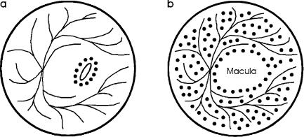
In the case of retinal holes, proper laser treatment prevents their further enlargement which could otherwise lead to retinal detachment. Laser surgery is performed by welding the retina to the underlying chorioidea within a narrow ring-shaped zone around the hole as shown in Fig. 4.3a. The attachment of the coagulated tissue is so strong that further tearing is usually suppressed. If necessary, however, the procedure can be repeated several times without severe complications.
Retinal detachment is often a consequence of undetected retinal holes or tears. It mainly occurs in myopic patients, since the vitreous body then induces an increased tensile stress to the retina. Moderate detachments are treated in a similar mode as retinal holes. In the case of a severe detachment, the treatment aims at saving the fovea or at least a small segment of the macula. This procedure is called panretinal coagulation and is illustrated in Fig. 4.3b. Unfortunately, laser treatment of retinal detachment is often associated with the formation of new membranes in the vitreous body, the retina, or beneath the retina. These complications are summarized by the clinical term proliferative retinopathy. A useful therapeutic technique for the dissection of such membranes was given by Machemer and Laqua (1978).
Fig. 4.3. (a) Placement of coagulation spots in the case of retinal holes or moderate detachments. (b) Placement of coagulation spots during panretinal coagulation
156 4. Medical Applications of Lasers
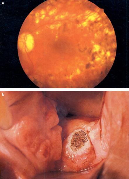
Fig. 4.4. (a) Panretinal coagulation in a diabetes patient performed with an argon ion laser (power: 200mW). (b) Vaporization of cervical tissue with a CO2 laser (power: 10W). Photographs kindly provided by Dr. Burk (Heidelberg) and Dr. Kurek (Heidelberg)
4.1 Lasers in Ophthalmology |
157 |
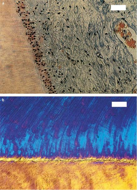
Fig. 4.5. (a) Histologic section of a human tooth after exposure to 16000 pulses from a Nd:YLF laser (pulse duration: 30ps, pulse energy: 500μJ, bar: 50μm). The junction of dentin (left) and pulp (right) is shown which was located next to the application site. (b) Polarized microscopy of a human tooth slice after exposure to 16000 pulses from the same laser (bar: 50μm). The junction of dentin (top) and enamel (bottom) is shown which was located next to the application site