

Laser-Tissue Interactions Fundamentals and Applications - Markolf H. Niemz
.pdf98 3. Interaction Mechanisms
much controversy. Andrew et al. (1983) and Brannon et al. (1985) claimed UV ablation to be exclusively of thermal character, whereas Garrison and Srinivasan (1985) tended to attribute it to photochemical e ects. Today, it is well accepted that photoablation – or the synonym ablative photodecomposition in the sense of UV ablation – shall be considered as an interaction mechanism of its own that can certainly be distinguished from pure photochemical and thermal processes described in Sects. 3.1 and 3.2. However, due to the dissociation of molecules during photoablation, a chemical transition is involved which could also justify misleading terms like “photochemical ablation”. But, with respect to the historical sequence, the term “photochemical” should be reserved for pulse interactions with photosensitizers at long pulse durations. And only an ablation caused by UV photons should be regarded as photoablation9.
In order to distinguish photoablation or ablative photodecomposition from thermal interaction, we take a closer look once more at the energy level diagram shown in Fig. 3.33. In the case of photoablation, we concluded that the energy of a single UV photon is su cient to dissociate the former bound molecule AB. In thermal interactions, the situation is completely di erent. The photon energy is not high enough for the molecule to reach a repulsive state. The molecule is promoted only to a vibrational state within the ground level or to a rather low electronic state including any of its vibrational states. By means of nonradiative relaxation, the absorbed energy then dissipates to heat, and the molecule returns to its ground state. Hence, the crucial parameter for di erentiating these two mechanisms – photoablation and thermal interaction – is the photon energy or laser wavelength. Only if hν > 3.6eV, or in other measures λ < 350nm, is the single photon dissociation of C–C bonds enabled. Of course, several photons with hν < 3.5eV can be absorbed. Then, these photons add up in energy and may thus lead to a dissociated state, as well. However, during the time needed for such a multiphoton absorption process, other tissue areas become vibrationally excited, hence leading to a global increase in temperature and an observable thermal e ect (usually either vaporization or melting). If this e ect is associated with ablation, the whole process is called thermal decomposition and has to be distinguished from pure ablative photodecomposition. These statements hold true, unless ultrashort pulses with a pulse duration shorter than 100ps are used at pulse energies high enough to induce a localized microplasma. Then, even VISand IR-lasers can interact nonthermally. But the discussion of this mechanism shall be deferred to Sect. 3.4.
In reality, pure photoablation is only observed for the 193nm wavelength of the ArF excimer laser. Higher wavelengths are usually associated with a more or less apparent thermal component. Sutcli e and Srinivasan (1986)
9Actually, the term “photoablation” itself is not well defined. It only states that an ablation occurs which is caused by photons, but it does not imply any further details. Photoablation in the common sense, though, is a very precise ablation caused by UV photons.
3.3 Photoablation |
99 |
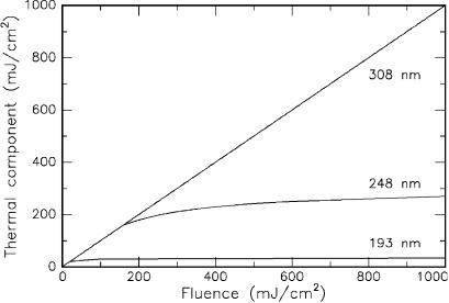
even postulated that the thermal component of the radiation from a XeCl excimer laser at 308nm is 100% thermal if its radiation is incident on PMMA. In Fig. 3.37, the thermal components of three excimer lasers are compared with each other. Obviously, each laser induces thermal e ects at low energy densities. In the cases of the ArF laser and the KrF laser, the slope of the curve strongly decreases above a certain threshold, i.e. the threshold of photoablation. Radiation from the XeCl laser, on the other hand, continues to act thermally even at higher energy densities.
Fig. 3.37. Thermal component of UV radiation from three di erent excimer lasers (ArF laser at 193nm, KrF laser at 248nm, and XeCl laser at 308nm) in PMMA. Data according to Sutcli e and Srinivasan (1986)
The data presented in Fig. 3.37 are only valid for the target material PMMA. They cannot be generalized to any biological tissue, since the absorption characteristics of PMMA and biological tissue are quite di erent. However, the bottom line of the above observation – i.e. that the significance of the thermal component increases with higher wavelengths – will be true in biological tissue, as well. Therefore, we can conclude that ArF excimer lasers are probably the best choice when aiming at photoablation. XeCl laser could be used for thermal decomposition, although CW lasers might do a similar job. KrF lasers o er both photoablative and thermal decomposition but are less useful in medical applications as we will discuss next.

100 3. Interaction Mechanisms
3.3.2 Cytotoxicity of UV Radiation
It has been argued that the application of UV radiation for photoablative purposes might induce mutagenic – and thus cytotoxic – e ects within cells. This statement has to be taken seriously, since laser surgery, of course, should not evoke new maladies when eliminating others. It is a fact that DNA strongly absorbs UV radiation, especially at approximately 240–260nm. And it is also well known that this radiation can cause mutagenic alterations of cells, e.g. exchanges of sister chromatids.
The e ect of UV radiation on cells and biological tissue is initiated by photochemical reactions of chromophores contained by them. Absorption of UV photons by DNA causes alterations in the chemical structure of the DNA molecule. The major chemical change is the formation of a dimer from two adjacent pyrimidine bases. Other products are also synthesized in the DNA that may have biological consequences. Cells are frequently able to repair dimers before any adverse responses occur. This is an indispensable mechanism of protection, since the DNA contains important genetic information. Thus, if these photoproducts are not repaired, erroneous information may be passed on to progeny cells when the cell divides. This event, finally, leads to the process of mutagenesis.
Several studies have been done in order to evaluate potential hazards from UV laser radiation. Usually, the e ects of one or two excimer lasers were compared to each other and to low-pressure Hg lamps. With these, most of the UV spectrum is covered. In Table 3.11, the wavelengths of commonly used UV sources are listed.
Table 3.11. Sources of UV radiation
Light source |
Wavelength (nm) |
|
|
ArF laser |
193 |
KrF laser |
248 |
Hg lamp |
254 |
Nd:YLF laser (4ω) |
263 |
Nd:YAG laser (4ω) |
266 |
XeCl laser |
308 |
XeF laser |
351 |
|
|
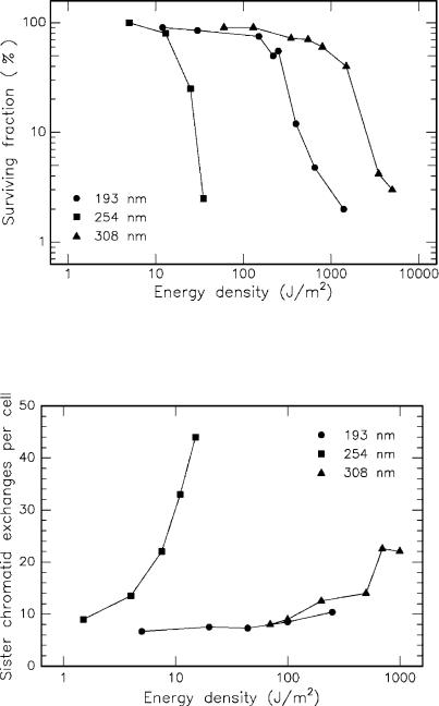
Cytotoxic e ects of UV radiation have been investigated by several groups, among them especially by Green et al. (1987), Kochevar (1989), and Rasmussen et al. (1989). In general, it can be concluded that the relative ability of excimer laser radiation to cause DNA defects decreases in the order of 248nm > 193nm > 308nm. Only in some cases was radiation at 193nm found to be less cytotoxic than at 308nm. In Fig. 3.38, the surviving fraction of Chinese hamster ovary (CHO) cells after exposure to three di erent light sources is shown as a function of incident energy density. Whereas the Hg
3.3 Photoablation |
101 |
Fig. 3.38. Surviving fraction of Chinese hamster ovary (CHO) cells after UV irradiation. Light sources: ArF laser (193nm), Hg lamp (254nm), and XeCl laser (308nm). Data according to Rasmussen et al. (1989)
Fig. 3.39. Sister chromatid exchanges per cell after UV irradiation. Light sources: ArF laser (193nm), Hg lamp (254nm), and XeCl laser (308nm). Data according to Rasmussen et al. (1989)
102 3. Interaction Mechanisms
lamp induces measurable damage at 15J/m2, it takes a few hundred J/m2 of excimer laser radiation to cause a similar e ect. When comparing the ArF laser at 193nm and the XeCl laser at 308nm, the latter is less cytotoxic.
In the same study, the exchange of sister chromatids was measured. These can either be repaired by the cell or, in severe cases, lead to irreversible defects. In the latter case, either cell necrosis or uncontrolled cell proliferation, i.e. certain types of cancer, can occur. The extent of sister chromatid exchange was investigated for the same UV sources and is shown in Fig. 3.39. All three radiations caused an increase in the incidence of sister chromatid exchange but with di erent e ectiveness. The required energy density at 254nm, for instance, is much less than at 193nm or 308nm. Moreover, the slope of the curve at 254nm is much steeper than the others. Thus, another proof is given that radiation from a Hg lamp can be considered as being more mutagenic than ArF or XeCl lasers. This is the main reason why there are no significant medical applications for KrF lasers, since its wavelength at 248nm almost coincides with that of Hg lamps.
We conclude that radiation from excimer lasers is less mutagenic than UV light from Hg lamps. This observation can probably be explained by the existence of proteins in the cell matrix which strongly absorb radiation at 193nm, before it reaches the cell nucleus containing the DNA. According to Green et al. (1987), about 60% of incident radiation is already absorbed by only 1μm of cytoplasm. Hence, the sensitive DNA inside the nucleus is shielded by the surrounding cytoplasm. However, potential risks should never be ignored when using ArF or XeCl lasers. Actually, a few altered cells might be enough to induce cancer in tissue. And, according to Rasmussen et al. (1989), there is some preliminary evidence that fluorescence emission around 250nm occurs following absorption at 193nm.
In general, the di culty in judging the severity of mutagenic e ects is due to the long follow-up periods during which maladies can develop. For instance, a haze inside the cornea is frequently observed within a few years after refractive corneal surgery has been performed with excimer lasers. The origin of this haze is yet unknown. It might well be attributed to cell alterations, even if corneal tumors do not occur.
3.3.3 Summary of Photoablation
• Main idea: |
direct breaking of molecular bonds by high- |
|
energy UV photons |
• Observations: |
very clean ablation, associated with audible |
|
report and visible fluorescence |
• Typical lasers: |
excimer lasers, e.g. ArF, KrF, XeCl, XeF |
• Typical pulse durations: |
10 ... 100ns |
• Typical power densities: |
107 ... 1010 W/cm2 |
• Special applications: |
refractive corneal surgery |
3.4 Plasma-Induced Ablation |
103 |
3.4 Plasma-Induced Ablation
When obtaining power densities exceeding 1011 W/cm2 in solids and fluids
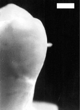
– or 1014 W/cm2 in air – a phenomenon called optical breakdown occurs. This e ect is demonstrated in Fig. 3.40. One 30ps laser pulse from a mode locked and amplified Nd:YLF laser was focused on an extracted human tooth. A bright plasma spark is clearly visible which is pointing toward the laser source. If several laser pulses are applied, a typical sparking noise at the repetition rate of the pulses is heard.
Fig. 3.40. Laser-induced plasma sparking on tooth surface caused by a single pulse from a Nd:YLF laser (pulse duration: 30ps, pulse energy: 1mJ, focal spot size: 30μm, bar: 1mm). With these parameters, a power density of about 5×10¹² W/cm2 is obtained
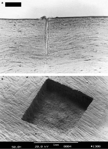
By means of plasma-induced ablation, very clean and well-defined removal of tissue without evidence of thermal or mechanical damage can be achieved when choosing appropriate laser parameters. The e ects of plasma-induced ablation on both soft and hard tissues are demonstrated in Figs. 3.41a–b. In one case, a sample of corneal tissue was ablated with a picosecond Nd:YLF
104 3. Interaction Mechanisms
Fig. 3.41. (a) Excision in a human cornea achieved with a picosecond Nd:YLF laser (pulse duration: 30ps, pulse energy: 200μJ, bar: 50μm). (b) Human tooth exposed to 16000 pulses from a picosecond Nd:YLF laser (pulse duration: 30ps, pulse energy: 1mJ, pattern size: 1×1mm2). Superficial cracking occurred as an artefact during the preparation for electron microscopy

3.4 Plasma-Induced Ablation |
105 |
laser by applying the pulses in a straight line pattern. The cross-sectional view shows a very precise excision without mechanical ruptures. In the other case, pulses from the same Nd:YLF laser were scanned over the surface of a tooth in a 1×1mm2 square pattern. In particular, the fairly smooth bottom of the cavity and the steep side walls are very significant. Further details on potential applications are given in Sects. 4.1 and 4.2.
Sometimes, plasma-induced ablation is also referred to as plasma-mediated ablation. It was recently investigated and discussed by Teng et al. (1987), Stern et al. (1989), and Niemz et al. (1991). Both synonyms express a generally well accepted interpretation that this kind of ablation is primarily caused by plasma ionization itself. This is in contrast to a more mechanical process called photodisruption which will be described in Sect. 3.5. The most important parameter of plasma-induced ablation10 is the local electric field strength E which determines when optical breakdown is achieved. If E exceeds a certain threshold value, i.e. if the applied electric field forces the ionization of molecules and atoms, breakdown occurs. The electric field strength itself is related to the local power density I by the basic electrodynamic equation
I(r,z,t) = |
|
1 |
ε0cE2 |
, |
(3.23) |
|
2 |
||||||
|
|
|
|
|||
where ε0 is the dielectric constant, and c is the speed of light. For picosecond pulses, typical threshold intensities of optical breakdown are 1011 W/cm2. Hence, the corresponding electric field amounts to approximately 107 V/cm. This value is comparable to the average atomic or intramolecular Coulomb electric fields, thus providing the necessary condition for plasma ionization. Within a few hundred picoseconds, a very large free electron density with typical values of 1018/cm3 is created in the focal volume of the laser beam. In general, plasma generation due to an intense electric field is called dielectric breakdown. The term optical breakdown especially emphasizes that UV, visible, and IR light are strongly absorbed by the plasma. The physical principles of breakdown have been investigated in several theoretical studies, e.g. by Seitz (1949), Molchanov (1970), Yablonovitch and Bloembergen (1972), Bloembergen (1974), Epifanov (1981), and Sacchi (1991).
Fradin et al. (1973b) have measured the intensity of ruby laser pulses transmitted through NaCl. They used a TEM00 mode ruby laser with pulse energies of 0.3mJ and pulse durations of 15ns which was focused with a lens of 14mm focal length. In Figs. 3.42a–c, the transmitted intensities are shown as a function of time. Obviously, the shape of the transmitted pulse is not altered in Fig. 3.42a, whereas the rear part of the pulse is strongly absorbed in Figs. 3.42b–c. Fradin et al. (1973b) concluded that the intensity as in Fig. 3.42a was just below the threshold of optical breakdown, while it was above in Fig. 3.42b, thereby damaging the NaCl sample. In Fig. 3.42c, breakdown was achieved at the very peak of the laser pulse.
10Throughout this book we will use the term plasma-induced ablation rather than plasma-mediated ablation, since the first term emphasizes its primary cause.

106 3. Interaction Mechanisms
Fig. 3.42. (a–c) Ruby laser pulses transmitted through NaCl. Data according to Fradin et al. (1973b)
The initiation of plasma generation can be two-fold and was described in detail by Puliafito and Steinert (1984). It was observed that either Q- switched pulses in the nanosecond range or mode locked laser pulses in the picosecond or femtosecond range can induce a localized microplasma. In Q- switched pulses, the initial process for the generation of free electrons is supposed to be thermionic emission, i.e. the release of electrons due to thermal ionization as shown in Fig. 3.43. In mode locked pulses, multi-photon ionization may occur due to the high electric field induced by the intense

3.4 Plasma-Induced Ablation |
107 |
laser pulse. In general, the term multi-photon ionization denotes processes in which coherent absorption of several photons provides the energy needed for ionization. Due to the requirement of coherence, multi-photon ionization is achievable only during high peak intensities as in picosecond or femtosecond laser pulses. Plasma energies and plasma temperatures, though, are usually higher in Q-switched laser pulses because of the associated increase in threshold energy of plasma formation. Thus, optical breakdown achieved with nanosecond pulses is often accompanied by nonionizing side e ects and will be deferred to Sect. 3.5.
Fig. 3.43. Initiation of ionization with subsequent electron avalanche. Reproduced from Puliafito and Steinert (1984) by permission. c 1984 IEEE
In either case, however, a few “lucky” electrons initiate an avalanche e ect, leading to the accumulation of free electrons and ions. A free electron absorbs a photon and accelerates. The accelerated electron collides with another atom and ionizes it, resulting in two free electrons, each of them with less individual kinetic energy than the initial electron. Again, these free electrons may absorb incoming photons, accelerate, strike other atoms, release two more electrons, and so forth. The basic process of photon absorption and electron acceleration taking place in the presence of an atom is called inverse Bremsstrahlung11. It is based on a free-free absorption, i.e. a transition, where a free electron is present in the initial and final states. In order to fulfill the conservation laws of energy and momentum, this event must necessarily take place in the electric field of an ion A+ or a neutral atom. The process is schematically written as
hν + e + A+ −→ e + A+ + Ekin .
11In ordinary Bremsstrahlung, the opposite e ect occurs, i.e. electrons are accelerated in the electromagnetic field of an atom, thereby emitting radiation.