

Laser-Tissue Interactions Fundamentals and Applications - Markolf H. Niemz
.pdf78 |
3. Interaction Mechanisms |
|
Table 3.6. Thermal e ects of laser radiation |
||
|
|
|
Temperature |
Biological e ect |
|
|
|
|
|
37◦C |
Normal |
|
45◦C |
Hyperthermia |
|
50◦C |
Reduction in enzyme activity, |
|
60◦C |
cell immobility |
|
Denaturation of proteins and collagen, |
|
|
80◦C |
coagulation |
|
Permeabilization of membranes |
|
|
100◦C |
Vaporization, |
> 100◦C |
thermal decomposition (ablation) |
|
Carbonization |
||
> 300◦C |
Melting |
|
|
|
|
In general, the exact temperature for the onset of cell necrosis is rather di cult to determine. As a matter of fact, it was observed that not only the temperature achieved but also the temporal duration of this temperature plays a significant role for the induction of irreversible damage. It is illustrated in Fig. 3.21 how the critical temperature and the corresponding temporal duration relate to each other if irreversible damage is meant to occur. The curve is derived from several empirical observations. In the example selected in Fig. 3.21, a temperature of 60◦C lasting for at least 6s will lead to irreversible damage.
Fig. 3.21. Critical temperatures for the occurrence of cell necrosis. Data according to Henriques (1947) and Eichler and Seiler (1991)

3.2 Thermal Interaction |
79 |
Areas in which the temperature reaches values higher than 60◦C are coagulated, and irradiated tissue cells become necrotic. Areas with maximum temperatures less than 60◦C are treated hyperthermically only, and the probability of cells staying alive depends on the duration and temporal evolution of the temperature obtained. For a quantitative approximation of the remaining active molecules and cells at a certain temperature level, Arrhenius’ equation is very useful:
|
C(t) |
|
t |
− |
|
dt ≡ −Ω , |
|
ln |
= −A |
exp |
|
(3.19) |
|||
C |
RT(t ) |
||||||
0 |
|
0 |
|
ΔE |
|
||
|
|
|
|
|
|
||
where C0 is the initial concentration of molecules or cells, C(t) is the concentration at a time t, A is Arrhenius’ constant, R is the universal gas constant, and ΔE and Ω are specific tissue properties. According to Welch (1984), the coe cient A can be approximated by
A kTh exp ΔSR ,
where ΔS is the activation entropy, k is Boltzmann’s constant, and h is Planck’s constant. But, according to Johnson et al. (1974), this relation has no simple significance.
The local degree of tissue damage is determined by the damage integral given in (3.19). The damage degree is defined as the fraction of deactivated molecules or cells given by
Dd(t) = C0 −C(t) = 1 −exp(−Ω) .
C0
Thus, by inserting an appropriate value of the tissue constant Ω, we are able to calculate the probable damage degree Dd(t) as a function of time t. This was performed by Weinberg et al. (1984) in the case of retinal tissue.
Unfortunately, experimental data for the two parameters A and ΔE are very di cult to obtain due to the inhomogeneity of most tissues and the uncertainty in measuring the surviving fraction. However, in Table 3.7 some values of various tissues are listed as found in di erent studies.
Table 3.7. Arrhenius’ constants of di erent tissues
Tissue |
A(s−1) |
ΔE (J/mol) |
Reference |
|
|
|
|
|
|
Retina |
1.0 × 1044 |
2.9 × 10⁵ |
Vassiliadis et al. (1971) |
|
|
|
|
|
Weinberg et al. (1984) |
Retina (T < 50◦C) |
4.3 |
× 1064 |
4.2 × 10⁵ |
Takata et al. (1977) |
Retina (T > 50◦C) |
9.3 |
× 10104 |
6.7 × 10⁵ |
Takata et al. (1977) |
Skin |
3.1 |
× 1098 |
6.3 × 10⁵ |
Henriques (1947) |
Liver |
1.0 |
× 1070 |
4.0 × 10⁵ |
Roggan and M¨uller (1993) |
|
|
|
|
|
80 3. Interaction Mechanisms
Laser radiation acts thermally if power densities ≥ 10W/cm2 are applied from either CW radiation or pulse durations exceeding approximately 1μs. Typical lasers for coagulation are Nd:YAG lasers or diode lasers. CO2 lasers are very suitable for vaporization and the precise thermal cutting of tissue. Carbonization and melting can occur with almost any type of laser if su cient power densities and exposure durations are provided.
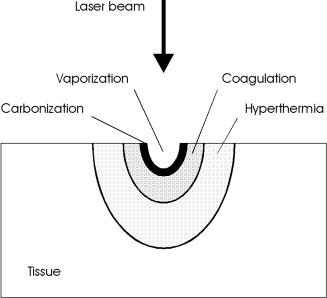
Frequently, not only one but several thermal e ects are induced in biological tissue, depending on the laser parameters. These e ects might even range from carbonization at the tissue surface to hyperthermia a few millimeters inside the tissue. In most applications, though, only one specific e ect is aimed at. Therefore, careful evaluation of the required laser parameters is essential. Reversible and irreversible tissue damage can be distinguished. Carbonization, vaporization, and coagulation certainly are irreversible processes, because they induce irrepairable damage. Hyperthermia, though, can turn out to be either a reversible or an irreversible process, depending on the type of tissue and laser parameters. Since the critical temperature for cell necrosis is determined by the exposure time as shown in Fig. 3.21, no well-defined temperature can be declared which distinguishes reversible from irreversible e ects. Thus, exposure energy, exposure volume, and exposure duration together determine the degree and extent of tissue damage. The coincidence of several thermal processes is illustrated in Fig. 3.22. The location and spatial extent of each thermal e ect depend on the locally achieved temperature during and after laser exposure.
Fig. 3.22. Location of thermal e ects inside biological tissue
3.2 Thermal Interaction |
81 |
3.2.4 Laser-Induced Interstitial Thermotherapy (LITT)
The possibility of localized tissue coagulation has formed the basis of a novel tumor treatment technique called laser-induced interstitial thermotherapy. It was recently introduced to the treatment of various types of tumors such as in retina, brain, prostate, liver, or uterus, and has already become a well established tool in minimally invasive surgery (MIS). Detailed descriptions and first clinical results are found in the papers by Svaasand et al. (1989), Ascher et al. (1991), Muschter et al. (1992), Roggan and M¨uller (1993), and Wallwiener et al. (1994), respectively. The most significant applications of LITT reside in the disciplines of gynecology and urology, e.g. the treatment of malignant tumors in the uterus and the treatment of benign prostatic hyperplasia (BPH) as described in Sects. 4.3 and 4.4.
The principal idea of LITT is to position an appropriate laser applicator inside the tissue to be coagulated, e.g. a tumor, and to achieve necrosis by heating cells above 60◦C. As stated in Table 3.6, denaturation of proteins and coagulation occur at these temperatures. Due to the associated coagulation of blood vessels, severe hemorrhages are less likely to occur than in conventional surgery. This is of particular importance in the case of tumors, because they usually are highly vascularized. Either Nd:YAG lasers at 1064nm or di erent types of diode lasers at 800–900nm are applied, since light deeply penetrates into tissue at these wavelengths. Thus, large volumes can be treated with temperature gradients not as steep as those associated with conventional thermotherapies based on heat conduction only. Typical parameters of the procedure are 1–5W of CW laser power for a period of several minutes and coagulation volumes with diameters of up to 40mm.
In order to achieve a safe LITT procedure, knowledge of the final damage zone is essential. Especially in neurosurgery, it is very important to prevent injury from adjacent healthy tissue and sensitive structures. The spatial extent of the damage zone primarily depends on laser power, laser exposure, geometry of the laser applicator, and on thermal and optical tissue properties. Since the optical penetration depth of laser light in the near infrared is very high, deeper zones are reached more easily by the mechanism of light scattering rather than by heat conduction. Therefore, laser applicators emitting their radiation through a scattering surface are favored compared to focusing optics. Actually, first applications of LITT had been performed using bare fibers which were directly guided into the tissue. The main disadvantage was the high power density at the end surface of the fiber, leading to immediate carbonization of adjacent tissue. Recently improved applicators scatter laser light isotropically into all spatial directions by means of a frosted (rough) surface as described by Roggan et al. (1995c).
The basic setup for LITT is illustrated in Fig. 3.23. The laser applicator usually consists of a flexible fiber and a transparent catheter through which the fiber is moved into the tissue. Frequently, fibers with typical diameters of 400μm are chosen. In order to ensure isotropic scattering at the interface

82 3. Interaction Mechanisms
of the applicator and tissue, the surface of the fiber is etched by a special technique after removing the cladding. Continuous emitting surfaces with active lengths up to 20mm can be manufactured. Depending on size and geometry of the treated volume, though, fibers with di erent active lengths should be available during surgery. Laser light is emitted from the distal end of the fiber on an active length which is approximately half the tumor size. The specially designed catheter protects the sensitive fiber from mechanical stress. Optional cooling of the catheter may help to prevent thermal damage to the fiber tip at even higher power densities. Only then can large tissue volumes be safely coagulated at a rather moderate gradient of temperature.
Fig. 3.23. Experimental setup for laser-induced interstitial thermotherapy. Laser radiation is applied to the tissue through an optical fiber. The fiber is placed inside the tissue by means of a specially designed, transparent catheter. Tissue necrosis occurs in selected coagulated volumes only
At the start of a LITT procedure, the catheter is placed inside the tumor assisted by qualified monitoring. According to Roggan et al. (1994), Eyrich et al. (2000), and Wacker et al. (2001), either ultrasound or magnetic resonance imaging (MRI) can be used for this purpose6. The fiber is then carefully guided through the catheter without actually being in contact with the tissue. In an accidental break of the fiber, glass splinters will remain inside the catheter without injuring the patient. After laser exposure, the fiber is either completely pulled back or only by its active length if several treatments are to be performed. Finally, i.e. if no further irradiation is required, the fiber is withdrawn. And, after a short period of temperature equalization,
6In MRI-controlled LITT treatments, a special marker is mounted at the distal end of the fiber which facilitates its localization.

3.2 Thermal Interaction |
83 |
the protecting catheter may be removed. The same target may be treated several times by LITT to increase the spatial extent of tissue necrosis. For large lesions, the distance between adjacent puncturing canals should not exceed 1.5mm to ensure an overlap of the coagulated tissue volumes.
The whole procedure can be performed either intraoperatively or percutaneously. In both cases, it should be preceded by suitable irradiation planning to obtain best surgical results. This can be achieved with appropriate computer simulations by considering a variety of input parameters. Most important among these are the position, extent, and topology of the diseased volume as well as optical and thermal tissue parameters, and the rate of blood perfusion. A choice of laser power and exposure duration should then be provided by the program. Detailed computer simulations for the laser treatment of liver tissue are illustrated in Figs. 3.24 and 3.25. They were obtained by applying the method of finite di erences to the inhomogeneous heat conduction equation (3.11). This is performed by dividing the tissue into consecutive shells and using a mathematical algorithm which takes into account the heat conduction from adjacent layers only. After a few recursions of this algorithm, a steady-state solution is obtained expressing the desired temperature distribution. Further details on the simulation technique can be found in the paper by Roggan and M¨uller (1993). From Fig. 3.24, we conclude that adequate computer simulations fit very well to measured data, whereas Fig. 3.25 proves that cooled laser applicators enable the coagulation of larger tissue volumes at comparable peak temperatures.
Fig. 3.24. Computer simulation and experimental results of LITT for liver tissue (laser power: 4W, surface of applicator: 125mm2). The switch-o time of the laser is indicated. Data according to Roggan and M¨uller (1993)

84 3. Interaction Mechanisms
Fig. 3.25. Comparison of the calculated final temperature distributions for di erent types of thermotherapies (conventional hot tip, LITT without fiber cooling, LITT with fiber cooling). In the case of fiber cooling, the peak temperature does not occur at the applicator surface which is located at the left end of all curves. Data according to Roggan and M¨uller (1993)
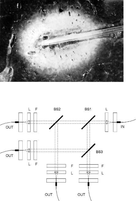
The propagation of laser light in biological tissue and its transformation to thermal energy due to absorption is governed by the optical properties of the tissue. In the model of thermal interaction as described above, only absorption was taken into account. During LITT, scattering also plays a significant role for reasons just stated. Especially during the process of coagulation, the optical properties of tissue are changed, leading to higher scattering but nearly the same absorption. Usually, this e ect is observable by a change in color of the irradiated volume, resulting in a reduced penetration depth. In Fig. 3.26, a sample of liver tissue is shown which was irradiated by using a standard LITT applicator as described above. The coagulated zone around the fiber is clearly visible.
For the treatment of very large tissue volumes with diameters of up to a few centimeters the application of several fibers is strongly recommended according to Klingenberg et al. (2000). The laser output is distributed among several fibers by optical beamsplitters as illustrated in Fig. 3.27. Each beamsplitter separates the incident laser beam into two laser beams with equal power. Using a total of three optical beamsplitters, a single input beam can be divided into four output beams. Focusing lenses are used to couple the laser power out of the input fiber and into the output fibers. Further adjustment of the laser power within each output beam can be achieved by implementation of optical filters.
3.2 Thermal Interaction |
85 |
Fig. 3.26. Liver tissue after exposure to a Nd:YAG laser, using a standard LITT fiber applicator (laser power: 5.5W, exposure duration: 10min). The coagulated volume visibly pales. Photograph kindly provided by Dr. Roggan (Berlin)
Fig. 3.27. LITT with four output fibers (BS: beamsplitter, F: filter, L: lens)
86 3. Interaction Mechanisms
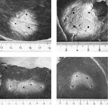
Depending on number and arrangement of the fibers, di erent geometries can be obtained for the coagulation of tissue. As shown in Fig. 3.28, the surgeon may choose among either spherical, ellipsoidal, or trapezoidal lesions. He can thus adjust the coagulated volume to the geometry of a tumor. Some indications, e.g. in brain surgery, require the treatment of tumors closely located to vital structures. In these cases, it is extremely important to avoid the coagulation of these organs at risk. This task can be achieved by applying trapezoidal lesions. Detailed discussion on this topic and the advantages of a multi-fiber treatment is provided by Klingenberg et al. (2000).
Fig. 3.28. Coagulations achieved in muscle tissue with several fibers (Nd:YAG laser, 4 W per fiber)
In general, two kinds of LITT applicators are distinguished: surface scatterers and volume scatterers. In surface scatterers, light is scattered only at the very surface of the applicator leading to a less homogeneous scattering profile as shown in Fig. 3.29. In volume scatterers, light is scattered by tiny scattering centers distributed throughout the whole volume of the applicator. Raw quartz is one of the basic materials used for volume scatterers. In raw quartz, the scattering centers consist of tiny gas bubbles.

3.2 Thermal Interaction |
87 |
Fig. 3.29. Scattering profiles of surface scatterer and volume scatterer, respectively
As stated above, scattering is the dominant mechanism by which light is homogeneously distributed inside the tissue. In Sect. 2.5, we have already become acquainted with the transport equation of radiation, (2.36), which takes scattering e ects into account. It was mentioned that exact analytical solutions to this integro-di erential equation do not exist. However, five approaches have been made to solve the equation:
–first-order scattering,
–Kubelka–Munk theory,
–di usion approximation,
–Monte Carlo simulations,
–inverse adding–doubling.
In principle, the same five approaches can be chosen when trying to estimate the temperature field within laser-irradiated tissue. Detailed computer calculations based on Monte Carlo simulations are found in the paper by Roggan and M¨uller (1995b). For a further characterization of each method, the reader is referred to Sect. 2.5.
3.2.5 Summary of Thermal Interaction
• Main idea: |
achieving a certain temperature which leads |
|
to the desired thermal e ect |
• Observations: |
either coagulation, vaporization, |
|
carbonization or melting |
• Typical lasers: |
CO2, Nd:YAG, Er:YAG, Ho:YAG, argon ion |
|
and diode lasers |
• Typical pulse durations: |
1μs... 1min |
• Typical power densities: |
10 ... 106 W/cm2 |
• Special applications: |
coagulation, vaporization, melting, |
|
thermal decomposition, |
|
treatment of retinal detachment, |
|
laser-induced interstitial thermotherapy |