

Laser-Tissue Interactions Fundamentals and Applications - Markolf H. Niemz
.pdfXIV
pictures taken with scanning electron microscopy, the editorial and production sta of Springer-Verlag for their care and cooperation in producing this book, and last but definitely not least all friends who spent some of their precious time in reading the manuscript.
In spite of great care and e ort on my part, I am fairly sure that some errors still remain in the book. I hope you will bring these to my attention for further improvements.
Heidelberg, |
|
February 1996 |
Markolf H. Niemz |
Table of Contents
1. |
Introduction . . . . . . . . . . . . . . . . . . . . . . . . . . . . . . . . . . . . . . . . . . . . . . |
1 |
||
|
1.1 |
Historic Review . . . . . . . . . . . . . . . . . . . . . . . . . . . . . . . . . . . . . . . . |
1 |
|
|
1.2 |
Goal of the Book . . . . . . . . . . . . . . . . . . . . . . . . . . . . . . . . . . . . . . . |
6 |
|
|
1.3 |
Outlook . . . . . . . . . . . . . . . . . . . . . . . . . . . . . . . . . . . . . . . . . . . . . . . |
7 |
|
2. |
Light and Matter . . . . . . . . . . . . . . . . . . . . . . . . . . . . . . . . . . . . . . . . . |
9 |
||
|
2.1 |
Reflection and Refraction. . . . . . . . . . . . . . . . . . . . . . . . . . . . . . . . |
10 |
|
|
2.2 |
Absorption . . . . . . . . . . . . . . . . . . . . . . . . . . . . . . . . . . . . . . . . . . . . |
15 |
|
|
2.3 |
Scattering . . . . . . . . . . . . . . . . . . . . . . . . . . . . . . . . . . . . . . . . . . . . . |
19 |
|
|
2.4 |
Turbid Media . . . . . . . . . . . . . . . . . . . . . . . . . . . . . . . . . . . . . . . . . . |
25 |
|
|
2.5 |
Photon Transport Theory . . . . . . . . . . . . . . . . . . . . . . . . . . . . . . . |
27 |
|
|
2.6 |
Measurement of Optical Tissue Properties . . . . . . . . . . . . . . . . . |
37 |
|
|
2.7 |
Questions to Chapter 2. . . . . . . . . . . . . . . . . . . . . . . . . . . . . . . . . . |
43 |
|
3. |
Interaction Mechanisms . . . . . . . . . . . . . . . . . . . . . . . . . . . . . . . . . . |
45 |
||
|
3.1 |
Photochemical Interaction . . . . . . . . . . . . . . . . . . . . . . . . . . . . . . . |
47 |
|
|
|
3.1.1 |
Photodynamic Therapy (PDT). . . . . . . . . . . . . . . . . . . . . |
49 |
|
|
3.1.2 |
Biostimulation . . . . . . . . . . . . . . . . . . . . . . . . . . . . . . . . . . . |
57 |
|
|
3.1.3 Summary of Photochemical Interaction. . . . . . . . . . . . . . |
58 |
|
|
3.2 |
Thermal Interaction . . . . . . . . . . . . . . . . . . . . . . . . . . . . . . . . . . . . |
58 |
|
|
|
3.2.1 |
Heat Generation . . . . . . . . . . . . . . . . . . . . . . . . . . . . . . . . . |
68 |
|
|
3.2.2 |
Heat Transport . . . . . . . . . . . . . . . . . . . . . . . . . . . . . . . . . . |
68 |
|
|
3.2.3 |
Heat E ects . . . . . . . . . . . . . . . . . . . . . . . . . . . . . . . . . . . . . |
77 |
|
|
3.2.4 |
Laser-Induced Interstitial Thermotherapy (LITT) . . . . |
81 |
|
|
3.2.5 Summary of Thermal Interaction . . . . . . . . . . . . . . . . . . . |
87 |
|
|
3.3 |
Photoablation. . . . . . . . . . . . . . . . . . . . . . . . . . . . . . . . . . . . . . . . . . |
88 |
|
|
|
3.3.1 |
Model of Photoablation . . . . . . . . . . . . . . . . . . . . . . . . . . . |
96 |
|
|
3.3.2 Cytotoxicity of UV Radiation . . . . . . . . . . . . . . . . . . . . . . |
100 |
|
|
|
3.3.3 |
Summary of Photoablation . . . . . . . . . . . . . . . . . . . . . . . . |
102 |
|
3.4 |
Plasma-Induced Ablation . . . . . . . . . . . . . . . . . . . . . . . . . . . . . . . . |
103 |
|
|
|
3.4.1 Model of Plasma-Induced Ablation . . . . . . . . . . . . . . . . . |
108 |
|
|
|
3.4.2 Analysis of Plasma Parameters . . . . . . . . . . . . . . . . . . . . . |
121 |
|
|
|
3.4.3 Summary of Plasma-Induced Ablation . . . . . . . . . . . . . . |
125 |
|
|
3.5 |
Photodisruption . . . . . . . . . . . . . . . . . . . . . . . . . . . . . . . . . . . . . . . . |
126 |
|
|
|
3.5.1 |
Plasma Formation . . . . . . . . . . . . . . . . . . . . . . . . . . . . . . . . |
131 |
XVI Table of Contents
3.5.2 Shock Wave Generation . . . . . . . . . . . . . . . . . . . . . . . . . . . 135 3.5.3 Cavitation . . . . . . . . . . . . . . . . . . . . . . . . . . . . . . . . . . . . . . . 143 3.5.4 Jet Formation. . . . . . . . . . . . . . . . . . . . . . . . . . . . . . . . . . . . 147 3.5.5 Summary of Photodisruption . . . . . . . . . . . . . . . . . . . . . . 149 3.6 Questions to Chapter 3. . . . . . . . . . . . . . . . . . . . . . . . . . . . . . . . . . 149
4. Medical Applications of Lasers. . . . . . . . . . . . . . . . . . . . . . . . . . . . 151 4.1 Lasers in Ophthalmology . . . . . . . . . . . . . . . . . . . . . . . . . . . . . . . . 152 4.2 Lasers in Dentistry . . . . . . . . . . . . . . . . . . . . . . . . . . . . . . . . . . . . . 181 4.3 Lasers in Gynecology . . . . . . . . . . . . . . . . . . . . . . . . . . . . . . . . . . . 201 4.4 Lasers in Urology. . . . . . . . . . . . . . . . . . . . . . . . . . . . . . . . . . . . . . . 207 4.5 Lasers in Neurosurgery . . . . . . . . . . . . . . . . . . . . . . . . . . . . . . . . . . 213 4.6 Lasers in Angioplasty and Cardiology . . . . . . . . . . . . . . . . . . . . . 221 4.7 Lasers in Dermatology . . . . . . . . . . . . . . . . . . . . . . . . . . . . . . . . . . 227 4.8 Lasers in Orthopedics . . . . . . . . . . . . . . . . . . . . . . . . . . . . . . . . . . . 232 4.9 Lasers in Gastroenterology. . . . . . . . . . . . . . . . . . . . . . . . . . . . . . . 237 4.10 Lasers in Otorhinolaryngology and Pulmology. . . . . . . . . . . . . . 241 4.11 Questions to Chapter 4. . . . . . . . . . . . . . . . . . . . . . . . . . . . . . . . . . 247
5. Laser Safety . . . . . . . . . . . . . . . . . . . . . . . . . . . . . . . . . . . . . . . . . . . . . . 249 5.1 Introduction . . . . . . . . . . . . . . . . . . . . . . . . . . . . . . . . . . . . . . . . . . . 249 5.2 Laser Hazards. . . . . . . . . . . . . . . . . . . . . . . . . . . . . . . . . . . . . . . . . . 249 5.3 Eye Hazards . . . . . . . . . . . . . . . . . . . . . . . . . . . . . . . . . . . . . . . . . . . 250 5.4 Skin Hazards . . . . . . . . . . . . . . . . . . . . . . . . . . . . . . . . . . . . . . . . . . 251 5.5 Associated Hazards from High Power Lasers . . . . . . . . . . . . . . . 253 5.6 Laser Safety Standards and Hazard Classification . . . . . . . . . . . 253 5.7 Viewing Laser Radiation . . . . . . . . . . . . . . . . . . . . . . . . . . . . . . . . 258 5.8 Eye Protection . . . . . . . . . . . . . . . . . . . . . . . . . . . . . . . . . . . . . . . . . 260 5.9 Laser Beam Calculations . . . . . . . . . . . . . . . . . . . . . . . . . . . . . . . . 262 5.10 Questions to Chapter 5. . . . . . . . . . . . . . . . . . . . . . . . . . . . . . . . . . 263
A. Appendix. . . . . . . . . . . . . . . . . . . . . . . . . . . . . . . . . . . . . . . . . . . . . . . . . 265
A.1 Medical Neodymium Laser System. . . . . . . . . . . . . . . . . . . . . . . . 265
A.2 Physical Constants and Parameters . . . . . . . . . . . . . . . . . . . . . . . 269
B. Solutions . . . . . . . . . . . . . . . . . . . . . . . . . . . . . . . . . . . . . . . . . . . . . . . . . 273
References . . . . . . . . . . . . . . . . . . . . . . . . . . . . . . . . . . . . . . . . . . . . . . . . . . . . 275
Index . . . . . . . . . . . . . . . . . . . . . . . . . . . . . . . . . . . . . . . . . . . . . . . . . . . . . . . . . 299
About the Author . . . . . . . . . . . . . . . . . . . . . . . . . . . . . . . . . . . . . . . . . . . . 307
1. Introduction
1.1 Historic Review
Since the first report on laser radiation by Maiman (1960), many potential fields for its application have been investigated. Among these, medical laser surgery certainly belongs to the most significant advances of our present century. Actually, various kinds of lasers have already become irreplaceable tools of modern medicine. Although clinical applications were first limited to ophthalmology – the most spectacular and today well-established laser surgery being argon ion laser coagulations in the case of retinal detachment – the fields of medical laser treatment have meanwhile considerably widened. Due to the variety of existing laser systems, the diversity of their physical parameters, and last but not least the enthusiasm of several research groups almost every branch of surgical medicine has been involved. This should not be interpreted as criticism, although much damage has been done in some cases – especially in the field of biostimulation – when researchers have lost orientation due to striving for new publications and success, and industries have praised laser systems that later turned out to be completely useless. In general, though, many really useful laser techniques have been developed and clinically established with the help of all kinds of scientists. These methods of treatment have been reconfirmed by other researchers and properly documented in a variety of well-accepted scientific journals. And, even with early laser applications primarily aimed at therapeutic results, several interesting diagnostic techniques have recently been added. Only some of them will be addressed in this book wherever appropriate, for instance diagnosis of tumors by fluorescence dyes and diagnosis of caries by spectroscopical analysis of laser-induced plasma sparks. However, the discussion of these diagnostic applications is not the main goal of the author, and the interested reader is referred to detailed descriptions found elsewhere.
From the historic point of view, lasers were first applied in ophthalmology. This was obvious, since the eye and its interior belong to the easiest accessible organs because of their high transparency. And it was only a few years earlier that Meyer-Schwickerath (1956) had successfully investigated the coagulative e ects of xenon flash lamps on retinal tissue. In 1961, just one year after the invention of the laser, first experimental studies were published by Zaret et al. (1961). Shortly afterwards, patients with retinal de-
21. Introduction
tachment were already being treated as reported by Campbell et al. (1963) and Zweng et al. (1964). At the same time, investigations were first carried out in dentistry by Goldman et al. (1964) and Stern and Sognnaes (1964). In the beginning, laser treatment was limited to the application of ruby lasers. Later on, other types of lasers followed. And, accordingly, clinical research extended within the disciplines of ophthalmology and dentistry.
Starting in the late 1960s, lasers were introduced to other medical disciplines, as well. And today, a large variety of laser procedures is performed all over the world. Most of them belong to the family of minimally invasive surgery (MIS), a novel term of our decade describing non-contact and bloodless surgical procedures. These two characteristics have mainly promoted the laser to being a universal scalpel and treatment aid. Many patients, and also surgeons as sketched in Fig. 1.1, believed in lasers as if they were some kind of wonder instruments. This attitude evoked misleading statements and unjustified hopes. Careful judgment of new developments is always appropriate, and not every reported laser-induced cure can be taken for granted until it is reconfirmed by independent studies. Laser-induced e ects are manifold as will be shown in this book. Most of them can be scientifically explained. However, the same e ect which might be good for a certain treatment can be disastrous for another. For instance, heating of cancerous tissue by means of laser radiation might lead to desired tumor necrosis. On the other hand, using the same laser parameters for retinal coagulation can burn the retina, resulting in irreversible blindness. Thermal e ects, in particular, tend to be irreversible if temperatures > 60◦C are achieved as will be shown in Sect. 3.2.
Fig. 1.1. Cartoon

1.1 Historic Review |
3 |
Laser systems can be classified as continuous wave (CW) lasers and pulsed lasers. Whereas most gas lasers and to some extent also solid-state lasers belong to the first group, the family of pulsed lasers mainly includes solid-state lasers, excimer lasers, and certain dye lasers. In Table 1.1, a list of medical laser types and two of their characteristic parameters are given: wavelength and pulse duration. The list is arranged with respect to the latter, since the duration of exposure primarily characterizes the type of interaction with biological tissue, as we will evaluate in Chap. 3. The wavelength is a second important laser parameter. It determines how deep laser radiation penetrates into tissue, i.e. how e ectively it is absorbed and scattered. Frequently, a third parameter – the applied energy density – is also considered as being significant. However, its value only serves as a necessary condition for the occurrence of a certain e ect and then determines its extent. Actually, it will be shown in Chap. 3 that all medically relevant e ects are achieved at energy densities between 1J/cm2 and 1000J/cm2. This is a rather narrow range compared to the 15 orders of magnitude of potential pulse durations. A fourth parameter – the applied intensity – is given as the ratio of energy density and pulse duration. For a detailed discussion of all these dependences, the reader is referred to Chap. 3. Each laser type listed in Table 1.1 is used for particular clinical applications as described in Chap. 4.
Table 1.1. List of some medical laser systems
Laser type |
Wavelength |
Typical pulse duration |
|
|
|
Argon ion |
488/514nm |
CW |
Krypton ion |
531/568/647nm |
CW |
He-Ne |
633nm |
CW |
CO2 |
10.6μm |
CW or pulsed |
Dye laser |
450–900nm |
CW or pulsed |
Diode laser |
670–900nm |
CW or pulsed |
Ruby |
694nm |
1–250μs |
Nd:YLF |
1053nm |
100ns–250μs |
Nd:YAG |
1064nm |
100ns–250μs |
Ho:YAG |
2120nm |
100ns–250μs |
Er:YSGG |
2780nm |
100ns–250μs |
Er:YAG |
2940nm |
100ns–250μs |
Alexandrite |
720–800nm |
50ns–100μs |
XeCl |
308nm |
20–300ns |
XeF |
351nm |
10–20ns |
KrF |
248nm |
10–20ns |
ArF |
193nm |
10–20ns |
Nd:YLF |
1053nm |
30–100ps |
Nd:YAG |
1064nm |
30–100ps |
Free electron laser |
800–6000nm |
2–10ps |
Ti:Sapphire |
700–1000nm |
10fs–100ps |
|
|
|
41. Introduction
Two recent laser developments have become more and more important for medical research: diode lasers and free electron lasers. Diode lasers can emit either CW or pulsed radiation and are extremely compact. Free electron lasers provide very short laser pulses but are huge machines which are driven by powerful electron accelerators and are available at a few selected locations only.
The progress in laser surgery can be primarily attributed to the rapid development of pulsed laser systems. As already mentioned above, it is the pulse duration which finally determines the e ect on biological tissue. In particular, thermal and nonthermal e ects may be distinguished. A rough approximation is the “1μs rule” stating that pulse durations > 1μs are often associated with measurable thermal e ects. At pulse durations < 1μs, on the other hand, thermal e ects usually become negligible if a moderate repetition rate is chosen (see Sect. 3.2 for further details). Without implementation of additional features, many lasers will either emit CW radiation or pulses with durations > 1μs. Investigations are thus limited to the study of potential thermal e ects. Only when generating shorter laser pulses do other types of interactions become accessible. Among these are very e cient ablation mechanisms such as photoablation, plasma-induced ablation, and photodisruption which take place on the nanosecond or picosecond scale. Today, even shorter pulses in the femtosecond range can be realized. But their clinical advantage is being disputed as will be explained when comparing the related mechanisms of plasma-induced ablation and photodisruption. Both of them originate from a physical phenomenon called optical breakdown. And, as will be shown in a theoretical analysis in Sect. 3.4, the threshold parameters of optical breakdown do not decrease any further when proceeding from picosecond to femtosecond pulses. In general, though, it can be summarized that the development of laser systems capable of providing shorter pulses has always evoked new and interesting applications, as well.
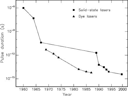
In Fig. 1.2, the progress in the development of pulsed laser systems is illustrated. In the case of solid-state lasers, two milestones were reached when discovering the technique of mode locking and when developing novel laser media with extremely large bandwidths as will be discussed below. These two events are characterized by two steps of the corresponding curve in Fig. 1.2. The other important group of lasers capable of providing ultrashort pulses consists of dye lasers. They were invented after the first solid-state lasers. Their progress was not so stepwise but proceeded smoothly. Several new techniques such as colliding pulse mode locking were developed which also lead to very short pulse durations comparable to those of solid-state lasers. However, medical applications of dye lasers will be rather limited because of their inconvenience and complicated maintenance. In contrast to long-living solid-state crystals, dyes need to be recirculated and exchanged on a regular basis which often disables a push-button operation.
1.1 Historic Review |
5 |
Fig. 1.2. Shortest achieved pulse durations with solid-state lasers and dye lasers
The very first laser was a ruby laser pumped with a xenon flash lamp. The output of such a laser is characterized by several spikes. Their overall duration is determined by the flash itself which is matched to the lifetime of the upper state of the laser transition, in ruby approximately 1ms. With the invention of Q-switching, pulses as short as 50ns could be obtained. Either mechanical devices (rotating apertures or laser mirrors) or optical devices (electrooptic or acoustooptic Pockels crystals) may serve as a Q- switch. In both cases, losses inside the resonator are kept artificially high until an extremely large inversion of the energy levels is achieved. Then, when removing the artificial losses, all energy stored in the laser medium is suddenly converted by means of stimulated emission. Even shorter pulses were obtained when initiating mode locking inside the laser cavity. During mode locking, a modulation of the electromagnetic field is induced by using either fast modulating crystals (active mode locking) or saturable absorbers (passive mode locking). By this means, the phases of all oscillating axial laser modes are forced to coincide, resulting in picosecond pulses. A typical representative is the Nd:YAG laser with an optical bandwidth of the order of 1nm. This bandwidth limits the shortest achievable pulse duration to a few picoseconds. Thus, the realization of femtosecond lasers mainly depended on the discovery of novel laser media with larger optical bandwidths. These were found in crystals such as Ti:Sapphire or Cr:LiSAF which currently led to the
61. Introduction
generation of laser pulses as short as 8.5fs according to Zhou et al. (1994). This duration is equivalent to a spatial pulse extent of a few wavelengths only. The most significant techniques of pulse generation are described in detail in the excellent book written by Siegman (1986).
1.2 Goal of the Book
The main goal of this book is to o er an interdisciplinary approach to the basics of laser–tissue interactions. It thus addresses all kinds of scientists, engineers, medical doctors, and graduate students involved in this field. Special emphasis is put on
–giving a detailed description of the physical background of potential interaction mechanisms between laser light and biological tissue,
–providing an updated review of clinical laser applications,
–including a chapter on laser safety.
In Chap. 2, mandatory prerequisites are given which are essential for understanding all the interaction mechanisms discussed in Chap. 3. Basic phenomena dealing with light and matter such as reflection, absorption, and scattering are explained by their physical roots. In each case, special attention is paid to their indispensable mathematical handling. The informed reader may well skip these sections and directly proceed with Sect. 2.5. In that section, when discussing photon transport theory, important tools will be derived which are of considerable importance in modern theoretical research. In order to solve the governing equation of energy transfer, either the Kubelka–Munk theory, the method of di usion approximation, or Monte Carlo simulations are most frequently used. All of them will be comprehensively reviewed in Sect. 2.5 and compared to each other along with their advantages and disadvantages. The interested reader, of course, should also consult the original works by Kubelka (1948), Metropolis and Ulam (1949), and the profound theory developed by Ishimaru (1978).
The main chapter of the book is Chap. 3. Whereas Chap. 2 focuses on how matter acts on light, here we will consider the opposite e ect, i.e. how light acts on matter. Starting with some general remarks and definitions, a general classification scheme is developed with the exposure duration being the main physical parameter. Five di erent types of interaction mechanisms are presented: photochemical interaction, thermal interaction, photoablation, plasma-induced ablation, and photodisruption. Each of them is thoroughly discussed including selected photographs and manifold illustrations. At the end of each section, a comprehensive statement is given summarizing in brief significant features of each interaction mechanism. Special recently developed techniques such as photodynamic therapy (PDT) or laser-induced interstitial thermotherapy (LITT) are explained according to the latest references.
1.3 Outlook |
7 |
Both of these techniques are concerned with the laser treatment of cancer, either photochemically or thermally, as an alternative to conventional methods which still remain unsatisfactory for a large group of patients. When discussing photoablation, potential risks originating from UV radiation will be surveyed. The di erentiation between plasma-induced ablation and photodisruption is emphasized and properly substantiated. Novel theoretical models are introduced describing the basic mechanism of plasma-induced ablation. They help to better understand the physical phenomena associated with optical breakdown and its threshold parameters.
In Chap. 4, the most important clinical applications are reviewed based on the latest results and references. Due to the historic sequence and their present significance, applications in ophthalmology, dentistry, and gynecology are considered first. In ophthalmology, various standard techniques are discussed such as coagulation of the retina, laser treatments of glaucoma, and fragmentation of the lens. The newest methods and results concerning refractive corneal surgery are presented, as well. In dentistry, special emphasis is put on di erent laser treatments of caries in comparison to conventional drills. In gynecology, various thermal e ects of laser radiation have recently been investigated. Major tasks and first clinical results are surveyed. Other disciplines of clinical importance follow as mentioned in the table of contents. In each case, experimental procedures and clinical results are discussed along with any complications arising or technical di culties. By means of specially selected photographs and artwork, it is intended to pass on clinical relevance and professional insight to the interested reader.
Finally, Chap. 5 comprises the latest standard of laser safety. It outlines a careful selection of essential guidelines published by the Laser Institute of America, Orlando, Florida. Meanwhile, most of them have been adapted by other governments, as well. A laser classification scheme is included which is commonly used all over the world. Moreover, important exposure limits are given to be taken into account when treating patients. In general, this chapter is meant to serve as a quick reference when operating lasers, but it might also be a useful guide for the inexperienced reader.
1.3 Outlook
It is interesting to observe that almost every new technique initially evokes a euphoric reaction among surgeons and patients. This period is often followed by indi erence and rejection when long-term e ects and limitations become obvious. Eventually, researchers agree on certain indications for applying the new technique which then leads to the final approval. One typical example for the occurrence of this sequence was the introduction of photodisruptive lasers to ophthalmology by Aron-Rosa et al. (1980).
At present, lasers have already contributed to the treatment of a wide variety of maladies. However, today’s clinical lasers and their applications