

Laser-Tissue Interactions Fundamentals and Applications - Markolf H. Niemz
.pdf228 4. Medical Applications of Lasers
Radiation from the argon ion laser is strongly absorbed by hemoglobin and melanin as already illustrated in Fig. 2.4. This laser is thus predestined for superficial treatments of highly vascularized skin. Apfelberg et al. (1978) and Apfelberg et al. (1979a) investigated laser-induced e ects on various abnormalities of the skin. The most frequent indications for the application of argon ion lasers are given by port wine stains (naevi flammei). Earlier methods of treating these malformations – e.g. cryotherapy, X-ray, or chemical treatment – had failed, and patients were advised to accept their misery. The idea of removing port wine stains with argon ion lasers has led to the most significant progress of lasers in dermatology so far. The treatment itself requires a lot of patience, since several sessions are necessary over a period of up to a few years. The faster the treatment is to come to an end, the higher is the probability for the occurrence of scarring. However, “patient” patients are usually rewarded with an acceptable outcome. In Figs. 4.62a–b, two photographs of the preand postoperative states of a laser-treated port wine stain are shown.
Treatment of port wine stains with argon ion lasers is usually performed in several sessions. First, a small test area of approximately 4mm2 is irradiated. During this test, a suitable laser power is determined by gradually increasing it until the skin visibly pales. According to Dixon and Gilbertson (1986) and Philipp et al. (1992), laser powers of 2–5W are applied during an exposure time of 0.02–0.1s. Immediately after laser exposure, inflammation of the skin frequently occurs. After four weeks, the test area is checked for recanalization and scarring. And after another four weeks, a second test area is treated. If both tests lead to acceptable results, the whole stain is exposed. Multiple exposures of the same area should be avoided in any case. Laser treatment may be repeated after a few years, but it is advisable to choose pulsed dye lasers for the second time. Haina et al. (1988) did not recommend treatment of patients up to 16 years of age, since otherwise severe scarring might occur. Laser radiation is usually applied by means of a flexible handpiece. In the treatment of facial stains, the eyes of both patient and surgeon must be properly protected. One disadvantage of treating port wine stains with argon ion lasers is that it is rather painful to the patient. Depending on the location and spatial extent of the stain, treatment is performed during either local or complete anesthetization.
Less painful and probably even more e cient is the treatment of port wine stains with dye lasers. Although quite expensive, these machines have recently gained increasing significance in dermatology, especially in the treatment of port wine stains and capillary hemangiomas. Detailed studies were reported by Morelli et al. (1986), Garden et al. (1988), and Tan et al. (1989). Frequently, Rhodamine dye lasers are used which emit radiation at wavelengths in the range 570–590nm. Typical pulse durations of 0.5ms and energy densities of 4–10J/cm2 have been recommended. About 20–60s after laser exposure, the color of the treated skin turns red, and after another few

4.7 Lasers in Dermatology |
229 |
Fig. 4.62. (a) Preoperative state of a port wine stain. (b) Postoperative state of the same stain after several treatments with an argon ion laser (pulse duration: 0.3s, power: 2.5W, focal spot size: 2mm). Photographs kindly provided by Dr. Seipp (Darmstadt)
230 4. Medical Applications of Lasers
minutes livid blue. Although pain is less pronounced as with argon ion lasers, patients frequently talk of triple pain perception: mechanical impact during the light flash, stabbing pain shortly afterwards, and finally a longer lasting heat wave within the skin. The irradiated area itself might be irritating for several days. One major advantage of treating port wine stains with dye lasers is that this procedure can be successfully performed among children as reported by Tan et al. (1989).
The basic mechanism by which pulsed laser radiation can cause selective damage to pigmented structures in vivo has been termed selective photothermolysis and was thoroughly described by Anderson and Parrish (1983). It requires the presence of highly absorbing particles, e.g. pigments of the skin. Extensive experimental and theoretical studies were recently performed by Kimel et al. (1994) and van Gemert et al. (1995). With their results, treatment of port wine stains will be further improved in the near future.
In dermatology, the CO2 laser is used for tissue vaporization. Compared to the conventional scalpel, it o ers the possibility of precise tissue removal without touching the tissue. Thus, feeling of pain is significantly reduced. External ulcers and refractory warts are common indications. In warts, however, deep lesions should be performed to reduce the probability of recurrence.
Recently, argon ion and CO2 lasers have also gained attention in e ciently removing tattoos. Clinical studies were reported by Apfelberg et al. (1979b) and Reid and Muller (1980). Today, ruby lasers are commonly used for tattoo removal as stated by Scheibner et al. (1990) and Taylor et al. (1990). Indeed, good results can be obtained, although they do depend on the dyes used in the tattoo. It is extremely important that all dye particles are removed during the same session. In Figs. 4.63a–b, two photographs are shown which prove the e ciency of laser-induced tattoo removal.
Radiation from the Nd:YAG laser is significantly less scattered and absorbed in skin than radiation from the argon ion laser. The optical penetration depth of Nd:YAG laser radiation is thus much larger. According to
Seipp et al. (1989), major indications for Nd:YAG laser treatments in dermatology are given by deeply located hemangiomas or semimalignant skin tumors. However, argon ion and CO2 lasers should never be replaced by Nd:YAG lasers when treating skin surfaces.
Dermatology is one of the few medical disciplines where biostimulative e ects of laser radiation have been reported. Positive stimulation on wound healing is one of the current topics of controversy as discussed in Sect. 3.1. A considerable number of papers has been published, but most of the results could not be reproduced, and initial claims could thus not be verified. Moreover, the principal mechanisms of biostimulation have not yet been understood. In general, one should be very careful when using laser radiation for such purposes, especially when applying so-called “soft lasers” with extremely low output powers which most probably do not evoke any e ect at all other than additional expenses according to Alora and Anderson (2000).

4.7 Lasers in Dermatology |
231 |
Fig. 4.63. (a) Preoperative state of a tattoo. (b) Postoperative state of the same tattoo after six complete treatments with an argon ion laser (pulse duration: 0.3s, power: 3W, focal spot size: 0.5mm). Photographs kindly provided by Dr. Seipp (Darmstadt)

232 4. Medical Applications of Lasers
4.8 Lasers in Orthopedics
Progress in surgical medicine is often related to an improved technique of performing osteotomies, i.e. bone excisions. Standard tools in orthopedics are saws, milling-machines and mechanical drills. All of them operate in contact mode and possibly induce severe mechanical vibrations and hemorrhage. It is thus straightforward to ask whether lasers might represent a considerable alternative in orthopedic surgery.
Bone fulfills three major functions: mechanical support of the body, protection of soft tissues, and supply of minerals and blood cells. The hardness of bone results from a complex structure of hydroxyapatite, water, soluble agents, collagen, and proteins. The chemical composition of bone is listed in Table 4.5. The high water content is responsible for strong absorption of infrared radiation. Therefore, CO2, Er:YAG, and Ho:YAG lasers are predestined for the e cient treatment of bone.
Table 4.5. Mean composition of human bone
Matter |
Percentage |
Constituent |
|
|
|
Anorganic |
50–60% |
Hydroxyapatite |
|
15–20% |
Water |
|
5% |
Carbonates |
|
1% |
Phosphates |
Organic |
20% |
Collagen |
|
1–2% |
Proteins |
|
|
|
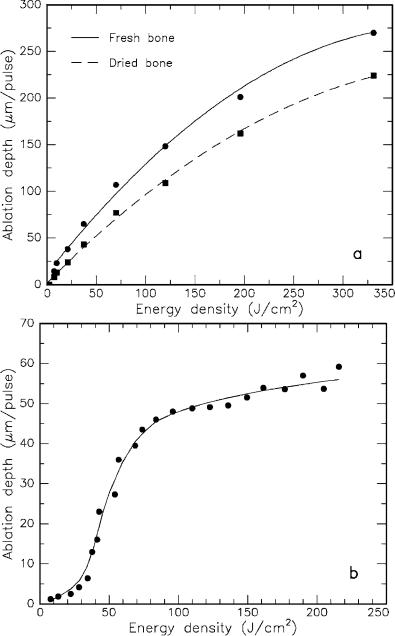
In the 1970s, Moore (1973), Verschueren and Oldho (1975), and Clayman et al. (1978) reported on osteotomies performed with CO2 lasers. Extensive studies on bone healing were published by Gertzbein et al. (1981) and Pao-Chang et al. (1981). All researchers agreed on a delayed healing process compared with conventional osteotomies. Thermal damage of the bone rim is exclusively made responsible for this time delay. Detailed data on the ablation characteristics were given by Kaplan and Giler (1984) and Forrer et al. (1993). The ablation curves of fresh and dried bone obtained with the CO2 laser are illustrated in Fig. 4.64a. From the above, we could conclude that CO2 lasers always evoke severe thermal side e ects in bone. This statement, however, is not generally true. Forrer et al. (1993) have also demonstrated the potential of CO2 lasers for bone ablation with very little thermal damage. When selecting the laser transition at 9.6μm, a pulse duration of 1.8μs, and an energy density of 15J/cm2, they found thermally altered damage zones of 10–15μm only. In this case, both wavelength and pulse duration play a significant role. First, the absorbance of bone at 9.6μm is higher than at 10.6μm. Second, shorter pulse durations tend to be associated with less thermal damage as already discussed in Sect. 3.2.
4.8 Lasers in Orthopedics |
233 |
Fig. 4.64. (a) Ablation curves of fresh and dried bone obtained with a CO2 laser (pulse duration: 250μs, wavelength: 10.6μm). Due to its higher water content, fresh bone is ablated more e ciently. Data according to Forrer et al. (1993). (b) Ablation curve of bone obtained with an Er:YAG laser (pulse duration: 180μs, wavelength: 2.94μm). Data according to Scholz and Grothves-Spork (1992)

234 4. Medical Applications of Lasers
In the 1980s, research focused on laser radiation at a wavelength of approximately 3μm which is strongly absorbed by water. For instance, Wolbarsht (1984) compared the e ects induced by CO2 lasers at 10.6μm and HF lasers10 at 2.9μm with each other. From his observations, he concluded that the latter wavelength is better suited for orthopedic applications. Similar results were published by Izatt et al. (1990). Unfortunately, though, HF lasers are very unwieldy machines. Walsh and Deutsch (1989), Nelson et al. (1989a), and Gonzales et al. (1990) reported on the application of compact Er:YAG lasers at a wavelength of 2.94μm. They stated that this radiation e ciently ablates both bone and cartilage. The ablation curve of bone obtained with the Er:YAG laser is illustrated in Fig. 4.64b.
Another promising laser in orthopedics is the Ho:YAG laser which emits at a wavelength of 2.12μm. Nuss et al. (1988), Charlton et al. (1990), and Stein et al. (1990) have investigated acute as well as chronic e ects of bone ablation with this laser. Its major advantage is that its radiation can be e - ciently transmitted through flexible fibers. However, thermal e ects are significantly enhanced compared to those induced by Er:YAG lasers at a wavelength of 2.94μm as observed by Romano et al. (1994). They found that thermal damage is extremely pronounced when applying 250μs pulses from a Ho:YAG laser. At an incident energy density of 120J/cm2, a thermal damage zone of roughly 300μm is determined. On the other hand, pulses from an Er:YAG laser are associated with very little thermal damage. At an energy density of 35J/cm2, a damage zone of only 12μm is estimated. The corresponding histologic sections are shown in Fig. 4.65a–b. In the case of the Er:YAG laser, a lower energy density was chosen to obtain a similar ablation depth as with the Ho:YAG laser. One potential application field of erbium lasers is microsurgery of the stapes footplate in the inner ear. This treatment belongs to the discipline of otorhinolaryngology, and it will therefore be addressed in Sect. 4.10.
Due to their high precision in removing tissues, excimer lasers have also been proposed for the ablation of bone material, e.g. by Yow et al. (1989). However, it was soon observed that their e ciency is much too low to justify their clinical application. Moreover, osteotomies performed with XeCl lasers at 308nm are associated with severe thermal damage as reported by Nelson et al. (1989b). As in the case of CO2 laser radiation, these thermal e ects are believed to be responsible for the manifest delay in healing of the laser-induced bone excisions.
An interesting approach to determine laser e ects on bone has recently been reported by Barton et al. (1995) and Christ et al. (1995). By using a confocal laser scanning microscope, they were able to analyze ablation rate and morphology as a function of incident pulse energy from a Ho:YAG laser. They concluded that scattering is a dominant factor in the interaction of Ho:YAG laser radiation and bone.
10 Hydrogen fluoride lasers.

4.8 Lasers in Orthopedics |
235 |
Fig. 4.65. (a) Histologic section of bone after exposure to a Ho:YAG laser (pulse duration: 250μs, energy density: 120J/cm2, bar: 100μm). (b) Histologic section of bone after exposure to an Er:YAG laser (pulse duration: 250μs, energy density: 35J/cm2, bar: 200μm). Photographs kindly provided by Dr. Romano (Bern)
236 4. Medical Applications of Lasers
Another discipline for laser applications within orthopedics is arthroscopy. Preliminary results regarding laser meniscectomy, i.e. the treatment of the meniscus, have already been reported by Glick (1981) and Whipple (1981) when using Nd:YAG and CO2 lasers, respectively. At that time, though, suitable delivery systems were not available. Moreover, the CW mode of these lasers led to unacceptable thermal damage. Katsuyuki et al. (1983) and Bradrick et al. (1989) applied Nd:YAG lasers in arthroscopic treatment of the jaw joint. Significant improvements were not achieved until O’Brien and Miller (1990) made use of specially designed contact probes consisting of ceramics. Limbird (1990) pointed out the necessity of blood perfusion measurements after surgery. Major limitations for all infrared lasers in arthroscopic surgery arise from the optical delivery system. Transmission through flexible fibers can be regarded as a mandatory requirement for an e cient surgical procedure. Therefore, CO2 lasers will never gain clinical relevance in arthroscopic treatments.
A new era of laser arthroscopy began with the application of holmium and erbium lasers. Trauner et a. (1990) reported promising results when using the Ho:YAG laser for the ablation of cartilage. Recently, Ith et al. (1994) have investigated the application of a fiber-delivered Er:YSGG laser emitting at a wavelength of 2.79μm. The transmittance of novel zirconium fluoride (ZrF4) fibers at this specific wavelength is satisfactory. Ith et al. (1994) have used fresh human meniscus from the knee joint which was obtained during surgery. They have observed a thermally damaged zone of 60μm when exposing the tissue in air to five laser pulses at a pulse energy of 53mJ and a pulse duration of 250μs. On the other hand, when exposing the tissue through water at a slightly higher energy of 65mJ, thermal damage extended to only 40μm close to surface and was even negligible elsewhere. In either case – whether exposed in air or through water – a crater depth of roughly 1mm was achieved. The surprising result of this study is that laser radiation at 2.79μm can be e ectively used for tissue ablation, although it should be strongly absorbed by surrounding water. Thus, Ith et al. (1994) concluded that light – after exiting the fiber – is guided through a water-vapor channel created by the leading part of the laser pulse. The period during which this channel is open was found to be dependent on the duration of the laser pulse. For pulse durations of 250–350μs, most of the laser energy is transmitted through the water-vapor channel to the target.
The experimental results mentioned above encourage the application of holmium and erbium lasers in arthroscopic surgery. Nevertheless, further investigations need to be performed regarding both thermal and mechanical side e ects associated with laser exposure. From today’s perspective, though, it is already obvious that arthroscopy belongs to those medical disciplines where minimally invasive techniques based on laser radiation will turn into unrenouncable surgical tools.
4.9 Lasers in Gastroenterology |
237 |
4.9 Lasers in Gastroenterology
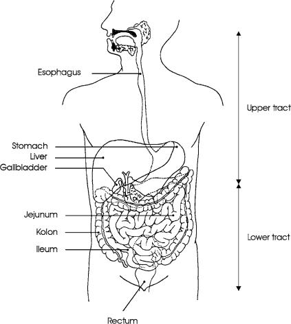
Gastrointestinal diseases primarily include ulcers and tumors of the esophagus, stomach, liver, gallbladder, and intestine. The intestine further consists of the jejunum, ileum, colon, and rectum. According to the position of these organs, the gastrointestinal tract is subdivided into an upper and a lower tract. Both tracts are schematically illustrated in Fig. 4.66. Most intestinal tumors are reported to occur inside the colon or the rectum.
Fig. 4.66. Cross-section of the upper and lower gastrointestinal tracts. The upper tract includes esophagus, stomach, liver, and gallbladder, whereas the lower tract primarily consists of the intestine