

Bioregenerative Engineering Principles and Applications - Shu Q. Liu
..pdf856 |
URINARY REGENERATIVE ENGINEERING |
|
||||
|
|
|
|
|
|
|
|
|
(A) |
|
(C) |
|
(E) |
|
|
|
|
|
|
|
(B) |
|
(D) |
|
(F) |
(G)
Glomerulus
Tubule
Polycarbonate membrane
Figure 20.3. Characterization of renal explants. (A,B) Cloned cells stained positively with synaptopodin antibody (A) and AQP1 antibody (B). (C) The allogeneic controls displayed a foreign-body reaction with necrosis. (D) Cloned explant shows organized glomeruli-like structures. Vascular tufts (v); visceral epithelium (arrow). H&E stain. (E) Organized tubules (arrows) were shown in the retrieved cloned explant. (F) Immunohistochemical analysis using factor VIII antibodies (black) identifies vascular structures. (G) There was a clear unidirectional continuity between the mature glomeruli, their tubules, and the polycarbonate membrane. Scale bars: 100 μm (B,D–F); 200 μm (A); 800 μm (C). (Reprinted by permission from Macmillan Publishers Ltd.: Robert P et al: Generation of histocompatible tissues using nuclear transplantation, Nature Biotechnol 20: 689–96, copyright 2002.)
of renal function, eventually leading to the loss of renal function and the accumulation of toxic and acidic substances in the blood. Chronic renal failure is associated with impaired metabolism and deteriorating functions of virtually all organ systems, resulting from cell injury by uremia or the retention of blood urea. Common diseases that induce chronic renal failure include glomerulonephritis, glomerulosclerosis, tubulointerstitial fibrosis, diabetes mellitus, and hypertension.
Glomerulonephritis is a glomerular and tubular disorder characterized by the injury of glomerular capillary endothelial cells and tubular epithelial cells, impaired glomerular

DISORDERS OF THE URINARY SYSTEM |
857 |
Monocytes/ macrophages
L
Cytokines
 Chemokines
Growth factors
|
GBM |
Endothelial |
Platelet |
injury/activation |
|
|
Mesangial |
|
|
cells |
|
Glomerular injury/proliferation |
Epithelial cell |
Inflammation |
|
|
|
Mesangial |
|
|
activation/ |
|
Mesangial cells/ |
proliferation |
Segmental mesangial sclerosis |
transformation |
|
mesangioblast |
|
|
|
|
|
|
Excessive ECM |
|
|
deposition |
|
|
ECM |
Global glomerulosclerosis |
Stretched podocyte |
Glormerulosclerosis |
|
|
Figure 20.4. Schematic representation of the stages of glomerulosclerosis. GBM = glomerular basement membrane; ECM = extracellular matrix. (Reprinted from Meguid El, Nahas A, Bello AK: Chronic kidney disease: The global challenge, Lancet 365:331–40, copyright 2005, with permission from Elsevier.)
filtration, and malfunctioned tubular processing of water, electrolytes, and waste substances. Glomerulosclerosis is a glomerular disorder characterized by glomerular endothelial injury, glomerular inflammatory reactions, mesangial cell proliferation, extracellular matrix deposition, and impairment of glomerular filtration (Fig. 20.4). Tubulointerstitial fibrosis is primarily a tubular disorder, characterized by tubular epithelial injury, epithelial cell apoptosis, inflammatory reactions, fibroblast proliferation, extracellular matrix deposition and fibrosis, tubular atrophy, and impairment of the tubular functions (Fig. 20.5). Diabetes mellitus, as discussed in Chapter 19, induces arteriosclerotic changes in the renal arteries, which influences glomerular filtration. Hypertension causes renal

 Excessive ECM deposition
DISORDERS OF THE URINARY SYSTEM |
859 |
urea or uremia. Urea is a waste product of protein metabolism and is removed from the kidneys. Chronic renal failure is always associated with uremia. Increased urea in the blood suppresses the activity of ATPases and the transport of electrolytes across the cell membrane, resulting in the imbalance of Na+ and K+. Such a disorder influences the resting and action potentials across the cell membrane and thus impairs cell functions. Uremia often induces a reduction in the responsiveness of cells to insulin, resulting in a decrease in the utilization of glucose by peripheral tissues and organs. The toxicity of urea also influences lipid metabolism. Uremia is commonly associated with hyperlipidemia and a decrease in high-density lipoproteins. The impairment of cellular metabolic functions in uremia is responsible for these changes. It is important to note that the kidneys have a high capacity of functional reserve. For a healthy person, one normal kidney is sufficient for the removal of toxins and the maintenance of the homeostasis. Thus, the clinical symptoms described above appear only when the majority of nephrons are destroyed in both kidneys.
Pathological examinations may demonstrate a number of structural changes in chronic renal failure, including a decrease in the density of nephrons, distortion of renal tubules, necrosis of glomerular endothelial cells and tubular epithelial cells, and formation of massive fibrous tissue. Chronic renal failure is often associated with complications in other systems, such as the cardiovascular, pulmonary, hematopoietic, nervous, and gastrointestinal systems. As in acute renal failure, fluid retention occurs commonly in chronic renal failure. Such a disorder can induce congestion, which further causes congestive heart failure and pulmonary edema. Uremia exerts toxic effects on the cardiomyocytes and pulmonary cells, facilitating the development of cardiac and pulmonary disorders. In the hematopoietic system, the toxic effect of urea on the bone marrow results in a reduction in the generation of hematopoietic cells. Uremia also induces hemolysis and a decrease in the number of red blood cells. In the nervous system, uremia induces neuronal injury, resulting in clinical symptoms such as drowsiness, insomnia, misjudgment, and loss of memory. In the gastrointestinal system, uremia causes epithelial cell injury and mucosal ulceration, resulting in disorders of digestion and absorption. Patients with chronic renal failure often express clinical symptoms such as anorexia, nausea, vomiting, and hiccups.
Treatment of Chronic Renal Failure [20.8]. Chronic renal failure is often associated with uremia and accumulation of metabolic wastes in the blood. Thus, dialysis or renal transplantation is necessary to control the blood concentration of urea and metabolic wastes. In addition, patients with chronic renal failure should be managed with the following approaches: (1) treatment of diseases that cause chronic renal failure; (2) management of complications in other systems, such as circulatory congestion, hypertension, heart failure, and pulmonary edema; (3) restriction of salt and water intake; (4) control of protein intake; and (5) control of the intake of drugs and chemicals that are excreted from the kidney. Molecular and cellular approaches described on page 850 can be used for the treatment of chronic renal failure. Once chronic renal failure reaches its end stage, dialysis and renal transplantation are the approaches of choice.
Acute Glomerulonephritis
Pathogenesis, Pathology, and Clinical Features [20.9]. Acute glomerulonephritis is a disorder characterized by rapid injury of glomerular capillaries and renal tubules, which
860 URINARY REGENERATIVE ENGINEERING
results in a reduction in glomerular filtration, water and salt retention, proteinuria, and hematuria. Acute glomerulonephritis is caused by several pathogenic factors, including bacterial infection (streptococci, pneumococci, syphilis, and meningococcemia), viral infection (hepatitis B, mumps, measles, varicella, and coxsackievirus), parasitic infection (malaria and toxoplasmosis), systemic lupus erythematosus, and vasculitis. Salt and water retention is due to reduced glomerular filtration and tubular dysfunction and can induce circulatory congestion, pulmonary edema, and hypertension. Proteinuria is due to the damage of glomerular endothelial cells and increased capillary permeability, resulting in protein transport from the blood to the renal tubules. Similarly, hematuria is a result of the damage of the glomerular endothelial cells, allowing red blood cell migration to the renal tubules.
Pathological examinations or biopsies often demonstrate several structural changes, including injury of glomerular endothelial cells and tubular epithelial cells, infiltration of leukocytes, and the presence of red blood cells in the renal tubules. A considerable fraction of patients may experience acute renal failure. In most cases, acute glomerulonephritis can be recovered when the causative factors are removed. However, acute glomerulonephritis may develop into chronic glomerulonephritis in a certain fraction of patients (see next section).
Treatment of Acute Glomerulonephritis [20.9]. Acute glomerulonephritis is often treated with several approaches, including: (1) administration of antibiotics for causative bacterial infection, (2) appropriate rest until clinical symptoms disappear, (3) restriction of water and salt intake, (4) restriction of protein intake, (5) administration of diuretics, and (6) administration of vasodilators in the presence of hypertension. In about half of the cases, the disorder can be self-cured without long-term clinical consequences.
It is important to point out that an ideal treatment for acute glomerulonephritis is to prevent the development of acute renal failure. It is obvious that the conventional approaches described above are merely supportive and cannot be used to achieve such a goal. Molecular regenerative engineering is thought an effective approach that prevents acute renal failure. The strategies of molecular therapy for acute glomerulonephritis are identical to those described on page 850 for acute renal failure. When acute renal failure occurs, the cellular approach described on page 852 can be used to reduce uremia and toxic effects.
Chronic Glomerulonephritis
Pathogenesis, Pathology, and Clinical Features [20.10]. Chronic glomerulonephritis is a disorder with slow, persistent, and progressive impairment of renal function, which is manifested by the presence of long-term proteinuria and hematuria, eventually leading to chronic renal failure. Acute renal disorders, such as acute glomerulonephritis and nephrotic syndrome, can lead to chronic glomerulonephritis. Chronic glomerulonephritis is associated with a number of pathological changes, including the proliferation of glomerular endothelial cells and tubular epithelial cells, excessive formation of extracellular matrix and fibrosis, glomerulosclerosis, and tubulointerstitial fibrosis. During the early stage, patients with chronic glomerulonephritis may not demonstrate apparent clinical symptoms. During the late stage, chronic glomerulonephritis inevitably develop into chronic renal failure, exhibiting typical symptoms of renal failure such as uremia, water and salt
DISORDERS OF THE URINARY SYSTEM |
861 |
retention, and complications in the cardiovascular, pulmonary, nervous, and gastrointestinal systems, as described on page 855.
Treatment of Chronic Glomerulonephritis [20.10]. Chronic glomerulonephritis is treated primarily with supportive approaches. A treatment with steroids may slow the progress of the disorder. Hypertension, if present, should be treated with vasodilators. It is necessary to administrate diuretics to reduce salt and water retention. Furthermore, the intake of water, salt, and protein should be controlled. In most cases of chronic glomerulonephritis, there exist various degrees of chronic renal failure. The treatment of chronic glomerulonephritis is similar to that of chronic renal failure. Molecular and cellular regenerative approaches described on page 850 and 852 can be used for the treatment of chronic renal glomerulonephritis.
Urinary Tract Obstruction
Pathogenesis, Pathology, and Clinical Features [20.11]. Urinary tract obstruction is the mechanical blockade of the urinary drainage system at a location from the renal calices to the urethra, often resulting in acute and chronic renal failure. A number of pathogenic factors can induce urinary tract obstruction. These factors include congenital abnormalities (ureteropelvic narrowing or obstruction, bladder neck narrowing and obstruction, and urethral valve stricture), inflammation, trauma, tumor, retroperitoneal fibrosis, aortic aneurysm, and kidney stone (inducing urine blockade within the kidney). Urinary tract obstruction can be effectively treated by surgical removal of the mechanical blockade and urinary tract reconstruction. Acute renal failure due to urinary tract obstruction can be recovered once the blockade is removed. However, chronic renal failure is usually irreversible. The clinical consequences of urinary tract obstruction are dependent on the degree of the mechanical blockade. Partial obstruction may not exert any influence on the renal functions. In contrast, complete urinary obstruction may induce acute renal failure within several days.
Conventional Treatment of Urinary Tract Obstruction [20.11]. The principle of treating urinary tract obstruction is to remove the factors that cause the obstruction. As discussed above, common causes for urinary obstruction are urinary tract tumors, congenital urinary tract constriction, compression by aortic aneurysms, trauma-induced scar formation, and inflammation-induced fibrosis. Surgical removal of tumors, congenital constriction, aortic aneurysm, and scars usually results in the restoration of renal functions within months if chronic renal failure is absent. When the urinary tract is severely damaged or a segment is removed due to tumors or other factors, it is necessary to reconstruct the urinary tract. This aspect is discussed in the following section. In addition, bacterial infection, if any, should be immediately controlled with antibiotics.
Cellular and Tissue Engineering. Severe urinary tract damage or loss can occur in several disorders, such as cancer, trauma, infection, and congenital abnormalities. In such cases, urinary tract reconstruction is often required to restore the function of the urinary tracts. There are two basic strategies for the reconstruction of the urinary tracts: (1) reconstruction with polymeric or extracellular matrix scaffolds and (2) reconstruction with cell-seeded matrix scaffolds. For the first strategy, a tubular scaffold can be used directly
862 URINARY REGENERATIVE ENGINEERING
for the reconstruction of a malfunctioned urinary tract. The host urinary tract is capable of regenerating necessary cells to cover the reconstructed segment. For the second strategy, selected cells can be seeded in a scaffold of extracellular matrix or synthetic materials. The seeded cells can proliferate and produce extracellular matrix, thus facilitating the formation of the urinary tract. A fundamental issue in urinary tract reconstruction is to find a biocompatible material that can be used to construct a suitable scaffold. Furthermore, the materials should be chemically stable in urine and resistant to inflammation. Several types of biomaterials exhibit characteristics suitable for the repair and reconstruction of the urinary tracts, including the ureter, bladder, and urethra. These materials are briefly described as follows.
Polymeric Biomaterials for Urinary Tract Reconstruction [20.12]. A number of synthetic polymers, including polyurethane, silicone, and their copolymers, have been used for the reconstruction of the urinary tracts. Polyurethane has been particularly used for the construction of the ureter stents. Although this material is mechanically flexible and suitable for the reconstruction of the urinary tracts, it often causes inflammatory reactions and ulceration. Silicone is an inert material relatively compatible with biological tissues. Silicone and its copolymers have been widely used for the repair and reconstruction of the urinary tracts.
Biodegradable polymeric materials have been recently used for the repair and reconstruction of the urinary tracts. Commonly used biodegradable materials include polyglycolic polymers and poly-L-lactic and poly-L-glycolic copolymers. By altering the concentrations of various ingredients, the rate of degradation can be controlled to suite specific types of tissue. The advantage of using biodegradable materials is that, with the gradual degradation of the materials, native cells and tissue can grow into the polymeric matrix and self-reconstruct the lost structures. In experimental studies, poly-L-lactic and poly-L-glycolic copolymers have been used to fabricate urethral stents for the restoration of the urethral function. For a copolymer composed of 80% L-lactide and 20% glycolic acid, the copolymer stents are biocompatible and mechanically flexible, and can degrade within several months. In preliminary clinical studies, the same type of copolymeric urethral stent has been used in human patients with prostate disorders. The stents can be used to maintain the function of the urethra and can be degraded within 6 months.
Polymeric materials have been used for constructing the urinary bladder in animal experiments. Autologous epithelial and smooth muscle cells can be collected from the urinary bladder and seeded in a bladder-shaped polymeric scaffold to construct a neobladder. Such a construct can be used to replace the bladder of the animal that donates the epithelial and smooth muscle cells (Fig. 20.6). In preliminary tests, the reconstructed bladder can function for nearly a year and demonstrates the capability of retaining urine (Fig. 20.7). Furthermore, the neobladder can integrate to the native bladder tissue and exhibits natural structure and mechanical properties. These investigations suggest a potential for reconstructing a disordered urinary bladder by using cell and tissue regenerative engineering approaches.
Metallic Materials for Urinary Tract Reconstruction [20.13]. Several types of metallic materials, including stainless steel and titanium and nickel alloys, have been used for the reconstruction of the ureter and urethra. Given the mechanical strength and durability, metallic materials are often used for long-term implantation. The material surface can be gradually covered with epithelial cells following implantation, which enhances the com-
DISORDERS OF THE URINARY SYSTEM |
863 |
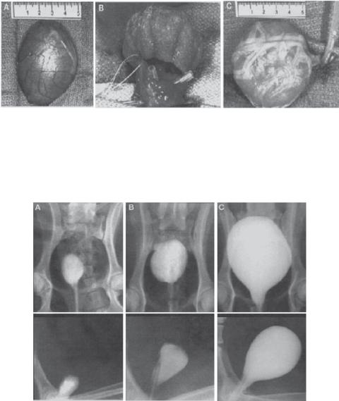
Figure 20.6. Surgical technique for cystectomy and preparation of implants: (A) the native canine bladder prior to trigone-sparing cystectomy; (B) the engineered neoorgan is anastomosed to the trigone; (C) the implant, decompressed by a transurethral and suprapubic catheter, is wrapped with omentum. (Reprinted by permission from Macmillan Publishers Ltd.: Oberpenning F et al: De novo reconstitution of a functional mammalian urinary bladder by tissue engineering, Nature Biotechnol 17:149–55, copyright 1999.)
Figure 20.7. Radiographic cystograms 11 months after subtotal cystectomy: (A) subtotal cystectomy without reconstruction; (B) polymer-only implant; (C) tissue-engineered neoorgan. (Reprinted by permission from Macmillan Publishers Ltd.: Oberpenning F et al: De novo reconstitution of a functional mammalian urinary bladder by tissue engineering, Nature Biotechnol 17:149–55, copyright 1999.)
patibility of the metal implant with the host system. Although the success rate is acceptable in some experimental and clinical studies, metal devices usually stimulate host inflammatory responses, cell proliferation, matrix production, and tissue fibrosis, resulting in implant occlusion. The administration of antiinflammatory agents and growth inhibitors may reduce the rate of occlusion.
864 URINARY REGENERATIVE ENGINEERING
Biological Materials for Urinary Tract Reconstruction [20.14]. Biological tissues, such as intestinal submucosa and bladder submucosa, have been used as materials for urinary tract reconstruction. Given the natural structure and mechanical properties, these tissues are preferred compared to synthetic polymers and metallic materials. In the lack of autogenous tissues, allogenic tissues can be used. A problem with allogenic tissues is acute immune rejection. To reduce such a problem, it is necessary to remove the cellular components, which stimulate host immune responses. Decellularized matrix tissues exhibit significantly reduced immunogenicity and rarely cause acute immune rejection. Allogenic intestinal or bladder specimens can be decellularized by treatment with NaOH, KOH, or detergents. The mucosa and muscular layers can be removed and the submucosa matrix can be used for constructing ureteric, urethral, and bladder substitutes. Experimental and clinical studies have demonstrated promising results, although further investigations are necessary to improve the performance of the substitutes.
BIBLIOGRAPHY
20.1. Anatomy and Physiology of the Urinary System
Guyton AC, Hall JE: Textbook of Medical Physiology, 11th ed, Saunders, Philadelphia, 2006.
McArdle WD, Katch FI, Katch VL: Essentials of Exercise Physiology, 3rd ed, Lippincott Williams & Wilkins, Baltimore, 2006.
Germann WL, Stanfield CL (with contributors Niles MJ, Cannon JG), Principles of Human Physiology, 2nd ed, Pearson Benjamin Cummings, San Francisco, 2005.
Thibodeau GA, Patton KT: Anatomy & Physiology, 5th ed, Mosby, St Louis, 2003.
Boron WF, Boulpaep EL: Medical Physiology: A Cellular and Molecular Approach, Saunders, Philadelphia, 2003.
Ganong WF: Review of Medical Physiology, 21st ed., McGraw-Hill, New York, 2003.
20.2. Pathogenesis, Pathology, and Clinical Features
Schneider AS, Szanto PA: Pathology. 3rd ed., Lippincott Williams & Wilkins, Philadelphia, 2006.
McCance KL, Huether SE: Pathophysiology: the biologic basis for disease in adults & children. 5th ed. Elsevier Mosby, St. Louis, 2006.
Porth CM: Pathophysiology: concepts of altered health states, 7th ed., Lippincott Williams & Wilkins, Philadelphia, 2005.
Frazier MS, Drzymkowski JW: Essentials of human diseases and conditions, 3rd ed., Elsevier Saunders, St. Louis, 2004.
Dai C, Yang J, Liu Y: Single injection of naked plasmid encoding hepatocyte growth factor prevents cell death and ameliorates acute renal failure in mice, J Am Soc Nephrol 13:411–422, 2002.
Liu Y, Tolbert EM, Lin L, Thursby MA, Sun AM et al: Up-regulation of hepatocyte growth factor receptor: An amplification and targeting mechanism for hepatocyte growth factor action in acute renal failure, Kidney Int 55:442–53, 1999.
BIBLIOGRAPHY 865
20.3. Growth Factor Genes
Yang J, Dai C, Liu Y: Systemic administration of naked plasmid encoding hepatocyte growth factor ameliorates chronic renal fibrosis in mice, Gene Ther 8(19):1470–9, 2001.
Mizuno S, Nakamura T: Prevention of neutrophil extravasation by hepatocyte growth factor leads to attenuations of tubular apoptosis and renal dysfunction in mouse ischemic kidneys, Am J Pathol 166(6):1895–905, 2005.
Bonventre JV, Siegel N, Rosen S, Portilla D, Venkatachalam M: Acute renal failure. I. Relative importance of proximal vs. distal tubular injury, Am J Physiol Renal Physiol 275:F623–32, 1998.
Edelstein CL, Ling H, Wangsiripaisan A, Schrier RW: Emerging therapies for acute renal failure, Am J Kidney Dis 30:S89–95, 1997.
Kelly KJ, Molitoris BA: Acute renal failure in the new millennium: Time to consider combination therapy, Semin Nephrol 20:4–19, 2000.
Hammerman MR, Miller SB: Therapeutic use of growth factors in renal failure, J Am Soc Nephrol 5:1–11, 1994.
Humes HD, MacKay SM, Funke AJ, Buffington DA: Acute renal failure: Growth factors, cell therapy, and gene therapy, Proc Assoc Am Physicians 109:547–57, 1997.
Hammerman MR, Safirstein R, Harris RC, Toback FG, Humes HD: Acute renal failure. III. The role of growth factors in the process of renal regeneration and repair, Am J Physiol Renal Physiol 279:F3–11, 2000.
Zarnegar R, Michalopoulos GK: The many faces of hepatocyte growth factor: From hepatopoiesis to hematopoiesis, J Cell Biol 129:1177–80, 1995.
Matsumoto K, Nakamura T: Emerging multipotent aspects of hepatocyte growth factor, J Biochem 119:591–600, 1996.
Vargas GA, Hoeflich A, Jehle PM: Hepatocyte growth factor in renal failure: Promise and reality, Kidney Int 57:1426–36, 2000.
Matsumoto K, Mizuno S, Nakamura T: Hepatocyte growth factor in renal regeneration, renal disease and potential therapeutics, Curr Opin Nephrol Hypertens 9:395–402, 2000.
Miller SB, Martin DR, Kissane J, Hammerman MR: Hepatocyte growth factor accelerates recovery from acute ischemic renal injury in rats, Am J Physiol 266:F129–34, 1994.
Kawaida K, Matsumoto K, Shimazu H, Nakamura T: Hepatocyte growth factor prevents acute renal failure and accelerates renal regeneration in mice, Proc Natl Acad Sci USA 91:4357–61, 1994.
Ueda T, Takeyama Y, Hori Y, Shinkai M, Takase K et al: Hepatocyte growth factor increases in injured organs and functions as an organotrophic factor in rats with experimental acute pancreatitis, Pancreas 20:84–93, 2000.
Liu Y, Tolbert EM, Lin L, Thursby MA, Sun AM et al: Up-regulation of hepatocyte growth factor receptor: An amplification and targeting mechanism for hepatocyte growth factor action in acute renal failure, Kidney Int 55:442–53, 1999.
Liu ML, Mars WM, Zarnegar R, Michalopoulos GK: Uptake and distribution of hepatocyte growth factor in normal and regenerating adult rat liver, Am J Pathol 144:129–40, 1994.
Kelley VR, Suhatme VP: Gene transfer in the kidney, Am J Physiol 276:F1–9, 1999.
Imai E, Isaka Y: New paradigm of gene therapy: Skeletal-muscle-targeting gene therapy for kidney disease, Nephron 83:296–300, 1999.
Yang J, Chen S, Huang L, Michalopoulos GK, Liu Y: Sustained expression of naked plasmid DNA encoding hepatocyte growth factor in mice promotes liver and overall body growth, Hepatology 33:848–59, 2001.