

Bioregenerative Engineering Principles and Applications - Shu Q. Liu
..pdf766 PULMONARY REGENERATIVE ENGINEERING
16.13. Pathogenesis, Pathology, and Clinical Features of Primary
Pulmonary Hypertension
Archer S, Rich S: Primary, pulmonary hypertension: A vascular biology and translational research “Work in progress,” Circulation 102(22):2781–91, 2000.
Moser KM, Fedullo PF, Finkbeiner WE, Golden J: Do patients with primary pulmonary hypertension develop extensive central thrombi? Circulation 91(3):741–5, 1995.
Rubin LJ: Pathology and pathophysiology of primary pulmonary hypertension, Am J Cardiol 75(3):51A–54A, Jan 1995.
Klinger JR, Hill NS: Right ventricular dysfunction in chronic obstructive pulmonary disease. Evaluation and management, Chest 99(3):715–23, 1991.
16.14. Pathogenesis, Pathology, and Clinical Features of Hypoxic
Pulmonary Hypertension
Fung YC, Liu SQ: Change of zero-stress state of rat pulmonary arteries in hypoxic pulmonary hypertension, J Appl Physiol 70:2455–70, 1991.
Liu SQ: Alterations in structure of elastic laminae of rat pulmonary arteries in hypoxic hypertension, J Appl Physiol 81:2147–55, 1996.
Liu SQ: Regression of hypoxic hypertension-induced changes in the elastic laminae of rat pulmonary arteries, J Appl Physiol 82:1677–84, 1997.
16.15. Nitric Oxide Synthase Gene
Gelband CH, Katovich MJ, Raizada MK: Current perspectives on the use of gene therapy for hypertension, Cir Res 87:1118, 2000.
Champion HC, Bivalacqua TJ, D’Souza FM, Ortiz LA, Jeter JR et al: Gene transfer of endothelial nitric oxide synthase to the lung of the mouse in vivo: Effect on agonist-induced and flow-medi- ated vascular responses, Circ Res 84:1422–32, 1999.
Champion HC, Bivalacqua TJ, Greenberg SS, Giles TD, Heistad DD et al: Gene transfer of endothelial nitric oxide synthase to the lung of the mouse in vivo: selective rescue of pulmonary hypertension in eNOS-deficient mice, Circulation 100:1–28, 1999.
Janssens SP, Bloch KD, Nong Z, Gerard RD, Zoldhelyi P et al: Adenoviral-mediated transfer of the human endothelial nitric oxide synthase gene reduces acute hypoxic pulmonary vasoconstriction in rats, J Clin Invest 98:317–24, 1996.
16.16. Prostaglandin I2 Synthase Gene
Geraci MW, Gao B, Shepherd DC, Moore MD, Westcott JY et al: Pulmonary prostacyclin synthase overexpression in transgenic mice protects against development of hypoxic pulmonary hypertension, J Clin Invest 103:1509–15, 1999.
Chevalier D, Cauffiez C, Bernard C, Lo-Guidice JM, Allorge D et al: Characterization of new mutations in the coding sequence and 5-prime-untranslated region of the human prostacyclin synthase gene (CYP8A1), Hum. Genet 108:148–55, 2001.
Miyata A, Hara S, Yokoyama C, Inoue H, Ullrich V et al: Molecular cloning and expression of human prostacyclin synthase, Biochem Biophys Res Commun 200:1728–34, 1994.
Nakayama T, Soma M, Watanabe Y, Hasimu B, Sato M et al: Splicing mutation of the prostacyclin synthase gene in a family associated with hypertension, Biochem Biophys Res Commun 297:1135–9, 2002.
BIBLIOGRAPHY 767
Wang LH, Chen L: Organization of the gene encoding human prostacyclin synthase, Biochem Biophys Res Commun 226:631–7, 1996.
Yokoyama C, Yabuki T, Inoue H, Tone Y, Hara S et al: Human gene encoding prostacyclin synthase (PTGIS): Genomic organization, chromosomal localization, and promoter activity, Genomics 36:296–304, 1996.
16.17. Preprocalcitonin-Related Peptide Gene
Champion HC, Bivalacqua TJ, Toyoda K, Heistad DD, Hyman AL et al: In vivo gene transfer of prepro-calcitonin gene-related peptide to the lung attenuates chronic hypoxia-induced pulmonary hypertension in the mouse, Circulation 101(8):923–30, 2000.
Kwan YW, Wadsworth RM, Kane KA: Effects of neuropeptide Y and calcitonin gene-related peptide on sheep coronary artery rings under oxygenated, hypoxic and simulated myocardial ischaemic conditions, Br J Pharmacol 99:774–8, 1990.
Tjen ALS, Ekman R, Lippton H, Cary J, Keith I: CGRP and somatostatin modulate chronic hypoxic pulmonary hypertension, Am J Physiol 263:H681–90, 1992.
Shimosegawa T, Said SI: Pulmonary calcitonin gene-related peptide immunoreactivity: Nerveendocrine cell interrelationships, Am J Resp Cell Mol Biol 4:126–34, 1991.
Springall DR, Polak JM: Calcitonin gene-related peptide and pulmonary hypertension in experimental hypoxia, Anat Rec 236:96–104, 1993.
Stevens TP, McBride JT, Peake JL, Pinkerton KE, Stripp BR: Cell proliferation contributes to PNEC hyperplasia after acute airway injury, Am J Physiol 272:L486–93, 1997.
Kusakabe T, Kawakami T, Powell FL, Ellisman MH, Sawada H et al: Distribution of substance P and calcitonin gene-related peptide immunoreactive nerve fibers in the trachea of chronically hypoxic rats, Brain Res Bull 39:335–9, 1996.
Champion HC, Bivalacqua TJ, Toyoda K, Heistad DD, Hyman AL et al: In vivo gene transfer of prepro-calcitonin gene-related peptide to the lung attenuates chronic hypoxia-induced pulmonary hypertension in the mouse, Circulation 101:923–30, 2000.
Amara SG, Arriza JL, Leff SE, Swanson LW, Evans RM et al: Expression in brain of a messenger RNA encoding a novel neuropeptide homologous to calcitonin gene-related peptide, Science 229:1094–7, 1985.
Hoovers JMN, Redeker E, Speleman F, Hoppener JWM, Bhola S et al: High-resolution chromosomal localization of the human calcitonin/CGRP/IAPP gene family members, Genomics 15:525–9, 1993.
ANATOMY AND PHYSIOLOGY OF THE LIVER |
769 |
ANATOMY AND PHYSIOLOGY OF THE LIVER
Structure [17.1]
The liver is located in the upper right abdominal cavity. This organ is composed of a large number of functional units, known as hepatic lobules. Each hepatic lobule is a cylindrical structure of several millimeters in length and about 1 mm in diameter, and is composed of hepatic cell plates and several tubular systems, including the central vein, portal vein, hepatic artery, lymphatic vessels, and bile ducts. The portal vein in each unit is a branch of the major hepatic portal vein and conducts blood from the gastrointestinal tracts into the central vein through a structure between the hepatic cell plates known as hepatic sinusoid. The central vein in each unit converges blood into the hepatic vein and then into the inferior vena cava. The hepatic artery in each unit is a branch of the major hepatic artery and supplies oxygenated blood to the hepatic unit. The lymphatic vessels collect and conduct excessive fluids from the interstitial tissue to the large lymphatic vessels and then to the vena cava. The bile ducts collect and conduct bile to larger bile ducts and then to the duodenum.
The liver consists of several types of cell, including the hepatocytes, Küpffer cells, Ito cells, epithelial cells, and endothelial cells. Hepatocytes are the largest cell population in the liver, are major constituents for the functional units of the liver, and are characterized by the expression of albumin (Chapter 17 opening figure). Küpffer cells are macrophages and are found in the hepatic sinusoids. These cells are responsible for the destruction and clearance of microorganisms present in the blood. Ito cells are found in the hepatic sinusoids and are responsible for the generation of extracellular matrix components. Epithelial cells are constituents of the bile ducts. Endothelial cells are found in blood vessels and lymphatic vessels and are responsible for the transport of molecules and electrolytes.
Functions [17.1]
The liver is a vital organ that conducts several essential functions, including nutrient metabolism, detoxification, blood filtration and storage, and bile excretion. The liver possesses a large capacity of functional reserve that is not used under physiological conditions. About one-third of the total liver is sufficient for the maintenance of metabolic homeostasis. The reserve capacity is developed during evolution for sudden changes in metabolic demand under unusual conditions, such as ingestion of a large amount of toxins, liver trauma, and liver infection. Here, the basic functions of the liver are briefly discussed.
Metabolism. The liver is responsible for the metabolism of the three major types of nutrient: carbohydrates, lipids, and proteins. Carbohydrates include glucose, galactose, and fructose. These substances are absorbed from the small intestine to the blood, and transported to and processed in the liver. Glucose is the most important carbohydrate for energy production. The liver is responsible for the control of a stable level of blood glucose at80 mg/dL. When the level of blood glucose is increased (for instance, immediately after a meal), the hepatocytes are able to synthesize glycogen, a glucose polymer, from glucose molecules via a process known as glycogenesis, thus reducing the blood glucose level. In contrast, when the level of blood glucose is decreased in the fasting state, the hepatocytes can hydrolyze glycogen to produce glucose via a process called glycogenolysis, bringing back the blood glucose level. Other types of carbohydrate, such as galactose and fructose,
770 LIVER REGENERATIVE ENGINEERING
can be converted to glucose in the liver, contributing to the accumulation of blood glucose. In addition, under conditions with a very low level of blood glucose, the liver is capable of converting amino acids and glycerol into glucose, a process called gluconeogenesis. The maintenance of the blood glucose level is essential for the function of vital organs and tissues, including the brain, heart, and skeletal muscles.
The liver conducts several functions related to lipid metabolism, including energy generation from fatty acids, synthesis of phospholipids and lipoproteins, and conversion of amino acids and carbohydrates to fatty acids. Fatty acids can be oxidized to form acetylcoenzyme A, which is further oxidized to generate energy. Hepatocytes are responsible for the synthesis of cholesterols, which are released to the blood, transported to cells in other organs, and used for the construction of cell membrane. Cholesterols are also used to constitute bile, which is released into the duodenum via the bile ducts. Hepatocytes can synthesize phospholipids, which are utilized to construct cell membranes. The liver is a major organ that synthesizes and processes lipoproteins, which are responsible for the transport of lipids and cholesterols between the blood, liver, and other organs. The liver is also responsible for the conversion of amino acids and carbohydrates to fatty acids, which are stored in the fat cells or adipocytes.
The liver is the most important organ in the body for protein metabolism. Hepatocytes conduct several protein-related metabolic functions, including the formation of urea, synthesis of proteins, formation of amino acids, and transamination and deamination of amino acids. Protein metabolism generates highly toxic ammonia, which is converted to urea in the hepatocytes. Urea is released into the blood and removed from the kidney. Hepatocytes can synthesize a number of plasma proteins, including albumin, fibrinogen, heparin, globulins, and blood coagulation factors. These factors play critical roles in many important cellular activities. Hepatocytes are capable of forming a number of amino acids, known as nonessential amino acids (meaning that it is not necessary to ingest these amino acids from diets). In addition, hepatocytes are able to carry out transamination and deamination of amino acids. Transamination is a process by which an amino group is transferred from an amino acid to a keto acid by an aminotransferase. After such a process, amino acids can participate in the metabolism of the citric acid cycle and generate energy. Deamination is a process by which the amino group of amino acids is removed by deaminases. Such a process generates ammonia, a waste product of protein metabolism.
Detoxification. The processes of metabolism as described above generate waste substances, most of which are toxic. In addition, various types of toxic substances, such as medicines and chemicals, may be ingested on a daily basis. Hepatocytes are able to chemically modify toxic substances, reducing their toxicity and rendering the substances less toxic and removable. For instance, biological agents, such as penicillin and ampicillin, are processed in the liver, released into the bile, and transported to the intestinal system, where the substances are removed. Excessive hormones, such as cortisol and estrogen, are modified and detoxified in the liver.
Blood Filtration and Storage. In the small intestine, bacterial and toxic particles can be transported into the blood during nutrient absorption. These toxic particles are transported to the liver through the portal vein. The Küpffer cells (specialized reticuloendothelial cells) can endocytose bacterial and toxic particles so that portal blood can be cleansed. In addition, the liver can serve as an organ for blood storage. Under physiological conditions, about 0.5 L of blood can be stored in the hepatic sinusoids and blood vessels. The
HEPATIC DISORDERS |
771 |
blood storage function is important when heart failure occurs. In such a case, excessive blood can be stored in the liver to prevent systemic edema.
Bile Excretion. Bile is produced by hepatocytes, released into the bile ducts, and transported into the duodenum. A human liver can produce about 1,000 ml bile per day. Bile is composed of bile salts, bilirubin, cholesterol, electrolytes, and water. The bile salts are produced from cholesterol and possess two important functions: (1) emulsifying fat diets in the small intestine into minute particles that can be digested by pancreatic enzymes and
(2) facilitating the transport and absorption of fat particles through the intestinal epithelial cells. Bilirubin is a metabolic product of hemoglobin, is produced in the hepatocytes, and is transported to the bile ducts and then to the duodenum, where the substance is removed. The accumulation of bilirubin, in the case of liver dysfunction, induces jaundice. Excessive cholesterol molecules are also removed through bile formation and excretion.
Liver Regeneration [17.2]
Organ regeneration is a process of organ self-reconstruction by cell differentiation and proliferation as well as matrix production following organ injury or partial removal. The capacity of regeneration varies among different organs. For instance, the brain and heart have very a low capacity of regeneration, whereas digestive organs are able to regenerate extensively following organ injury. In particular, the liver has a very high capacity of regeneration. It can completely regenerate itself when it is partially removed. No other organs in the human body have the regeneration capacity of the liver. Such a phenomenon has been documented since year the 1890. For more than a century, liver regeneration has fascinated physicians and scientists. For the past several decades, extensive investigations have been carried out for the mechanisms of liver regeneration. These investigations have provided a foundation for today’s liver regenerative engineering and reconstruction. See page 399 for detailed discussion about liver regeneration.
HEPATIC DISORDERS
Acute Viral Hepatitis and Liver Failure
Pathogenesis, Pathology, and Clinical Features [17.3]. Acute viral hepatitis is a liver infectious disorder induced predominantly by hepatitis A virus and hepatitis B virus. Hepatitis is one of the most popular diseases in the world. About 33% of the human population are infected by hepatitis viruses. This disorder is characterized by inflammatory reactions, edema, cell necrosis, and hyperplasia of Küpffer cells in the liver. The clinical manifestations and consequences of hepatitis vary widely, ranging from mild asymptomatic infections without any noticeable pathological changes in the liver to fatal acute liver failure with massive hepatocyte necrosis. Some patients can completely recover from acute hepatitis, whereas others exhibit persistent infections, which eventually develop into chronic hepatitis and cirrhosis. The outcome of the disease is largely dependent on the responsiveness or sensitivity of the immune system as well as the age of individual patients. The occurrence of chronic hepatitis from the population with acute hepatitis decreases with age. While chronic hepatitis may be found in about 30% of children at the age of <5, the disorder may be found in only about 2% of the adult population with acute hepatitis. The mechanisms for the age related epidemiology remain poorly understood.
772 LIVER REGENERATIVE ENGINEERING
Hepatitis A virus is a virus ( 26 nm in diameter) that invades the digestive system and blood of humans. This virus is often transmitted via the oral route. Poor hygiene and overpopulation are factors that facilitate the transmission of the virus. Hepatitis A virus can be found in the liver, bile, and stools from patients who carry the virus. Antibodies against hepatitis virus A (IgM class) can be detected in the serum of patients and is often used for the diagnosis of the disease. Hepatitis B virus is transmitted via several routes, including oral ingestion, blood infusion, intimate contact, and perinatal transmission. In patients with hepatitis B, viral antigens and corresponding antibodies can be detected in almost all body fluids, including the saliva, tears, serum, gastric fluid, and urine. The presence of hepatitis B antigens and antibodies indicates the infection of the virus.
The pathogenic mechanisms of hepatocyte injury and necrosis in response to the stimulation of hepatitis virus A and B remain poorly understood. It has been thought that the host cell-initiated immune responses may play a role in the development of infectious reactions. The invasion of hepatitis viruses stimulates the host immune system to produce antibodies, which form complexes with the viral antigens. These complexes may sensitize cytotoxic T cells, which recognize hepatitis antigens as well as certain host hepatic molecules that are similar to the viral antigens in structure. The cytotoxic T cells may in turn attack the host liver cells, inducing cell injury and necrosis.
In acute infection of hepatitis A and B viruses, pathological examinations often reveal several changes. These include infiltration of mononuclear cells into the parenchyma of the liver, hepatocyte degeneration and necrosis, and edema, which are often associated with hepatocyte proliferation. Immunohistochemical examinations demonstrate the presence of hepatitis viral antigens in the cytoplasm and plasma membrane of hepatocytes. In severe cases, massive hepatocyte necrosis occurs, resulting in acute hepatic atrophy and failure. Acute liver failure is accompanied with rapid jaundice, imbalance of fluid electrolytes, and accumulation of toxins, which cause symptoms such as anorexia, vomiting, fever, fatigue, and headache. Hepatitis induced by hepatitis viruses may contribute to the development of hepatoma. The incidence of hepatoma in patients with hepatitis is considerably higher than that of the general population.
Conventional Treatment [17.4]. Viral hepatitis can be effectively prevented by vaccination with specific vaccines. The effectiveness of vaccination can usually reach about 95%. However, once hepatitis occurs, there are few effective approaches for the treatment of the disorder. For patients with symptoms such as nausea and vomiting, bed rest may help to relieve the symptoms. Patients should avoid taking drugs that are metabolized and reduced in the liver. Hypoglycemia and imbalance of fluids and electrolytes, if any, should be corrected immediately via venous infusion of glucose and physiological fluids. Proteinrich diets should be limited to reduce the workload for the liver. Most patients can be self-cured without clinical consequences.
Certain drugs have been developed and used for suppressing the activities of hepatitis viruses and treat hepatitis. These drugs are primarily nucleoside analogs, including lamivudine and adefovir dipivoxil, which are analogues for deoxycytidine and deoxyadenosine, respectively. These nucleoside analogues can integrate into the viral genome during DNA replication, stop viral DNA elongation, and suppress viral amplification. Thus, a treatment with these nucleoside analogues reduces pathological changes in hepatitis.
In the case of acute liver failure or liver atrophy with complete loss of liver function, it is necessary to conduct allogenic liver transplantation. Since allogenic liver cells induce
HEPATIC DISORDERS |
773 |
immune rejection responses, it is necessary to administrate immune suppressors for protecting the transplanted liver from acute rejection. Although liver cells have high capacity of regeneration, it is impossible to generate a new liver within a short period. Furthermore, in acute liver failure, almost all hepatocytes are necrotic or injured. It is difficult to collect sufficient healthy hepatocytes that can be used for liver regeneration.
Molecular Regenerative Therapies. The strategies of molecular treatment are similar among acute and chronic hepatitis as well as cirrhosis, including suppressing viral activities, inhibiting inflammatory reactions, preventing fibrosis, and enhancing hepatocyte proliferation. Several approaches have been developed and used to achieve these goals. These include gene transfer, antisense oligonucleotide delivery, and genetic vaccination.
Suppression of Viral Activities [17.5]. Certain types of cytokines exert antiviral effects. A typical cytokine that inhibits the activity of hepatitis viruses is interferon α. As described above, a treatment with interferon α results in reduced activities of hepatitis viruses and improved hepatic function in chronic hepatitis. However, interferon α protein undergoes rapid degradation. It is difficult to induce long-term effects by protein delivery. The transfer of the interferon α gene represents a potential approach for overcoming such a problem. Experimental investigations have demonstrated that the transfer of the interferon α gene into the liver can induce a sustained increase in the level of interferon α. Such a gene transfer approach results in a reduction in fibrogenesis, improvement of hepatic function, and prevention of the development of hepatoma.
Another approach used for the suppression of the viral activities is to deliver antisense oligodeoxynucleotides or genes that encode oligodeoxynucleotides specific to viral mRNA. The delivered or expressed oligodeoxynucleotides bind to viral mRNAs and render the mRNAs incapable of translating necessary proteins, thus suppressing viral activities and replication. During the end stage of cirrhosis, few functional hepatocytes may be found in the liver. In such a case, the therapeutic oligodeoxynucleotides or genes can be used to transfect functional hepatocytes in vitro. The transfected hepatocytes can then be transplanted into the liver. These cells may become virus-resistant cells and may proliferate and repopulate the liver.
Genetic vaccination is an approach by which viral antigen genes are constructed and delivered into the host cells. The gene products, once expressed, may sensitize the host immune cells and induce antibody generation, rendering the cells prepared for further virus invasion. Sensitized cytotoxic T cells can suppress viral activities. A typical viral antigen gene is the gene that encodes the nucleocapsid protein of the woodchuck hepatitis virus. This gene has been used for immunization in animal models. Interestingly, the cotransfer of certain types of cytokine, such as interleukin-12, is required for the activation of the transferred antigen gene. The expression of the viral antigen gene alone does not effectively protect the hepatocytes from virus attacks.
Enhancement of Hepatocyte Proliferation [17.6]. Chronic hepatitis and cirrhosis are both associated with the loss of hepatocytes, a critical pathological change that causes functional deficiency of the liver. Thus, the enhancement of hepatocyte proliferation is an essential strategy for the treatment of these liver disorders. Several growth factors, including hepatocyte growth factor (HGF), interleukin-6 (IL6), epidermal growth factor (EGF), transforming growth factor (TGF)α, and keratinocyte growth factor (KGF) have been
774 LIVER REGENERATIVE ENGINEERING
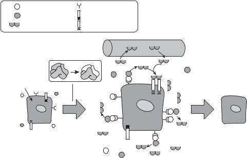
shown to stimulate hepatocyte proliferation. Several other factors, such as insulin, insulinlike growth factors, vasopressin, angiotensin, norepinephrine, and glucagon, can enhance the activity of the growth factors listed above, thus enhancing hepatocyte proliferation. Among these factors, HGF is one of the most potent growth factors that stimulates hepatocyte proliferation and inhibits hepatocyte apoptosis (Fig. 17.1). In addition, HGF exerts inhibitory effects on the activation of Ito cells, the expression of procollagen genes, and the expression of TGFβ, and thus suppressing hepatic fibrogenesis and the progression of cirrhosis. These observations suggest that the HGF gene is a potential therapeutic candidate gene for the treatment of chronic hepatitis and cirrhosis. Experimental investigations have shown that the transfer of the HGF gene into the liver results in an increase in the expression of HGF. The expression of HGF is associated with a number of changes, including (1) tyrosine phosphorylation of the HGF receptor, (2) an elevation in the expression of proliferative cellular nuclear antigen (PCNA), (3) enhancement of hepatocyte proliferation, (4) facilitation of angiogenesis, (5) a decrease in the expression of TGF-β,
(6) an reduction in hepatocyte apoptosis; and (7) improvement of hepatic structure and function.
Another hepatic growth promoter is hepatopoietin or augmenter of liver regeneration (ALR), which is a 30-kDa homodimeric protein (see Table 17.1 for characteristics of
uPA |
uPA receptor |
Pro-HGF |
c-Met |
Mature HGF |
Activated c-Met |
Blood vessel
PHx
Hepatocyte
G0 |
G1/S |
Intact |
|
extracellular |
Matrix |
matrix |
degradation |
Figure 17.1. Proposed model for the role of HGF in liver regeneration. Rapid upregulation of the uPA receptor leads to activation of uPA within 5 min after PHx. This initiates a protease cascade causing degradation of the scant extracellular matrix surrounding hepatocytes and releasing, among others, matrix-bound inactive pro-HGF. uPA activates pro-HGF into the mature active form. Active HGF is released in the blood and stimulates hepatocyte DNA synthesis by an endocrine or paracrine mechanism by binding to the c-Met receptor. (Reprinted with permission from Michalopoulos GK, DeFrances MC; Liver regeneration, Science 276:60–6, copyright 1997 AAAS.)
