

Bioregenerative Engineering Principles and Applications - Shu Q. Liu
..pdfHEPATIC DISORDERS |
777 |
hepatopoietin). This protein has been shown to stimulate hepatocyte proliferation and protect liver cells from injury and apoptosis. The transfer of the hepatopoietin gene into animal model of cirrhosis results in a reduction in the progression of hepatic fibrosis and improvement of hepatic structure and function. Genes that encode other hepatic growth promoters can also be used for therapeutic purposes.
Suppression of Inflammatory Reactions [17.7]. Inflammatory reactions in chronic hepatitis and cirrhosis often induce fibrogenesis, which prevents hepatocyte regeneration and deteriorates the function of the liver. Thus, one of the strategies for the treatment of chronic hepatitis and cirrhosis is to suppress inflammation. Interleukin (IL)10 is known as an anti-inflammatory cytokine, which suppresses the activity of proinflammatory cytokines. This factor is produced in several types of cells, including lymphocytes, monocytes, and macrophages (see page 634 for the characteristics of IL10). Genetically induced deficiency of IL10 is associated with enhanced hepatic fibrosis and monocyte infiltration in animal models. The transfer of IL10 gene into the liver cells induces a reduction in the activity of the collagen gene promoter, leukocyte infiltration, and fibrosis in experimental models of liver cirrhosis. Thus, the IL10 gene can be considered a potential gene for the treatment of human chronic hepatitis and cirrhosis.
Inhibition of Fibrosis [17.8]. Hepatic fibrosis is often promoted by certain factors. Transforming growth factor (TGF)β is one of such factors. TGFβ stimulates the transformation of Ito cells to fibroblast-like cells, which produce excessive extracellular matrix components, including collagen and fibronectin, and contribute to hepatic fibrosis. Thus, the blockade of the TGFβ signal transduction pathway may exert an inhibitory effect on hepatic fibrosis. One approach to reduce the activity of TGFβ is to modify the structure of the TGFβ receptor. Experimental investigations have demonstrated that the transfer of a truncated dominant-negative TGFβ receptor gene into the liver of experimental cirrhosis results in the inhibition of TGFβ activity, which is associated with a reduction in the production of collagen and fibronectin, monocyte infiltration, activity of the Ito and Küpffer cells, and hepatic fibrosis (Fig. 17.2). The hepatic function is improved accordingly. The truncated TGFβ receptor can compete for the TGFβ ligands with the wild-type TGFβ receptor present in the cells, but cannot transmit the TGFβ signal to the intracellular signaling pathways, thus reducing the effect of TGFβ. A large quantity of the truncated TGFβ receptor gene is usually required to achieve therapeutic effectiveness.
Enhancing the Activity of Telomerase [17.9]. Telomerase is a complex enzyme composed of RNA and two protein subunits and is responsible for the maintenance of telomere integrity and function. Telomere is a cap structure for eukaryotic chromosomes, and consists of a TTAGGG-rich DNA sequence and catalytic protein enzymes. This structure plays a critical role in the maintenance of the stability and function of chromosomes and in the regulation of DNA synthesis and cell mitosis. Hepatic cirrhosis is associated with reduced length and altered function of telomere, which is thought to contribute to the progression of hepatic cell apoptosis and fibrosis. These changes are attributed to alterations in the activity of the telomerase. Thus, the enhancement of the telomerase activity by telomerase gene transfer into the liver may improve the integrity of telomere and reduce hepatocyte apoptosis and hepatic fibrosis. Experimental investigations have provided evidence that supports such a possibility.
778 LIVER REGENERATIVE ENGINEERING
A
B
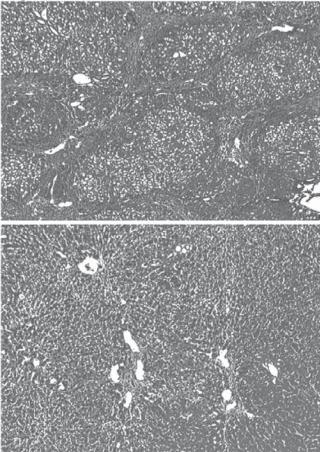
Figure 17.2. Histological micrographs of the rat liver treated with dimethylnitrosamine (DMN), a substance causing persistent liver fibrosis, and a dominant-negative type II TGFβ receptor gene. Rats were infused once via the portal vein with saline (A), or adenoviruses containing a dominantnegative type II TGFβ receptor gene (AdCAT βTR) (B). Both groups of rats were then treated with DMN for 3 weeks. Liver sections were examined histologically by Masson trichrome staining (×200). Note the formation of fibrotic structure in saline-treated liver, but not in the liver transfected with the dominant-negative type II TGFβ receptor gene. (Reprinted with permission from Qi Z et al: Proc Natl Acad Sci USA 96:2345–9, copyright 1999 National Academy of Science USA.)
Cell and Tissue Regenerative Engineering [17.10]. The goal of hepatic cell and tissue regenerative engineering is to replace malfunctioned hepatocytes or augment the function of an injured liver by using an engineered liver construct containing necessary liver cells. In principle, a liver construct can be established by assembling liver cells into a liver scaffold, which provides an appropriate environment for liver cell survival, proliferation, and differentiation. Essential criteria are that a liver construct should be implantable and able to conduct essential liver functions. There are a number of issues that should be taken into account for liver reconstruction. These include (1) selection, culture, and manipulation of liver cells; (2) fabrication of liver scaffolds; (3) maintenance of cell viability and
HEPATIC DISORDERS |
779 |
functions; (4) implantation of liver constructs; and (5) test of liver functions. These issues are discussed as follows.
Selection, Culture, and Manipulation of Liver Cells [17.11]. A critical issue for liver reconstruction is the selection of cell types. Ideally, a liver construct should contain all necessary hepatic cells. The liver construct should be assembled into the form of the natural liver. However, it is difficult to construct a realistic liver with available technologies. The current liver constructs are mostly extracellular matrixor polymer-based scaffolds containing selected liver or stem cells. Several types of liver cells have been used for such a purpose, including adult hepatocytes, genetically modulated hepatocytes, and hepatoma cells. Stem cells and hepatic progenitor cells derived from the embryo, fetus, and adult bone marrow are also candidates for liver regeneration.
Hepatocytes are the primary choice for liver regeneration since these cells can proliferate rapidly and conduct necessary functions immediately following implantation. Healthy autogenous hepatocytes from the host patient are ideal candidates for liver construction. Such cells do not induce acute immune rejection, which is the most serious problem in cell and tissue transplantation. However, patients who need liver reconstruction may not possess sufficient functional hepatocytes. In such a case, hepatocytes from a close relative may be considered. Other choices of hepatocyte sources may include allogenic and xenogenic livers. Allogenic cells are those collected from different individuals of the same species. Xenogenic cells are from different species. Hepatocytes from pigs are often used for constructing artificial livers in experimental models. Obviously, allogenic and xenogeneic cells induce acute immune rejection responses. Transplanted hepatocytes will be attacked by host immune cells, resulting in cell apoptosis and rejection. Immune suppressor agents should always be used to prevent immune reactions. Since hepatocytes have a high capacity of regeneration, a small biopsy sample of hepatocytes may generate a sufficient number of cells within relatively short period.
Genetically modified immortal hepatic cell lines have been used for liver reconstruction in experimental models. A major feature of these cell lines is that cells are immortalized and can survive in an engineering system, which is difficult to achieve by using primary hepatocytes or stem cells. Immortalized cell lines can be established by viral transformation. For instance, the introduction of simian virus 40 into cultured hepatocytes can transform the cells into an immortal form, which still exhibits certain characteristics of the hepatocytes, such as the generation of albumin and process of bilirubin. Another approach for cell immortalization is to coculture and transform hepatocytes with a different cell type from a different species. An example is the transformation of human hepatocytes by coculturing with the rat liver epithelial cells. The transformed hepatocytes exhibit not only immortal properties, but also certain hepatic characteristics such as the generation of albumin and α-fetoprotein. Hepatoma cell lines may also be considered for the construction of an artificial liver, since these cells are able to survive and keep certain hepatic characteristics. An example is the human hepatoblastoma C3A cell line. Experimental studies have demonstrated that this type of cells can survive in animal transplantation models for a longer time compared to primary cell lines and can produce hepatic proteins. Overall, cell lines are potential cell candidates for liver reconstruction. However, there is a risk of introducing cancers to the host system. In addition, the transformed cells may lose hepatic functions. Further investigations are necessary to clarify these issues.
Stem and progenitor cells derived from the embryo, fetus, and adult bone marrow are potential sources for regenerating functional liver cells, repairing an injured liver, and
780 LIVER REGENERATIVE ENGINEERING
reconstructing a malfunctioned liver. Embryonic stem cells are capable of differentiating to all specified cell types. Under an appropriate condition, these cells can differentiate to liver cells and thus can be used for liver reconstruction. Fetal stem and progenitor cells can also be used to regenerate liver cells and reconstruct malfunctioned liver. However, the use of embryonic and fetal cells remains an ethically debating issue, which will likely last for a long time. Alternatively, the adult bone marrow stem and progenitor cells can be used to regenerate liver cells. Several investigations have demonstrated that bone marrow cells can transform to liver cells when the bone marrow cells are delivered to the liver (Fig. 17.3). While the mechanisms of cell transformation remains a research topic, the fusion of bone marrow cells into liver cells has been considered a potential mechanisms for bone marrow cell-based liver regeneration.
Fabrication of Liver Scaffolds and Maintenance of Cell Viability and Function [17.12]. To construct an artificial liver, it is necessary to assemble liver cells in a scaffold. Biological extracellular matrix and synthetic polymers have been used for the construction of such a scaffold. Extracellular matrix components, such as various types of collagens, are natural polymeric materials, which serve as cell substrates and participate in the regulation of cell activities, including cell adhesion, proliferation, and migration. Collagen matrix has been used extensively for the construction of tissue scaffolds. Biodegradable polymers have also been synthesized and used for such a purpose. A unique feature for this type of material is that the scaffold can be gradually degraded in the host system and replaced with cells and natural extracellular matrix.
Several issues should be considered for the construction of hepatic scaffolds:
1.The selected material should be compatible with seeded cells and should not influence the survival and function of the cells. When synthetic polymers are used, biological molecules can be used to coat the surface to which cells attach. A polymeric material should be always tested for toxic effects before being used for constructing tissue scaffolds.
2.A scaffold should be constructed with an appropriate form and structure, factors that may influence the cell function and performance.
3.It is necessary to establish a circulatory system, which introduces blood to the cells seeded in a tissue scaffold.
4.Transplanted cells are subject to an environment that is not natural in a reconstructed liver. Cell apoptosis often occurs within a short period of cell transplantation. It is always a challenge to maintain cell viability when cells are transplanted into a host system in vivo. Thus, cell survival stimulators, such as hepatic growth factor and insulin or their genes, should be applied to liver constructs. These factors play an important role in regulating the survival and proliferation of transplanted liver cells.
Various forms of liver constructs have been established in experimental models. These include scaffolds based on extracellular matrix and polymeric materials, hollow fibers, and microcarriers. These constructs can be used to encapsulate liver cells for transplantation. Experimental investigations and clinical trials have demonstrated the feasibility of using the liver constructs for the treatment of liver failure. It should be noted that all forms of liver constructs established to date are relatively small with respect to the natural liver
HEPATIC DISORDERS |
781 |
A B
C D E
F G
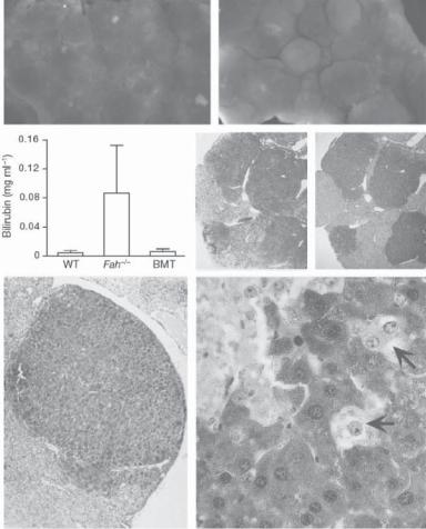
Figure 17.3. Formation of functional fumarylacetoacetate hydrolase-positive (Fah+) liver nodules in Fah−/− mice by transplantation of Fah+/+ wildtype bone marrow cells. (A, B) Gross liver specimens from transplant recipients showing embedded (A) and protruding (B) nodules photographed on a dissecting microscope. (C) Total serum bilirubin levels (mean +/−s.d., n >= 4) from wildtype (WT) mice, Fah−/− mice maintained without NTBC for >4 weeks, and Fah−/− mice after wildtype bone marrow transplantation (BMT). (D, E) Serial liver sections from transplant recipients stained with haematoxylin and eosin (D) or with an anti-Fah antibody to stain expressing cells brown (E– G). Images were photographed with ×2.5 (D, E), ×10 (F), or ×40 (G) objectives. Arrows indicate the locations of nonexpressing cells at the edge of an Fah+ nodule. These observations show that transplanted Fah+/+ wildtype bone marrow cells can engraft to the liver and differentiate into hepatocytes with fah function in transgenic Fah−/− mice. (Reprinted by permission from Macmillan Publishers Ltd.: Vassilopoulos G, Wang PR, Russell DW: Nature 422:901–4, copyright 2003.)
because of difficulties in the construction of a vascular system. These models are briefly discussed as follows.
For the scaffold model, hepatocytes can be seeded in an extracellular matrix or polymeric scaffold, which provides a substrate for cell attachment and assembly. The cellseeded scaffold can be then enclosed within a semipermeable membrane system
782 LIVER REGENERATIVE ENGINEERING
(Fig. 17.4). Several types of material, including polysaccharide hydrogels, hydroxyethyl methacrylate-methyl methacrylate matrix, calcium alginate, and collagen matrix, have been used for constructing liver scaffolds. The semipermeable membrane can be constructed with polymeric materials, such as cellulose and polysulfone. This membrane separates the enclosed hepatocytes from the host tissue and thus prevents leukocyte infiltration and immune rejection responses, when allogenic or xenogenic cells are used. At the same time, the semipermeable membrane allows the release of proteins produced by the enclosed liver cells from the scaffold to the surrounding tissue. The semipermeable membrane can also restrain the transplanted cells within the scaffold, preventing potentially harmful effects, such as carcinogenesis when immortal cells are used.
A liver construct can be implanted into the abdominal cavity of the host. Ideally, the liver construct should be connected to the host circulatory system so that liver-produced proteins can be release into the blood (see next section). However, it is often difficult to establish blood circulation in an artificial liver. Alternatively, multiple small liver scaffolds can be constructed and implanted into the abdominal cavity without connecting to the host circulatory system. When the scaffolds are sufficient small, oxygen and nutrients can diffuse to the cells seeded in the scaffolds.
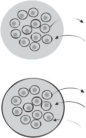
A. Matrix-based implant
Soluble factors
Oxygen & nutrients
T- and B-cells
B. Encapsulated implant
Soluble factors
Oxygen & nutrients
X
T- and B-cells
Figure 17.4. Types of implantable devices with hepatocytes. (A) Cells are in an open matrix, which is often biodegradable. Surrounding tissue, including blood vessels, can grow into the implanted matrix. This provides no protection from the immune system from the host. (B) Cells are encapsulated so that they are protected by a barrier that prevents immune cells and factors from reaching the cells while allowing small (usually <50 kDa) metabolites to transport from the capsule to surrounding tissue. Metabolite transport to and from the nearest vascular bed is chiefly by diffusion and may be adversely affected by the presence of a fibrotic layer, which often develops around such implants. (Reprinted from Chan C et al: Hepatic tissue engineering for adjunct and temporary liver support: Critical technologies, Liver Transplant 10:1331–42, copyright 2004 by permission of John Wiley & Sons, Inc.)
HEPATIC DISORDERS |
783 |
For the microcarrier model, polymeric materials can be used to construct bead-like carriers. The carriers can be coated with extracellular matrix molecules, such as collagen and fibronectin, which enhance cell attachment and survival. Hepatocytes can be collected and cultured on the carrier beads. When cells reach confluence, the carriers can be implanted into the abdominal cavity. In this model, the transplanted cells are directly exposed to the host system. Immune rejection reactions will occur when allogenic and xenogeneic cells are used.
For the hollow fiber model, polymeric hollow fibers can be filled with a gel of extracellular matrix component, such as collagen, mixed with hepatocytes (Fig. 17.5). The collagen gel serves as a matrix for the attachment and assembly of the seeded hepatocytes.
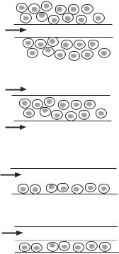
A
Oxygen
Nutrients
B
Oxygen
Nutrients
Oxygen
Nutrients
C
Oxygen
Nutrients
D
Oxygen |
|
Nutrients |
Membrane |
Figure 17.5. Common bioreactor designs for bioartificial livers. (A) Hepatocyte aggregates on microcarriers are placed on the outside of hollow fibers. Oxygenated plasma is flown through the hollow fibers. (B) Hepatocyte aggregates in a supporting matrix are inside hollow fibers and oxygenated plasma is flown outside the hollow fibers. (C) Similar to panel A, although separate hollow fibers are used to deliver hepatocyte culture medium, plasma, and oxygen into the system. Circle with O2 is a hollow fiber perpendicular to the plane of the paper. (D) Hepatocyte aggregates are in a supporting matrix next to hollow fibers that deliver oxygen. Oxygenated plasma is flown in the space outside of the hollow fibers and percolates through the matrix–hepatocyte network. (E) Hepatocytes are seeded as a monolayer on the bottom surface of a flat plate and placed within a parallel-plate flow chamber. Oxygenated plasma is flown directly above the cells. (F) System is similar to that shown in panel E, except that oxygen is delivered through a permeable membrane directly above the flow channel with the hepatocytes. (Reprinted from Chan C et al: Hepatic tissue engineering for adjunct and temporary liver support: Critical technologies, Liver Transplant 10:1331–42, copyright 2004 by permission of John Wiley & Sons, Inc.)
784 LIVER REGENERATIVE ENGINEERING
When the fibers are small, oxygen and nutrients can easily diffuse to the enclosed hepatocytes. By using porous polymeric materials with an appropriate pore size, the fiber wall can prevent the invasion of host immune cells and immune rejection responses. Multiple hollow fibers can be grouped and assembled into a large tubular device, which can be used for implantation. Host blood can be introduced to the interfiber spaces via vascular anastomoses (see next section), ensuring sufficient oxygen and nutrient supplies. Alternatively, hepatocytes can be cultured on the exterior surface of semipermeable hollow fibers. Multiple fibers can be assembled within a larger tubular device. Upon implantation, host blood can be introduced into the lumens of the hollow fibers via vascular anastomoses. The hollow fiber model gives a large surface area for molecular diffusion, ensuring efficient release of proteins produced by transplanted hepatocytes. The hollow fiber device can be implanted into the abdominal cavity of the host. Experimental studies have demonstrated the feasibility and usefulness of this model.
Implantation of Liver Constructs [17.13]. Various methods can be used to implant liver constructs, depending on the form of the construct. Microcarriers and capsules can be directly implanted into the abdominal cavity. Since these devices are small, oxygen and nutrients can diffuse from the abdominal serous fluid into the device. For large devices that require blood supply, such as the hollow fiber liver construct, an artery and a vein should be selected and anastomosed to the blood circulatory system of the liver construct. It is important to note that the artery and vein selected for such a purpose should be rich in collateral circulation, so that the use of the blood vessels for the liver construct does not influence blood supply to the distal tissues of the host system. In the abdominal cavity, the small and large intestines are supplied by the superior and inferior mesenteric arteries, which are connected by collateral arteries through the intestinal system. There are also collateral blood vessels for the mesenteric veins. The blockade of the inferior mesenteric artery and vein does not significantly influence the blood supply to the intestines. Thus, the inferior mesenteric artery and vein can be used as a blood supplying system for an implanted liver construct. A common problem for anastomoses with a nonvascular structure is blood coagulation within the liver construct as well as thrombosis and intimal hyperplasia within the anastomotic blood vessels. These pathological changes often result in obstruction of the blood circulation within the implanted liver construct. A persistent administration of anticoagulants and anti-proliferative agents is necessary for preventing thrombosis and intimal hyperplasia.
Testing Liver Function [17.13]. It is important to test the function and durability of the implanted liver construct. There are a number of parameters that are used for testing the liver function. These include the blood concentration of albumin, aminotransferases (aspartate aminotransferases and alanine aminotransferases), clotting factors, and ammonia. Albumin is produced by hepatocytes and its blood concentration is a useful index for the assessment of the liver function. The normal serum level of albumin is 3.5– 5 g/dL. A significant decrease in the albumin concentration compared to normal controls suggests insufficient function or malfunction of the liver construct. Aspartate aminotransferases and alanine aminotransferases are two enzymes that catalyze the transfer of the γ-amino group from aspartate and alanine to the γ-keto group of ketoglutarate, forming oxaloacetic acid and pyruvic acid. The normal blood level of these enzymes is about 40 IU. Under physiological conditions, these enzymes are degraded in the liver. An increase in the blood level of these enzymes suggests insufficient hepatic function. The liver
HEPATIC DISORDERS |
785 |
synthesizes several blood coagulation factors, including coagulation factor I (fibrinogen), II (prothrombin), V, VII, IX, and X. A reduction in the blood concentrations of these factors indicates insufficient hepatic function. Ammonia is a waste product generated by protein metabolism and is transformed into urea in the liver. An elevation in the blood concentration of ammonia strongly suggests deficiency of the hepatic function. Thus, these proteins and substances can be measured and used for assessing the function of an implanted liver construct.
Chronic Hepatitis and Cirrhosis
Pathogenesis, Pathology, and Clinical Features [17.3]. Chronic hepatitis is a disorder caused by various pathogens, including hepatitis viruses and chemical toxins, and characterized by continuous inflammatory reactions, hepatocyte necrosis, and fibrosis in the liver. Persistent pathological changes may eventually lead to cirrhosis and liver failure. About 30% of patients with chronic hepatitis have a history of hepatitis B infection. These patients often exhibit positive hepatitis B antigens in their serum. The clinical manifestations of chronic hepatitis vary from mild asymptomatic illness to liver failure. The mechanisms for such a wide range of changes remain poorly understood.
The pathogenesis of chronic hepatitis has been hypothetically related to immune responses in the liver. In many cases, hepatic lesions are associated with T-cell infiltration and activation. Antibodies against host components have been detected in some patients. Other types of autoimmune disorders, such as diabetes and thyroiditis, can be found in some patients with chronic hepatitis. The administration of corticosteroids, which are used to treat autoimmune disorders, is effective for the treatment of chronic hepatitis. These observations support the hypothesis that chronic hepatitis may be induced by autoimmune reactions in the liver.
Chronic hepatitis is usually diagnosed on the basis of biopsy examinations. Typical pathological changes include inflammatory reactions characterized by the presence of dense mononuclear cells, hepatocyte necrosis in peripheral regions of liver lobules, excessive formation of fibrous extracellular matrix, and regeneration of hepatic lobules. A large fraction of patients (up to 50%) are associated with cirrhosis (see next paragraph for details). These pathological changes usually develop within 1–2 years.
Cirrhosis is a hepatic disorder characterized by massive fibrosis, structural distortion, formation of regenerative nodules, and deterioration in the function of the liver. When most hepatocytes are replaced with fibrous tissue, the liver loses its function and dies. A number of factors contribute to the development of cirrhosis. These include alcohol toxicity, hepatitis viral infection, obstruction of biliary ducts, and right heart failure. These factors induce inflammatory reactions in the liver in association with excessive production of extracellular matrix components. Several types of liver cells, including the Kupffer cells, Ito cells, and natural killer cells, can release various cytokines and inflammatory mediators, which may further enhance inflammatory reactions. A typical inflammatory mediator is transforming growth factor β (TGFβ), which stimulates hepatic fibrogenesis and accelerates cirrhosis. Here, the role of alcohol toxicity, hepatitis viral infection, obstruction of biliary ducts, and right heart failure in regulating the development of cirrhosis is briefly discussed.
Alcohol toxicity is a common cause of cirrhosis. Long-term exposure to alcohol induces various changes in the liver. In certain cases, extensive fatty acid deposition occurs in hepatocytes due to the impairment of fatty acid oxidation, leading to fat accumulation in