

Bioregenerative Engineering Principles and Applications - Shu Q. Liu
..pdf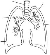
738 PULMONARY REGENERATIVE ENGINEERING
in response to the stimulation of inhaled particles. The ciliated epithelial cells are characterized by the presence of cilia at the cell surface. The cilia conduct periodic movements and are responsible for propelling and removing mucus and inhaled particles. In the middle layer, there is a series of C-shaped cartilage rings which alternate with connective/muscular tissue. The cartilage appears in the anterior and side wall, but not in the posterior wall of the trachea. The cartilage structures protect the trachea from collapsing.
The trachea is divided into the left and right bronchi, which enter the left and right lungs, respectively. The structure and cellular components of the left and right bronchi are similar to those of the trachea. Each bronchus is further divided into about 19 generations of bronchi with a graded decrease in diameter. The smallest airways are defined as bronchioles and the last generation is called terminal bronchioles. There are a large number of terminal bronchioles in the lung. The function of the bronchial tree is to conduct gases into and out of the lung. The structure of the small bronchi is different from that of the trachea and major bronchi. A major difference is that bronchi after the second generation do not contain cartilage rings. In addition, the density of goblet and ciliated epithelial cells reduces gradually as the airway diameter decreases. All bronchi contain smooth muscle cells, which control the diameter of the airways by contraction and relaxation.
The terminal bronchi are connected to the alveolar ducts, a structure mixed with tubular terminal bronchioles and alveoli. Alveoli are clusters of thin-walled, connected membrane sacs with an average size about 200 μm. There are about 300–400 million alveoli in the lung. The primary function of the alveoli is gas exchange between the blood and alveolar air. The wall of each alveolus is composed of a monolayer of alveolar epithelial cells on each side of the wall and a dense network of capillaries constituted with a monolayer of endothelial cells. There are two types of alveolar epithelial cells: type I and II epithelial cells. Type I cells are squamous thin epithelial cells that covers about 90% of the alveolar surface, where gas exchange takes places. Type II cells are specially differentiated epithelial cells which produce and secret surfactant, a mixture of lipids and proteins that spreads over the alveolar surface. The function of the surfactant is to reduce the surface tension of the alveolar wall at the interface between the air and the cell surface, ensuring even expansion and reduction of the alveoli through the entire lung during inspiration and expiration, respectively.
The Vascular System. The vascular system is composed of pulmonary arteries, capillaries, and veins. The pulmonary arteries originate from the right ventricle and are organized into a tree-like tubular system with a graded decrease in vessel diameter. Pulmonary arteries at each generation are arranged together with airways and pulmonary veins of the same generation. These arteries conduct deoxygenated blood from the right ventricle to the alveolar capillaries. A pulmonary capillary is a tube-like structure composed of a monolayer of endothelial cells about 6–10 μm in diameter and a basement membrane around the endothelial cells. The wall of alveoli contains a dense network of capillaries, where gas exchange occurs: oxygen diffuses from the alveolar air into the blood, and carbon dioxide diffuses from the blood to the alveolar air. The pulmonary veins are a tree-like tubular system, composed of multiple generations of veins and organized in parallel to the arterial system. The pulmonary veins conduct oxygenated blood from the capillaries to the left atrium.
The Lymphatic System. The pulmonary lymphatic system is a network of multiple generations of lymphatic vessels distributed around the airways and within the parenchymal tissue. The alveolar wall does not contain lymphatic vessels. The primary function of the lymphatic
ANATOMY AND PHYSIOLOGY OF THE RESPIRATORY SYSTEM |
739 |
system is to collect and remove excessive fluid from the parenchymal tissue. Because of the lack of lymphatic vessels at the alveolar level, the lung is susceptible to pulmonary edema, a disorder with excessive fluid in the parenchymal tissue and alveolar space. Pulmonary edema reduces the rate of gas exchange across the alveolar wall and is often found in patients with left heart failure, which induces an increase in blood pressure in the alveolar capillaries and excessive fluid transport from the blood to the alveolar space.
Pulmonary Function [16.1]
Gas Ventilation and Exchange. Gas ventilation is a cyclic process that includes an inspiration and an expiration phase. During the inspiration phase, fresh air is moved into the lung, whereas during the expiration phase exhaust air is removed from the lung. Gas ventilation is accomplished by coordinated action of a number of pulmonary constituents, including the airways, alveoli, pleural cavity, chest wall, skeletal muscles, and diaphragm. During the inspiration phase, the inward air flow is driven by a pressure gradient from the nasal cavity to the alveoli. The pressure gradient is generated by the contraction of inspiratory skeletal muscles, including the scalenes, pectoralis minor, external intercostals, and diaphragm. The contraction of these muscles increases the volume of the thoracic cavity. Because the pleural cavity is a closed negative system, the expansion of the thoracic cavity induces simultaneous expansion of the lung, resulting in a pressure gradient from the nasal cavity to the alveoli. During the expiration phase, the inspiratory muscles relax and the rib cage recoiled back, resulting in a decrease in the thoracic cavity and the volume of the lung. The volumetric change establishes an adverse pressure gradient from the alveoli to the nasal cavity, which removes the exhaust air from the lung.
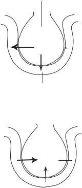
Gas exchange is a process of gas diffusion across the alveolar epithelial and endothelial cells, involving oxygen and carbon dioxide. Oxygen diffuses from the alveolar air space into the blood, whereas carbon dioxide diffuses from the blood to the alveolar space. The driving force for gas diffusion is the gradient of partial gas pressure, which is defined as the pressure contributed by a specified type of gas in a mixture of multiple gases. The total air pressure is about 760 mm Hg at the sea level. The oxygen partial pressure (Po2) is about 155 mm Hg in the air, about 105 mm Hg in the alveolar space, and about 40 mm Hg in the deoxygenated venous blood in the alveolar capillary network (Fig. 16.2). The difference of Po2 is about 65 mm Hg across the alveolar wall. This partial pressure difference drives oxygen diffusion from the alveolar space to the blood. Since oxygen diffusion is very efficient across the alveolar wall and the capillaries are fairly long, the capillary blood can be saturated with oxygen within the first half of the capillary length. Oxygenated blood is conducted to the pulmonary veins, left heart, and the arterial system.
At the same time, carbon dioxide diffuses from the blood to the alveolar space based on a gradient of partial pressure. The partial pressure of CO2 is about 45 mm Hg in the deoxygenated venous blood and about 40 mm Hg in the alveolar space. The difference of carbon dioxide partial pressure across the alveolar wall drives CO2 diffusion from the blood to the alveolar space. During expiration, the exhaust air containing a higher concentration of CO2 is removed from the lung.
Ratio of Air Ventilation to Blood Perfusion. The rate of air ventilation in a region of the lung or in the entire lung is proportional to the rate of blood perfusion within the same region. The ratio of air ventilation to blood perfusion is about 0.75 under physiological conditions. The pulmonary system intends to maintain this ratio within a narrow range around 0.75 under pathological conditions. For instance, when a bronchus is partially
740 |
PULMONARY REGENERATIVE ENGINEERING |
|||||
|
|
|
|
Alveolus |
||
|
Arterial end |
|
|
|
Venous end |
|
|
Po = 40 mm Hg |
|
|
|
Po2 = 104 mm Hg |
|
|
2 |
Po |
2 |
= 104 mm Hg |
||
|
|
|||||
|
|
|
|
|
|
|
|
|
|
|
O2 |
||
|
|
Pulmonary capillary |
||||
|
|
|
|
Alveolus |
||
|
Arterial end |
|
|
|
Venous end |
|
|
|
|
||||
|
Pco |
= 45 mm Hg |
|
|
|
Pco2= 40 mm Hg |
|
2 |
Pco2= 40 mm Hg |
||||
|
|
|||||
|
|
|
|
CO2 |
||
|
|
|
||||
|
|
|
|
|||
|
|
Pulmonary capillary |
||||
Figure 16.2. Schematic representation of the structure of the airways and alveoli. Based on bibliography 16.1.
blocked with a tumor, the rate of air ventilation is accordingly reduced in the alveolar system distal to the blocked bronchus, resulting in a reduction in the ventilation-to-perfu- sion ratio. Reduced oxygen or hypoxia in the under-ventilated region enhances the contractility of the arterial smooth muscle cells and induces arterial constriction, reducing the rate of blood perfusion into the region. Such a response brings up the ventilation-to- perfusion ratio toward the physiological level. With such a mechanism, the ventilation-to- perfusion ratio is maintained at a relatively constant range. The physiological significance of such a mechanism is to redistribute blood volume to well-ventilated regions and to ensure a sufficient volume of fully oxygenated blood.
Control of Gas Ventilation. There are two types of respiratory movement: involuntary and voluntary. The involuntary movement is rhythmic and responsible for the maintenance of the basal level of air ventilation. Such a movement is controlled by the respiratory center located in the medulla oblongata of the brainstem. In the case of increased physical exercise, which demands more oxygen in the skeletal muscle system and the heart, the respiratory center increases the contraction frequency and depth of the respiratory muscles, resulting in an increase in air ventilation and oxygen supply to the alveoli. At the same time, the heart beating rate and cardiac contractility are also increased under the influence of activated cardiovascular control center, resulting in an increase in blood supply to the lung. The increased blood supply can be fully oxygenated because of enhanced air
742 PULMONARY REGENERATIVE ENGINEERING
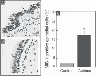
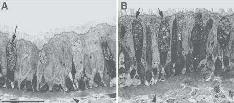
Figure 16.4. Airway cell proliferation in control and asthma. Cell proliferation was detected by an antibody (MIB1) directed against the Ki-67 antigen, a marker for proliferating cells. The black color indicates positive MIB1 staining in the asthmatic epithelial cells (A) and healthy controls (B).
(C) The graph shows MIB1-positive cells (mean ± SD) of three healthy controls and four asthmatics. Note that some fields in asthmatic airways show more than 80% MIB1-positive cells. Arrows show positive cells. (Reprinted with permission from Comhair SA et al: Superoxide dismutase inactivation in pathophysiology of asthmatic airway remodeling and reactivity, Am J Pathol 166:663–74, copyright 2005.)
proliferation (Fig. 16.5), and persistently increased responsiveness and contractility of airway smooth muscle cells in response to the stimulation of allergens, environmental factors, and pharmacological agents. Typical asthmatic allergens include air-borne particles derived from plants (e.g., grass pollens, ragweed pollens, birch pollens, mountain cedar pollens, and peanuts), animals (cat and dog fur dusts), and microorganisms (bacteria and viruses). Examples of environmental factors include air pollutants, chemicals, metal particles, dusts, and polymer particles. Pharmacological agents that cause asthma include antibiotics, aspirin, and β-adrenergic antagonists. In addition, viral infection of the lung, physical exercise, and emotional stress can initiate asthmatic attacks. All these factors can cause an increase in the reactivity of the airway smooth muscle cells, resulting in airway constriction and a reduction in the rate of ventilation. Asthmatic attacks are usually transient, lasting for a period from minutes to hours. The clinical consequence of asthma is dependent on the degree of airway constriction and oxygen deficiency. Severe airway constriction is often life threatening. Asthma occurs frequently in children and young adults. Asthmatic patients often have a family history of allergic diseases.
The pathogenic mechanisms of asthma vary, depending on the factors that cause asthma. Allergic asthma, one of the most common types, is related to enhanced immune
PULMONARY DISORDERS |
743 |
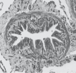
Figure 16.5. Inflammatory changes in asthmatic airways in rhesus monkeys. Histopathological comparison of epithelial morphology in the intrapulmonary bronchi of sensitized (B) and nonsensitized (A) rhesus monkeys with house dust mite (Dermatophagoides farinae). Note that the asthmatic airway was associated with inflammatory cell infiltration, epithelial hypertrophy, and mucous goblet cell hyperplasia (arrows). Scale bar: 20 μm. (Reprinted with permission from Schelegle ES et al: Allergic asthma induced in rhesus monkeys by house dust mite (Dermatophagoides farinae), Am J Pathol 158:333–41, copyright 2001.)
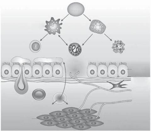
responses of lymphocytes exposed to allergens. In patients genetically susceptible to asthma, a primary exposure to an allergen in the airway system leads to the activation of the antigen-presenting dentritic cells (Fig. 16.6). These cells present the inhaled antigen to CD4+ T-helper cell precursors in the bronchial lymph nodes, potentially inducing the differentiation of the precursor cells into two types of cells: type 1 and type 2 T-helper cells. The fate of the differentiation is dependent on the presence of dominant cytokines. When interleukin (IL)12 is dominant, the precursor cells are induced to differentiate into type 1 T-helper cells. In contrast, in the presence of dominant IL4, the precursor cells are differentiated into type 2 T-helper cells. The type 1 T-helper cells can produce interferon- β, IL2, and Tumor necrosis factor (TNF)α. These factors exert inhibitory effects on asthmatic activities. In contrast, the type 2 T-helper cells produce IL4, IL5, and IL9, which promote asthmatic activities.
The type 2 T-helper cell cytokines can stimulate B lymphocytes to produce IgE antibodies against the allergen. These antibodies can attach to the surface of mast cells. A secondary exposure to the same allergen induces reaction of the allergen with the IgE antibodies attached to the mast cells, resulting in antibody activation. The activated antibodies in turn stimulate mast cells to release inflammatory factors, such as histamine, prostaglandins, and bradykinin, causing the contraction of airway smooth muscle cells. These inflammatory factors also cause mucosal edema, an increase in mucus secretion, and infiltration of leukocytes, especially eosinophils. IgE and allergens can bind to eosinophils, inducing the release of major basic protein (MBP) from the eosinophils. MBP can cause airway injury and inflammation. All these pathological changes contribute to the reduction in the luminal area of the bronchi. As asthma attacks continue, chronic inflammatory reactions may occur, inducing persistent leukocyte infiltration, mucosal thickening, and airway constriction.
744 |
PULMONARY REGENERATIVE ENGINEERING |
|
|
|
|
Allergen |
|
|
Macrophage/ |
|
|
|
dendritic cell |
|
Mast cell |
|
|
Eosinophil |
|
|
TH2 |
|
Neutrophil |
|
Mucus plug |
|
Epithelial shedding |
|
|
|
|
|
|
Nerve |
|
|
|
activation |
Airway |
|
|
|
|
|
|
|
epithelium |
|
|
|
Subepithelial |
|
|
|
fibrosis |
|
|
|
Fibroblast |
|
|
|
Sensory nerve |
|
Mucus |
|
activation |
|
|
|
|
|
hypersencretion |
Plasma leakage |
|
|
Hyperplasia |
Edema |
Cholinergic |
|
Vasodilatation |
|
reflex |
|
New vessels |
|
|
|
(angiogenesis) |
|
|
|
|
|
Bronchoconstriction |
|
Airway |
|
Hypertrophy/hyperplasia |
|
smooth muscle |
|
|
Subcutaneous layer
Figure 16.6. Pathogenesis of asthma. Several inflammatory cell types=s are recruited to and/or activated in the airways, releasing a variety of inflammatory mediators that have acute effects on the airway, such as bronchoconstriction, plasma leakage, vasodilatation, mucus secretion, sensory nerve activation, and cholinergic reflex-induced bronchoconstriction. These acute changes are followed with structural remodeling, resulting in subepithelial fibrosis, increased numbers of blood vessels and mucus-secreting cells, and increased thickness of airway smooth muscle, and airway hyperplasia and hypertrophy. (Reprinted by permission from Macmillan Publishers Ltd.: Barnes PJ: New drugs for asthma, Nature Revs Drug Discov 3:831–44, copyright 2004.)
Asthma is associated with several pathological changes. At the cellular and tissue level, there often exist epithelial cell detachment, airway mucosal edema, eosinophil infiltration, subepithelial fibrosis, basal lamina thickening, and smooth muscle hypertrophy and hyperplasia (Fig. 16.4). In addition, goblet cell hyperplasia occurs (Fig. 16.5), inducing mucus overproduction and secretion. All these changes contribute to the reduction in the airway diameter. In asthma, the sensory nerve endings are sensitized to a certain extent, contributing to the elevation of the smooth muscle tone. In severe cases, an apparent change is lung overexpansion due to airway obstruction and air retention in the alveoli.
Experimental models of asthma [16.3]. Experimental asthma can be induced by sensitizing rats or mice by administration of allergens. A common allergen used in asthma
PULMONARY DISORDERS |
745 |
induction is ovalbumin. For primary allergen sensitization, a mixture of ovalbumin (10– 100 μg) and aluminum hydroxide 1 mg in 0.2–1 ml saline can be prepared and injected into the peritoneal cavity of an animal three times at day 0, 7, and 14. The animal can be rechallenged by exposure to ovalbumin aerosol later. The presence of asthma can be assessed by measuring pathological changes, such as airway mucosal edema, smooth muscle hypertrophy, epithelial cell detachment, eosinophil infiltration, mucus overproduction, basal lamina thickening, and airway constriction.
Conventional Treatment of Asthma [16.4]. Several approaches have been developed and used to treat asthma, including the removal of the causative factors, immunotherapy, and drug therapy. The removal of the causative factors is an effective approach for the prevention of asthma. However, it is usually difficult to find the cause of asthma. Even though the causative factors are known, it is impossible to remove them completely from the environment.
Allergen immunotherapy is an effective method for the treatment of asthma. The hypothetical basis for immunotherapy is that the introduction of a selected allergen via a parenteral route may activate regulatory T lymphocytes, which inhibit Th2 lymphocytes and thus reduces the production of IgE antibodies by B lymphocytes. These changes lead to reduced activity of the immune system to subsequently inhaled allergens. To carry out immunotherapy, allergens specific to a patient should be identified, prepared, and used for therapeutic purposes.
Several types of drugs have been used to treat asthma. These include bronchodilators and glucocorticoids. Bronchodilators include β-adrenergic agonists and anticholinergic agents. β-adrenergic agonists, such as epinephrine, isoproterenol, and resorcinols, activate the β-adrenergic receptor of the airway smooth muscle cells, inducing airway dilation and relieving the symptoms of asthma. Anticholinergic agents, such as atropine sulfate, atropine methylnitrate, and ipratropium bromide, can be used to suppress acetylcholine, a substance that stimulates airway smooth muscle contraction, and thus to induce airway dilation. Airway inhalation is an effective method for the delivery of these agents. However, most bronchodilators stimulate cardiac activities and should be used with caution for patients with cardiac diseases. Glucocorticoids are hormones produced in the cortex of the adrenal gland and can be used to suppress inflammatory reactions. Since inflammation occurs in asthma and contributes to the obstruction of bronchi, glucocorticoids are often used to reduce inflammation.
Molecular Therapies for Asthma [16.5]. Based on the pathogenic mechanisms of asthma, two molecular therapeutic strategies have been developed: (1) suppressing inflammatory reactions and mucus secretion, and (2) inducing airway dilation. The suppression of airway inflammation can be achieved by the transfection of target cells with antiinflammatory cytokine genes and the glucocorticoid receptor gene, administration with inhibitors to inflammatory transcription factors and kinases, and the blockade of inflammatory cytokines and cytokine receptors (Fig. 16.7). Airway dilation can be achieved by transferring the β2-adrenergic receptor gene. These approaches are described as follows.
Suppression of Asthmatic Changes by Administration of Antiinflammatory Cytokine Genes, Antibodies, and Inhibitors [16.6]. There are several approaches for inhibiting allergic responses and asthmatic changes. These include the transfection of target cells with antiinflammatory cytokines or their genes, administration with antibodies against