

Bioregenerative Engineering Principles and Applications - Shu Q. Liu
..pdf796 LIVER REGENERATIVE ENGINEERING
Jauregui HO, Mullon CJP, Solomon BA: Extracorporeal artificial liver support, in Principles of Tissue Engineering, ed. R Lanza R, Langer W, Landes, Austin, TX, 1997, pp 463–79.
Sielaff TD, Chari RS, Cattral MC: Extracorporeal support of the failing liver, Curr Opin Crit Care 4:125–30, 1998.
Roberts EA, Letarte M, Squire J, Yang SY: Characterization of human hepatocyte lines derived from normal liver tissue, Hepatology 19:1390–99, 1994.
Schumacher IK, Okamoto T, Kim BH, Chowdhury NR, Chowdhury JR et al: Transplantation of conditionally immortalized hepatocytes to treat hepatic encephalopathy, Hepatology 24:337–43, 1996.
Pfeifer AMA, Cole KE, Smoot DT, Weston A, Groopman JD et al: Simian virus 40 large tumor antigen-immortalized normal human liver epithelial cells express hepatocyte characteristics and metabolize chemical carcinogens, Proc Natl Acad Sci USA 90:5123–7, 1993.
Woodworth C, Secott T, Isom H: Transformation of rat hepatocytes by transfection with simian virus 40 DNA to yield proliferating differentiated cells, Cancer Res 46:4018–26, 1986.
Wilson JM, Jefferson DM, Chowdhury R, Movikoff PM, Johnston DE et al: Retrovirus-mediated transduction of adult hepatocytes, Proc Natl Acad Sci USA 85:3014–18, 1988.
Nyberg SL, Remmel RP, Mann HJ, Peshwa MV, Hu WS et al: Primary hepatocytes outperform Hep G2 cells as the source of biotransformation functions in a bioartificial liver, Ann Surg 220:59–67, 1994.
Sussman NL, Chong MG, Koussayer T, He DE, Shang TA et al: Reversal of fulminant hepatic failure using an extracorporeal liver assist device, Hepatology 16:60–5, 1992.
Sussman NL, Gislason GT, Conlin CA, Kelly JH: The hepatic extracorporeal liver assist device: Initial clinical experience, Artif Organs 18:390–6, 1994.
Sussman NL, Kelly JH: Improved liver function following treatment with an extracorporeal liver assist device, Artif Organs 17:27–30, 1993.
Wang X, Montini E, Al-Dhalimy M, Lagasse E, Finegold M et al: Kinetics of liver repopulation after bone marrow transplantation, Am J Pathol 161(2):565–74, 2002.
Alvarez-Dolada M, Pardal R, Garcia-Verdugo JM, Kike JR, Lee HO et al: Fusion of bone marrowderived cells with Purkinje neurons, cardiomyocytes and hepatocytes, Nature 425:968–73, 2003.
Vassilopoulos G, Russell DW: Cell fusion: An alternative to stem cell plasticity and its therapeutic implications, Curr Opin Genet Dev 13:480–5, 2003.
Vassilopoulos G, Wang PR, Russell DW: Transplanted bone marrow regenerates liver by cell fusion, Nature 422:901–4, 2003.
Petersen BE, Bowen WC, Patrene KD, Mars WM, Sullivan AK et al: Bone marrow as a potential source of hepatic oval cells, Science 284:1168–70, 1999.
Lagasse E, Connors H, Al-Dhalimy M, Reitsma M, Dohse M et al: Purified hematopoietic stem cells can differentiate into hepatocytes in vivo, Nat Med 6:1229–34, 2000.
17.12. Fabrication of Liver Scaffolds and Maintenance of Cell Viability and Function
Sauer IM, Obermeyer N, Kardassis D et al: Development of a hybrid liver support system, Ann NY Acad Sci 944:308–19, 2001.
Bhandari RN, Riccalton LA, Lewis AL et al: Liver tissue engineering: a role for co-culture systems in modifying hepatocyte function and viability, Tissue Eng 7:345–57, 2001.
Miyoshi H, Ehashi T, Ema H et al: Long-term culture of fetal liver cells using a three-dimensional porous polymer substrate, ASAIO J 46:397–402, 2000.
Rhim JA, Sandgren EP, Degen JL et al: Replacement of diseased mouse liver by hepatic cell transplantation, Science 263:1149–52, 1994.
BIBLIOGRAPHY 797
Sussman NL, Gislason GT, Conlin CA, Kelly JH: The hepatic extracorporeal liver assist device: Initial clinical experience, Artif Organs 18:390–6, 1994.
Demetriou AA, Rozga J, Podesta L, Lepage E, Morsiani E et al: Early clinical experience with a hybrid bioartificial liver, Scand J Gastroenterol 108:111–7, 1995.
Rozga J, Williams F, Ro MS, Neuzil DF, Giorgio TD et al: Development of a bioartificial liver: Properties and function of a hollow-fiber module inoculated with liver cells, Hepatology 17:258– 65, 1993.
Rozga J, Holzman MD, Ro MS, Griffin DW, Neuzil DF et al: Development of a hybrid bioartificial liver, Ann Surg 217:502–11, 1993.
Wells GD, Fisher MM, Sefton MV: Microencapsulation of viable hepatocytes in HEMA-MMA microcapsules: A preliminary study, Biomaterials 14:615–20, 1993.
Eisa T, Sefton MV: Towards the preparation of a MMA-PEO block copolymer for the microencapsulation of mammalian cells, Biomaterials 14:755–61, 1993.
Koebe HG, Pahernik SA, Thasler WE, Schildberg FW: Porcine hepatocytes for biohybrid artificial liver devices: A comparison of hypothermic storage techniques, Artif Organs 20:1181–90, 1996.
Dixit V, Darvasi R, Arthur M, Lewin K, Gitnick G: Cryopreserved microencapsulated hepatocytes: Transplantation studies in Gunn rats, Transplantation 55:616–22, 1993.
Landry J, Bernier D, Ouellet C, Goyette R, Marceau N: Spheroidal aggregate culture of rat liver cells: histotypic reorganization, biomatrix deposition, and maintenance of functional activities, J Cell Biol 101:914–23, 1985.
Peshwa MV, Wu FJ, Sharp HL, Cerra FB, Hu WS: Mechanistics of formation and ultrastructural evaluation of hepatocyte spheroids, In Vitro Cell Dev Biol 32:197–203, 1996.
Wu FJ, Peshwa MV, Cerra FB, Hu WS: Entrapment of hepatocyte spheroids in a hollow fiber bioreactor as a potential bioartificial liver, Tissue Eng 1:29–40, 1995.
Wu FJ, Friend JR, Mann HJ, Remmel RP, Cerra FB et al: Hollow fiber bioartificial liver utilizing collagen entrapped porcine hepatocyte spheroids, Biotechnol Bioeng 52:34–44, 1996.
Catapano G, Di Lorenzo MC, Della Volpe C, De Bartolo L, Migliaresi C: Polymeric membranes for hybrid liver support devices: the effect of membrane surface wettability on hepatocyte viability and functions, J Biomater Sci Polym Ed 7:1017–27, 1996.
Lin P, Chan WC, Badylak SF, Bhatia SN: Assessing porcine liver-derived biomatrix for hepatic tissue engineering, Tissue Eng 10(7–8):1046–53, 2004.
17.13. Implantation of Liver Constructs and Test of Liver Functions
Rahaman MN, Mao JJ: Stem cell-based composite tissue constructs for regenerative medicine, Biotechnol Bioeng 91(3):261–84, 2005.
Kulig KM, Vacanti JP: Hepatic tissue engineering, Transpl Immunol 12(3–4):303–10, 2004.
Selden C, Hodgson H: Cellular therapies for liver replacement, Transpl Immunol 12(3–4):273–88, 2004.
Allen JW, Bhatia SN: Engineering liver therapies for the future, Tissue Eng 8(5):725–37, 2002.
Balis UJ, Yarmush ML, Toner M: Bioartificial liver process monitoring and control systems with integrated systems capability, Tissue Eng 8(3):483–98, 2002.
Yarmush ML, Toner M, Dunn JC, Rotem A, Hubel A et al: Hepatic tissue engineering. Development of critical technologies, Ann NY Acad Sci 13(665):238–52, 1992.
Bhatia SN, Yarmush ML, Toner M: Controlling cell interactions by micropatterning in co-cultures: Hepatocytes and 3T3 fibroblasts, J Biomed Mater Res 34(2):189–99, 1997.
ANATOMY AND PHYSIOLOGY OF THE GASTROINTESTINAL SYSTEM |
799 |
ANATOMY AND PHYSIOLOGY OF THE GASTROINTESTINAL SYSTEM
Structure [18.1]
The gastrointestinal system includes the esophagus, stomach, small intestine, and large intestine. The primary functions of the gastrointestinal system are food ingestion, digestion, and absorption, as well as waste product elimination. The esophagus is a tubular organ that is extended from the pharynx to the stomach. The function of the esophagus is food transport from the mouth to the stomach. The esophagus is composed of four distinct tissue layers: mucosa, submucosa, muscular layer, and adventitia. The mucosa consists of stratified epithelial cells and a subendothelial connective tissue layer. The submucosa is a connective tissue layer, composed of fibroblasts, extracellular matrix (collagen fibers, elastic fibers, and proteoglycans), blood vessels, nerves, and small glands. The gland cells can secret mucus, which serves as a lubricant for food ingestion. The muscular layer is composed of striated muscular cells in the upper esophagus and smooth muscle cells near the stomach. There are two layers of muscular cells: the inner layer with circumferentially aligned muscular cells and the outer layer with longitudinally aligned muscular cells. In addition, the muscular layer contains extracellular matrix and nerves. The nerves control the movement of the esophagus. The adventitia is a thin layer composed of fibroblasts, extracellular matrix, and a squamous epithelium on the outer surface. The epithelium is also known as visceral peritoneum in gastrointestinal organs.
The stomach is located in the upper left abdominal cavity and is responsible for the primary digestion of ingested foods. As other digestive tracts, the stomach is composed of four layers: mucosa, submucosa, muscular layer, and adventitia. The structure of these layers is similar to that of the esophagus with several exceptions. First, the inner surface of the stomach is lined with a monolayer of columnar epithelial cells, instead of stratified cells. The epithelial cells of the stomach are specially differentiated cells. Some of these cells line the stomach surface and produce mucus. Others form gastric glands, which produce hydrochloric acid, pepsinogen, and mucus. The acidic environment (pH 1–3) helps to digest foods. Pepsinogen is the precursor of the enzyme pepsin, which cleaves proteins into peptides for further digestion. Pepsinogen can be converted into pepsin when it is released into the stomach under the action of hydrochloride acid and pepsin. Second, the muscular layer contains three sublayers of smooth muscle cells: oblique, circumferential, and longitudinal muscular sublayers. The reinforced muscular structure enhances gastric movement and sufficient food digestion. The stomach undergoes constant movements, which help to mix and digest foods.
The small intestine is a tubular organ that is located in the abdominal cavity and is extended from the stomach to the large intestine. The small intestine is composed of three segments: the duodenum, jejunum, and ileum. At the tissue level, the small intestine consists of the four standard layers: the mucosa, submucosa, muscular layer, and adventitia. The mucosa and partial submucosa form numerous folds about 1 mm in length, also known as villi, on the surface of the small intestine. The villi are covered with columnar epithelial cells. These cells form cell membrane projections called microvilli. The formation of villi and microvilli increases the total surface area of the small intestine, which facilitates nutrient absorption. There are epithelial goblet cells and submucosa gland cells. These cells produce mucus. The muscular layer of the small intestine contains circumferential and longitudinal smooth muscle cells. These cells conduct constant movements to move and mix ingested foods. The structure is similar among the duodenum, jejunum, and
800 GASTROINTESTINAL REGENERATIVE ENGINEERING
ileum. The functions of the small intestine are food digestion and absorption. A number of digestive enzymes, including those for digesting proteins, polysaccharides, and lipids, are secreted from the pancreas into the small intestine. Enzymes for protein digestion include trypsin, chymotrypsin, and carboxypeptidase. Polysaccharides are digested by amylases, and lipids are digested by lipases. In addition, the pancreas secrets deoxyribonucleases and ribonucleases, which break down DNA and RNA, respectively.
The large intestine is a digestive tract that is extended from the ileum to the anus. The large intestine is usually divided into several segments, including the colon, rectum, and anal canal. The general structure of the large intestine is similar to that of the other digestive tracts. The large intestine possesses a large number of goblet cells which secret mucus. The primary function of the large intestine is to remove food wastes. Water is absorbed in the large intestine. However, little nutrient absorption takes place in the large intestine. The large intestine undergoes constant movements, which help to move the food remains.
Nutrient Digestion and Absorption [18.1]
The ingested foods contain three major types of nutrient: protein, carbohydrate, and lipid. All these nutrients are digested in the stomach and small intestine, and absorbed in the small intestine. Proteins from ingested meats and plants are first digested in the stomach by pepsin, which cleaves proteins into polypeptides. Remaining proteins and digested polypeptides are moved into the small intestine, where they are further digested into tripeptides, dipeptides, and amino acids by peptidases. Amino acids and short peptides are absorbed into the epithelial cells by an energy-consuming process involving sodium cotransport. The absorbed short peptides are digested into amino acids in the epithelial cells. The amino acids are transported into the blood of the portal vein and used for protein synthesis in different types of cell.
Ingested foods contains polysaccharides (starches and glycogen), disaccharides (sucrose and lactose), and monosaccharides (glucose). Polysaccharides are digested by salivary and pancreatic amylases into disaccharides and monosaccharides in the stomach and small intestine. Disaccharides are further digested into monosaccharides at the epithelial surface. Monosaccharides are absorbed into the epithelial cells of the small intestine by an active transport mechanism involving the co-transport of sodium and released into the intestinal capillaries.
Lipids include cholesterol, triglycerides, phospholipids, and steroids. Ingested lipids are emulsified in the small intestine by bile compounds produced by the liver and are digested by pancreatic lipases. Digested dietary lipids form small particles known as micelles. The lipid molecules diffuse into the epithelial cells according to the gradient across the cell membrane. Within the epithelial cell, lipids form chylomicrons (80–500 nm in diameter) by conjugating with apoproteins. The chylomicrons are transported into the blood and are further digested by bloodborne lipases, releasing chylomicron remnants and free fatty acids (triglycerides). The chylomicron remnants are taken up by hepatocytes in the liver through receptor-mediated endocytosis, digested in the lysosomes of the hepatocytes. Free cholesterol molecules are released from the chylomicron remnants and stored in the hepatocytes as cholesteryl esters. The cholesterol molecules are either excreted into the bile or used to form very-low-density-lipoproteins (VLDL, diameter 30–80 nm) in the hepatocytes. The VLDL molecules are released into the blood and digested by lipoprotein lipases, resulting in the formation of intermediate-density-lipoproteins (IDL) after releas-
GASTROINTESTINAL DISORDERS |
801 |
ing a fraction of fatty acids. The IDL is digested by lipoprotein lipases into low-density- lipoproteins (LDL), which usually circulate in the blood for 1–2 days and constitute the major reserve of plasma cholesterol (60–70% of the total cholesterol pool). The cholesterol molecules of LDL can be taken up by cells and used for the construction of cell membranes. The promotion of cell intake of cholesterol reduces the plasma LDL and cholesterol level, a beneficial process for reducing the risk of atherogenesis. With the release of the majority of cholesterol molecules, LDL is converted to high-density-lipoproteins (HDL), which consist of apoproteins and residual cholesterol molecules and can form complexes with cholesterols and fatty acids, contributing to the clearance of plasma lipids.
GASTROINTESTINAL DISORDERS
Peptic Ulcer
Pathogenesis, Pathology, and Clinical Features [18.2]. Peptic ulcer is a disorder that occurs primarily in the duodenum and stomach and is characterized by the presence of round or oval ulcerative lesions with various sizes. The lesions include injury and detachment of epithelial cells, necrosis and fibrosis of mucosa and submucosa, and infiltration of inflammatory cells. These pathological changes are possibly induced by the corrosive effect of acid and pepsin. Duodenal ulcer is often found in the proximal segment of duodenum near the stomach. About 10% of the human population is affected by duodenal ulcers some times during the lifespan. The incidence of duodenal ulcer is higher than that of gastric ulcer. Duodenal ulcer is a chronic disorder with a high rate of recurrence and is often found in patients about 50 years old. A large fraction of patients experience reoccurring duodenal ulcers within a period of 2–3 years. Gastric ulcer occurs in the stomach and exhibit similar pathological changes as found in the duodenal ulcer. This type of ulcer is often found in patients about 60 years old.
The pathogenesis of duodenal and gastric ulcers is related to several factors:
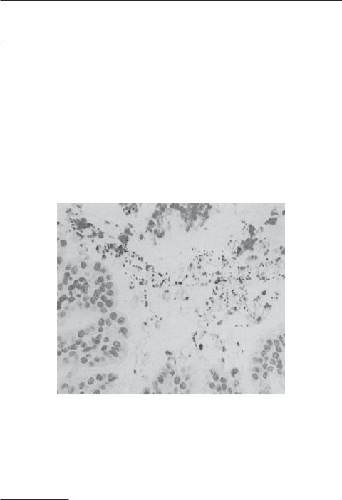
1.A type of bacteria known as Helicobacter pylori (H. pylori) can cause inflammatory reactions and peptic ulcers in the stomach of mammalian animals and humans (Chapter 18 opening figure).
2.Acid and pepsin secreted by the stomach epithelial cells may exert a corrosive effect on the duodenal and gastric epithelial cells. Some ulcer patients are associated with increased secretion of acid and pepsin.
3.A reduction in the mucosal resistance to the corrosion of acid and pepsin contributes to the development of duodenal and gastric ulcers.
4.An increase in the release of gastrin, a molecule that stimulates the secretion of hydrochloric acid in the stomach, is another potential factor that contributes to the development of duodenal and gastric ulcers. Even though the level of gastrin may be normal, the gastric epithelial cells in ulcer patients often exhibit increased responsiveness to gastrin stimulation, enhancing the secretion of hydrochloric acid. Fourth, hereditary factors may also play a role in the development of duodenal and gastric ulcers. Ulcer patients often have a familial history of ulcer. In addition, cigarette smoking is associated with increased incidence of duodenal and gastric ulcer, although smoking does not alter acid secretion. The mechanisms of smoking-related ulcers remain poorly understood.
802 GASTROINTESTINAL REGENERATIVE ENGINEERING
Patients with duodenal and gastric ulcers often experience epigastric burning pain. When a blood vessel is corroded and damaged, hemorrhage may occur. The severity of hemorrhage is dependent on the size of the damaged blood vessel. In general, ulcers do not significantly influence food digestion and absorption, since ulcers are usually small.
Experimental Models of Gastrointestinal Ulcers [18.3]. Gastric and duodenal ulcers can be induced by application of acid and pepsin to the stomach or duodenum of animal models. Here, the duodenum is used to demonstrate the experimental procedures. An animal can be anesthetized by peritoneal injection of sodium pentobarbital at a dose of 50 mg/kg body weight. The upper abdominal skin can be sterilized with 75% alcohol, Betadine (povidone-iodine), and 75% alcohol again. The abdominal cavity can be opened at a location in the upper middle area and the duodenum is identified. A segment of the duodenum can be isolated by applying a pair of intestinal clamps. A mixture of hydrochloric acid and pepsin can be injected into and kept in the duodenum for a desired period. The concentration of acid and pepsin and the treatment time should be determined by conducting a series of experiments with graded concentrations and different treatment times. The clamps can be released and the abdominal wounds closed after the treatment. At scheduled times following the surgery, specimens can be collected from the duodenum and used for pathological examinations.
Conventional Treatment [18.2]. There are several general approaches for the treatment of gastrointestinal ulcers (see Table 18.1 for a list of therapeatic proteins used to treat peptic ulcer). These include the administration of acid-reducing agents, protective coating agents, and diet control. Commonly used acid-reducing agents include antacids, anticholinergic agents, antagonists to histamine H2 receptor, and prostaglandins. The administration of antacid agents, such as sodium bicarbonate and calcium carbonate, results in direct neutralization of the acidic content in the stomach and duodenum, suppressing ulcer formation and progression. Acetylcholine is a neurotransmitter that promotes the production and secretion of gastric acid. A treatment with anticholinergic agents, such as atropine and atropine derivatives, can reduce the secretion of gastric acid. The H2 receptor of histamine mediates histamine-induced secretion of gastric acid. Antagonists of the histamine H2 receptor, such as cimetidine, suppress the activity of the receptor and reduce the secretion of gastric acid. Prostaglandins exert an inhibitory effect on gastric acid secretion in the stomach and enhance the capability of gastric mucosal resistance to acid corrosion. Stomach mucosal coating agents, such as sucralfate (polyaluminum hydroxide salt of sucrose sulfate), can adhere to the mucosal surface and protect the mucosa from acid and pepsin corrosion. In addition, the control of acidic diet intake reduces the progression of gastrointestinal ulcers. In the case of severe hemorrhage, it is necessary to remove the ulcerative tissue via surgery.
Molecular Regenerative Engineering. The principle of molecular engineering or therapy for peptic ulcer is to promote tissue recovery from ulcerative injury. Although the reduction of acid secretion in the stomach is the most important approach for treating peptic ulcer, there are no proteins or genes available for such a purpose. Several genes, including the serum response factor, platelet-derived growth factor, vascular endothelial growth factor, and angiopoietin-1 genes, have been identified and used in animal models for enhancing recovery from peptic ulcer-induced injury. These genes encode mitogenic factors that stimulate cell proliferation and migration, thus enhancing the recovery from

GASTROINTESTINAL DISORDERS |
805 |
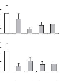
Vascular Endothelial Growth Factor (VEGF) [18.6]. As platelet-derived growth factor, VEGF stimulates cell proliferation and migration (see page 600 for the characteristics of VEGF). This growth factor has been shown to particularly regulate the development, survival, and proliferation of the vascular endothelial cells. In experimental models of peptic ulcer, this growth factor enhances angiogenesis as well as the proliferation and migration gastrointestinal epithelial cells. Thus, the overexpression of the VEGF gene may potentially improve the healing of peptic ulcer (Fig. 18.1).
Angiopoietin-1 [18.7]. Angiopoietin-1 is a protein that regulates angiogenesis (see Table 18.1 for the characteristics of angiopoietin-1). Local delivery of angiopoietin-1 expression vectors results in up-regulation of the angiopoietin-1 gene and an increase in angiogenic activities. Since angiogenesis is required for the regeneration of ulcerative tissue, the overexpression of the angiopoietin-1 gene can enhance ulcer healing.
Gastrointestinal Cancers
Pathogenesis, Pathology, and Clinical Features [18.8]. Gastrointestinal cancers are malignant tumors found in the mucosal, submucosal, and muscular layers of the gastrointestinal tracts. Gastrointestinal cancers are divided into several types, including carcinoma, leiomyosarcoma, and lymphoma based on the cellular origin of the tumor. As cancers in other organs, these cancers are highly metastatic.
Gastrointestinal carcinoma is a form of cancer originated from the mucosal epithelial cells. This form of cancer is more common in men than in women. It is often found in people about 60 years old or older. The etiology of gastrointestinal carcinoma remains poorly understood. Hereditary and dietary factors may play a role in the development of the disease. In addition, the presence of several disorders, such as atrophic gastritis and intestinal metaplasia, may enhance the development of gastrointestinal carcinoma. Pathological examinations usually reveal several forms of carcinoma, including ulcerative, superficial, and infiltrative carcinomas. Ulcerative carcinomas are cancers that appear similar to ulcers at the surface. Superficial carcinomas are those found within the epithelial layer. Infiltrative carcinomas are those that invade the deep layers. At the cellular level, carcinoma cells are characterized by the enlargement of cell nuclei and an increase in cell density. During the end-stage, carcinoma cell metastasis or infiltration into deeper layers can be found. A cell proliferation assay often demonstrates an increase in the rate of cell proliferation.
Gastrointestinal leiomyosarcoma is a form of cancer originated from the submucosal smooth muscle cells. This type of cancer is not as common as gastrointestinal carcinoma and accounts for about 3% of total gastrointestinal cancers. The tumor is usually spherical in shape with necrosis at the center. Gastrointestinal lymphoma is a type of cancer originated from the lymphoid tissue. This type of cancer accounts for about 5% of gastrointestinal cancers. The etiological and pathological features of gastrointestinal leiomyosarcoma and lymphoma are similar to those of gastrointestinal carcinomas.
Conventional Treatment. Several conventional approaches, including surgical removal, chemotherapy, and radiotherapy, have been developed and used for the treatment of cancer. These approaches will be discussed in Chapter 25 in detail.
Molecular Therapy [18.9]. A number of molecular strategies have been developed and used for the treatment of cancers. These include the up-regulation of tumor suppressor