

Bioregenerative Engineering Principles and Applications - Shu Q. Liu
..pdf746 |
PULMONARY REGENERATIVE ENGINEERING |
|
|
|
|
|
Transcription factor |
|
|
|
Transcription |
|
|
|
|
factor inhibitor |
|
Cytokine |
|
|
|
|
|
|
|
|
|
gene |
Immune cell |
|
|
Antisense |
|
|
|
|
oligonucleotide |
Synthesis |
of origin |
|
|
mRNA |
inhibitor |
|
|
|
|
|
|
|
|
|
||
Blocking |
Cytokine |
|
|
Soluble |
monoclonal |
|
|
|
cytokine |
|
Cytokine |
|||
antibody |
|
|
receptor |
|
|
receptor |
|
||
|
|
|
|
|
|
|
|
|
|
|
|
antagonist |
|
|
|
Cytokine receptor |
|
|
|
|
|
|
|
Kinase |
Signal transduction |
Target immune cell |
|
inhibitor |
|||
|
Transcription
factor inhibitor Cytokine gene
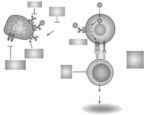
Figure 16.7. Strategies for inhibiting proinflammatory cytokines in asthma. These include inhibition of cytokine synthesis (e.g., corticosteroids), inhibition of transcription factors regulating cytokine expression (e.g., calcineurin inhibitors or decoy oligonucleotides), inhibition of secreted cytokines with blocking antibodies [e.g., antiinterleukin (IL)5 antibody] or soluble receptors (e.g., soluble IL4 receptors), blocking cytokine receptors (e.g., chemokine receptor antagonists), blocking signal-transduction pathways (e.g., p38 mitogen-activated protein kinase inhibitors) or transcription factors activated by cytokines (e.g., STAT6 inhibitors). (Reprinted by permission from Macmillan Publishers Ltd.: Barnes PJ: New drugs for asthma, Nature Revs Drug Discov 3:831–44, copyright 2004.)
inflammatory factors, and administration with inflammation inhibitors (Fig. 16.8). There are a number of antiinflammatory cytokines, including interleukin (IL)10, interleukin-12, and interferon-γ. Interleukin-10 is a cytokine that suppresses inflammatory reactions in the airway. In patients with asthma, the expression of the interleukin-10 gene is reduced compared to the normal population. In experimental models, the administration of inter- leukin-10 significantly suppresses inflammatory reactions in response to the stimulation of allergens. Immunotherapeutic approaches often induce upregulation of the interleukin10 gene. These observations suggest that interleukin-10 or its gene can be used as therapeutic agents for the treatment of asthma.
|
|
|
|
|
|
|
|
|
|
|
|
|
PULMONARY DISORDERS |
747 |
||
|
A |
|
|
|
|
|
|
|
|
|
|
|
|
|
|
|
|
|
Anti-IgE |
|
|
|
|
|
|
|
|
|
Allergen |
|
|
||
|
|
|
|
|
|
|
|
|
|
|||||||
|
|
|
|
|
|
|
Anti-IL-4 |
|
|
|
|
|
|
|
|
|
Mast |
|
|
|
|
Anti-IL13 |
|
|
|
|
|
|
|
|
|||
|
|
|
|
|
|
|
|
|
|
|
|
|
|
|||
cell |
|
|
|
|
IL-4, |
|
|
|
Antigen- |
|
||||||
|
|
|
|
|
|
|
|
|
|
|
||||||
|
|
|
|
|
|
|
IL-13 |
|
|
|
|
|||||
|
|
|
|
|
|
|
|
|
|
presenting cell |
|
|||||
|
|
|
|
|
|
|
|
|
|
|
|
|
|
|
||
|
|
|
|
IgE |
|
CD23 |
|
|
|
|||||||
|
|
|
|
|
|
|
|
B |
|
|
|
|
|
|
|
|
Cl– channel |
|
|
|
|
|
|
Anti-CD23 |
|
E Co-stimulation |
|
||||||
|
|
|
|
|
|
|
|
|
|
|||||||
|
|
|
|
|
|
|
|
|
|
|
|
|||||
|
|
|
|
|
|
|
|
|
|
|
|
MHC II |
|
|||
|
D |
|
|
|
|
|
|
|
|
|
|
B7.2 |
|
|
||
|
Syk kinase |
|
|
|
|
|
|
|
||||||||
|
|
|
|
|
|
|
|
|
TCR |
CD28, |
Anti-B7.2 |
|||||
|
|
|
inhibitor |
|
|
|
|
|
|
|||||||
|
|
|
|
|
|
|
|
|
ICOS |
Anti-CD28 |
||||||
C |
|
|
|
|
|
|
|
|
|
|
|
|
|
|||
|
|
|
|
|
|
|
|
|
|
|
|
|
||||
|
|
|
|
|
|
|
|
|
|
|
|
CTLA4 |
Anti-ICOS |
|||
Cromones |
|
|
|
|
|
|
|
|
|
|
|
|||||
|
|
|
|
|
|
|
|
|
|
|
|
|
CTLA4-Ig |
|||
|
Furosemide |
|
|
|
|
F |
IFN-γ |
|
|
|
|
|||||
|
|
|
|
|
|
|
|
|
|
|
||||||
|
|
|
|
|
|
|
|
|
|
|
|
|
||||
|
|
|
|
|
|
|
|
IL-12 |
|
|
|
TH2 |
|
|
||
|
|
|
|
|
|
|
|
IL-18 |
|
|
|
|
|
|
||
|
|
|
|
|
|
|
|
|
|
|
|
|
IL-4, IL-13 IL-5 |
|
|
|
|
|
|
|
|
|
|
|
|
|
|
|
|
Inflammation |
|
|
|
Figure 16.8. Strategies for inhibiting allergic responses underlying asthma. Immunoglobulin E (IgE) can be inhibited by the antibody omalizumab (A) and low-affinity IgE receptors by anti-CD23
(B). Mast cells can also be blocked by cromones and furosemide (C), probably acting on a chloride channel and by inhibitors of Syk kinase, which inhibit the signal-transduction pathways activated by IgE receptors (D). Antigen presentation can be blocked by inhibitors of costimulatory molecules (E), including B7.2, CD28, inducible costimulatory molecule (ICOS), and cytotoxic T-lymphocyte antigen-4 (CTLA4). TH2 cells can also be directly inhibited by interferon-γ (IFNγ), interleukin (IL)12 and IL18 (F). (Reprinted by permission from Macmillan Publishers Ltd.: Barnes PJ: New drugs for asthma, Nature Revs Drug Discov 3:831–44, copyright 2004.)
IL12 is a cytokine that suppresses the production of asthma-promoting cytokines from the type 2 T-helper cells, thus exerting an inhibitory effect on asthmatic activities, such as mucus secretion, inflammation, and eosinophil infiltration. Experimental investigations have demonstrated that the transfer of the IL12 gene into the lung of ovalbuminor dust mite-sensitized animals results in an increase in the level of interferon-γ, an anti-asthmatic cytokine, and a reduction in the activity of the type 2 T-helper cells, eosinophil infiltration, and inflammatory reactions. The cotransfer of IL12 with IL18, another anti-asthmatic cytokine, elicits a synergistic inhibitory effect on asthmatic changes. The IL12 and IL18 genes are potential candidates for the treatment of human asthma. Furthermore, the activation of nitric oxide synthase type 2 is required for the activity of IL12. Thus a cotransfer of the nitric oxide synthase type 2 gene with the IL12 gene may enhance the anti-asthmatic effect of IL12. The characteristics of IL12 and IL18 are presented in the following table.
748 PULMONARY REGENERATIVE ENGINEERING
Interferon-γ is a cytokine that suppresses inflammatory reactions and inhibits the production and secretion of asthma-promoting cytokines, including IL4 and IL5 (note that these cytokines stimulate the activation and migration of eosinophils and secretion of inflammatory agents such as IgE immunoglobulins), from the type 2 T-helper cells, and thus reduce asthmatic activities (see Table 16.1 for characteristics of interferon-γ). In several experimental investigations, interferon-γ gene has been transferred into the lung of asthmatic animals by using virus-mediated gene transfer approaches. These investigations have demonstrated that the overexpression of the interferon-γ gene is associated with a reduction in the density of eosinophils and mucus secretion, resulting in a decrease in asthmatic symptoms. Clinical investigations have demonstrated similar results. Interferon- γ and its gene have been proven effective antiinflammatory molecules that can be used for the treatment of asthma.
In addition to the administration of antiinflammatory cytokines and their genes, the inflammatory cytokines can be suppressed by local delivery of antisense oligonucleotides or small interfering RNA specific to these cytokines. As discussed on pages 448 and 449, antisense oligonucleotides or small interfering RNA (siRNA) are short nucleotide sequences, can be synthesized based on the sequence of target mRNA, and can be used as therapeutic agents. A selected agent can be delivered via inhalation to the airways. These short nucleotide sequences can be taken up by airway epithelial cells. Experimental investigations have demonstrated that either oligonucleotides or small siRNA can effectively suppress inflammatory reactions in the airways. Other potential therapeutic approaches are presented in Fig. 16.8.
Characteristics of asthma-related interleukins are listed in Table 16.1.
Suppression of Inflammatory Reactions by Transferring the Glucocorticoid Receptor Gene [16.7]. Glucocorticoid is a hormone produced in the cortex of the adrenal glands. It interacts with the glucocorticoid receptor and suppresses inflammatory reactions, such as leukocyte infiltration, the production of asthmatic cytokines, and mucus secretion. The glucocorticoid receptor, also known as GCR and nuclear receptor subfamily 3 group C member 1 (NR3C1), is a nuclear receptor of 777 amino acids and about 86 kDa. This receptor is expressed in a variety of cell and tissue types, including leukocytes, cardiomyocytes, brain, lung, kidney, skeletal muscle, liver, pancreas, intestine, eye, skin, and osteoblasts. The primary functions of this receptor are to interact with glucocorticoid and act as a transcription factor to induce the expression of antiinflammatory genes. Thus the activation of glucocorticoid receptor results in the suppression of inflammatory reactions. In vitro investigations by using human airway cells have demonstrated that the overexpression of the glucocorticoid receptor gene results in the suppression of several transcriptional factors, such as activator protein 1 and nuclear factor κB, which induce inflammatory reactions. Further investigations are necessary to verify these antiinflammatory activities in vivo.
Inducing Bronchodilation by Transferring Bronchodilator Genes and Proteins [16.8]. There are several types of bronchodilator proteins, including the β2-adrenergic receptor and atrial natriuretic peptide (ANP). The β2-adrenergic receptor of the airway smooth muscle cells interacts with epinephrine and induces smooth muscle relaxation and airway dilation, an effective approach for the relief of asthmatic symptoms. The transfection of the β2-adrenergic receptor gene into the airway smooth muscle cells can up-regulate

750 PULMONARY REGENERATIVE ENGINEERING
the expression of the receptor and enhance the activity of the bronchodilator epinephrine. Experimental investigations have demonstrated the effectiveness of this approach for the treatment of asthma. Atrial natriuretic peptide (ANP) serves as a potent bronchodilator. In experimental investigations, this peptide can protect the airways from chemical-induced bronchoconstriction. Blood delivery and local application of atrial natriuretic peptide are effective therapeutic approaches for the treatment of asthma.
Cystic Fibrosis
Pathogenesis, Pathology, and Clinical Features [16.9]. Cystic fibrosis is a fatal, autosomal, inherited disease which involves multiple systems, including the pulmonary, cardiovascular, gastrointestinal, skeletal, and reproductive systems. Cystic fibrosis in the pulmonary system is characterized by the presence of profound inflammation, bronchitis, bronchopneumonia, lung abscesses, atelectasis, pulmonary hypertension, cor pulmonale, and congestive heart failure. The pulmonary involvement is a major cause of death in cystic fibrosis. Cystic fibrosis is a common disease in the Caucasian population and the occurrence is about 1 in 2500. This disorder is often found in children.
The pathogenesis of cystic fibrosis is related to the mutation of a gene that encodes the cystic fibrosis transmembrane conductance regulator (CFTR), also known as cystic fibrosis transmembrane conductance regulator ATP binding cassette, MRP7, ABC35, and ABCC7 (1480 amino acids and 169 kDa). CFTR is a member of the superfamily of ATP-binding cassette (ABC) transporters and is found in the epithelial membrane of the lung and intestine. It serves as a cAMP-regulated chloride channel. CFTR mediates the transport of chloride as well as other ions such as sodium and HCO3− (Fig. 16.9). The mutation of the CFTR gene induces the disorder of ion transport across the epithelial cells, resulting in a reduction in chloride secretion and an increase in sodium absorption. These changes influence the function of epithelial cells and contribute to the development of cystic fibrosis. There are several types of genetic alterations, which may influence the function of the CFTR protein: null expression, mutation, and regulatory disorder of CFTR. CFTR gene mutation is the most common cause of cystic fibrosis. A typical mutation is the deletion of the 508 phenylalanine. In the lung of cystic fibrosis, the concentration of interleukin-10, an antiinflammatory cytokine, is reduced compared to that in normal people. This change also contributes to inflammatory reactions in cystic fibrosis.
In cystic fibrosis, pathological examinations often exhibit excessive growth of bronchial goblet cells and increased secretion of mucus. Inflammatory reactions are found around bronchi with increased leukocyte infiltration. These changes usually lead to partial or complete obstruction of the bronchi. Patients with cystic fibrosis are often associated with infection by bacteria, such as Pseudomonas aeruginosa and E. coli, viruses, and fungi. It is often difficult to remove these micro-organisms from the airways because of the functional impairment of the ciliated epithelial cells. As the disorder develops, extensive fibrosis in the parenchyma and bronchi occurs (Fig. 16.10), which is associated with irreversible bronchial distortion and obstruction, increased stiffness of parenchyma, and reduced ventilation. Fibrous disorders also occur in the alveolar wall, resulting in the impairment of oxygen exchange. Patients usually die of oxygen deficiency due to impaired gas ventilation and exchange.
Experimental Models of Cystic Fibrosis [16.10]. Experimental cystic fibrosis can be induced by introducing P. aeruginosa into the airways of the rat, mouse, or other animals.
PULMONARY DISORDERS |
751 |
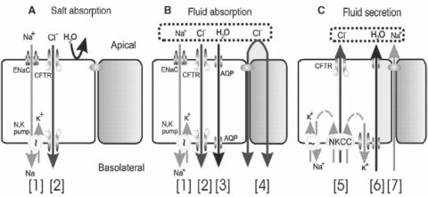
Figure 16.9. CFTR’s multiple roles in fluid and electrolyte transport. (A) Salt absorption. In the sweat duct, high apical conductance for Na+ [1] and Cl− [2] and relatively low water conductance allows salt to be reabsorbed in excess of water (hypertonic absorption), leaving a hypotonic luminal fluid. In the sweat duct CFTR is the only available anion conductance pathway, and when it is lost in CF the lumen quickly becomes highly electronegative and transport virtually ceases, resulting in high (similar to plasma) luminal salt (B). Fluid absorption. In epithelia with high water permeability (3) relative to electrolyte permeability water will absorbed osmotically with salt to decrease the volume of luminal fluid. If no other osmolytes or forces are present, the salt concentration will remain unchanged. If water-retaining forces are present, permeant electrolytes can be reduced preferentially. The consequences of eliminating CFTR depend on the magnitude of such forces, the relative magnitude of alternate pathways for transepithelial anion flow (4), and how CFTR affects other ion channels. The high-salt and low-volume hypotheses differ on each of these points.
(C) Anion-mediated fluid secretion. Secreting epithelia lack a significant apical Na+ conductance. Basolateral transporters such as NKCC move Cl− uphill into the cell; it then flows passively into the lumen via CFTR [5], K+ exits basolaterally, Na+ flows paracellularly and water follows transcellularly. Elimination of CFTR eliminates secretion. (Reprinted with permission from JJ: Wine The genesis of cystic fibrosis lung disease, J Clin Invest 103:309–12, 1999.)
Agarose beads can be used as carriers for bacterial delivery. To prepare P. aeruginosa- containing agarose beads, P. aeruginosa can be isolated from the airway of human cystic fibrosis patients, mixed with an agarose solution, and grown to near saturation. The bacterial and agarose solution is added to a bath of mineral oil at temperature 50ºC, stirred for several minutes, and placed on ice for several minutes. Agarose beads form in the oil bath when temperature reduces. The agarose beads can be washed in 0.5% deoxycholic acid in phosphate buffered saline (PBS, pH 7.4) and subsequently in PBS. The diameter of the beads can be controlled by altering the stirring speed and can be measured by microscopy.
To introduce the constructed P. aeruginosa-agorose beads into the airways of an animal, the animal can be anesthetized by intraperitoneal injection of sodium pentobarbital (50 mg/kg body weight). After sterilization, the trachea is cut open, and a volume of 50 μL bead slurry is instilled into the left and right bronchi. The tracheal wound is closed with one to two suture stitches (10-O nylon suture for mice or rats). At a scheduled time (e.g., 3 days after surgery), samples can be collected from the airways for detecting the presence of the bacterium. Mucus and lung tissue samples can be also collected for the
752 PULMONARY REGENERATIVE ENGINEERING
(A) |
|
(B) |
(C) |
|
(D) |
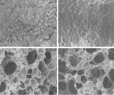
Figure 16.10. Morphological changes in the lung of cystic fibrosis. (A) Scanning electron micrographs of the surface of the respiratory epithelium from a terminal bronchiole in an 11-month-old wildtype mouse. Note the numerous ciliated and nonciliated cells. (B) Terminal bronchiole from a Cftr−/− littermate. Respiratory epithelium is encrusted in mucus-like material. (C) Alveoli from the wildtype animal. (D) Alveoli from the affected animal. Distal airways were caked with mucuslike material. Original magnifications: ×1000 (A,B); ×650 (C,D). (Reprinted with permission from Durie PR et al: Characteristic multiorgan pathology of cystic fibrosis in a long-living cystic fibrosis transmembrane regulator knockout murine model, Am J Pathol 164:1481–93, copyright 2004.)
detection and analysis of cytokines by biochemistry. Structural changes in the airways can be assessed by microscopy. It is important to note that this model is merely a model of P. aeruginosa infection, which often occurs in human patients with cystic fibrosis and exhibits certain features of cystic fibrosis. However, it is not a realistic cystic fibrosis model induced by the mutation of the cystic fibrosis transmembrane regulator gene.
Conventional Treatment of Cystic Fibrosis [16.11]. Cystic fibrosis can be treated with approaches which enhance the function of CFTR and relieve the symptoms of the disorder. Several types of chemical compound, including phenylbutyrate, 8-cyclopentyl-1,3-dipro- pylxanthine (CPX), and genistein have been tested in previous investigations. These agents may serve as chaperones that promote the deployment of CFTR to the cell membrane. Agents that stimulate chloride secretion (e.g., uridine triphosphate) and inhibit sodium absorption (e.g., amiloride) have also been considered and tested for the treatment of cystic fibrosis. However, few conventional approaches are available for the removal of the causative factors of the disorder.
Cystic fibrosis is associated with life-threatening alterations, such as bronchial constriction and obstruction by inflammatory cells and mucus. A critical issue for treating cystic fibrosis is to relieve bronchial obstruction. Bronchodilators, such as epinephrine, are often
PULMONARY DISORDERS |
753 |
used to achieve such as goal. As discussed on page 750 of this chapter, cystic fibrosis is often associated with bacterial infection. In such a case, antibiotics should be given to control infection. In addition, glucocorticoids are often used to control inflammatory reactions in cystic fibrosis. In the end-stage of cystic fibrosis, it is usually necessary to carry out lung transplantation. However, the long-term survival of lung transplants is often disappointing.
Molecular Engineering [16.12]. Cystic fibrosis can be genetically treated with two approaches: to remove the causative factors and reduce symptoms. Since CFTR gene mutation and deletion are major causes of cystic fibrosis, one strategy for the treatment of cystic fibrosis is to restore the structure and function of the CFTR gene. For the last two decades, numbers of investigations have been carried out to test the effectiveness of CFTR gene transfer into the respiratory system of cystic fibrosis. Limited clinical trials have been carried out. These investigations have shown promising results. However, it is often difficult to achieve long-term therapeutic effects by gene transfer. Furthermore, the effectiveness of the CFTR gene transfer, which requires viral gene-carriers, is limited by local immune reactions to the viral factors.
Another approach is to reduce inflammatory reactions, which often occur in cystic fibrosis. Inflammation is a major factor that contributes to the obstruction of bronchi in cystic fibrosis. As for the treatment of asthma, the overexpression of the glucocorticoid receptor gene by gene transfer is a potential therapeutic approach for reducing inflammatory reactions in cystic fibrosis. Since bronchial constriction and obstruction are major causes of cystic fibrosis-associated symptoms, the transfer of genes encoding bronchodilator proteins is a potential approach for the treatment of cystic fibrosis. A candidate gene is the β2-adrenergic receptor gene. The upregulation of the β2-adrenergic receptor induces bronchial dilation and relieves airway obstruction.
Pulmonary Hypertension
Pathogenesis, Pathology, and Clinical Features
Primary Pulmonary Hypertension [16.13]. Primary pulmonary hypertension, also known as idiopathic pulmonary hypertension, is a disorder characterized by an increase in pulmonary arterial blood pressure. The term “primary” or “idiopathic” is used because the pathogenic causes of pulmonary hypertension are poorly understood. There are other types of pulmonary hypertension, such as hypoxiaand pulmonary embolism-induced hypertension. Primary pulmonary hypertension is diagnosed only if these types of pulmonary hypertension are excluded.
Although the cause of primary pulmonary hypertension remains unknown, there are several hypothetical mechanisms for the pathogenesis of the disorder. One of the possible causes is disordered regulation of the arterial basal tone or contractility. In pulmonary hypertension, the basal tone of the pulmonary arteries is increased compared to that in the general population. The contribution of altered arterial contractility is supported by the observation that a treatment with vasodilators reduces pulmonary arterial blood pressure. However, the exact cause of disordered arterial contractility remains poorly understood. Another possible cause of primary pulmonary hypertension is the obstruction of pulmonary arteries by unrecognized thromboemboli. Such an obstruction induces elevation in the resistance to the right ventricle, stimulating the right ventricle to increase its contractility. As a result, pulmonary hypertension occurs.
754 PULMONARY REGENERATIVE ENGINEERING
In pulmonary hypertension, pathological examinations exhibit several changes in the structure of pulmonary arteries. These include intimal hyperplasia and wall hypertrophy of pulmonary arteries, a reduction in the density of small pulmonary arteries and capillaries, thickening of endothelial cells and basement membrane of capillaries, and occasionally atherosclerotic plaques in large pulmonary arteries. Pulmonary hypertension is often associated with enlarged right atrium and hypertrophied right ventricle. These changes are attributed to the increase in the workload of the right ventricle resulting from pulmonary hypertension. When the right ventricle is unable to overcome the resistance of the pulmonary arteries and eject sufficient blood into the pulmonary arteries, excessive blood is retained in the right atrium, resulting in the enlargement of the right atrium. The treatment for primary pulmonary hypertension is similar to that for hypoxic pulmonary hypertension (see next section).
Hypoxic Pulmonary Hypertension [16.14]. Hypoxia can induce rapid contraction of smooth muscle cells in the pulmonary arteries of mammals, resulting in acute pulmonary hypertension. Hypoxia is defined as a reduction in the oxygen concentration (<21%). The level of atmospheric oxygen changes in proportion to the altitude. An elevation of 1000 m in altitude is associated with a reduction of about 2% in oxygen concentration. The oxygen level at the altitude 5000 m is about 10%. The exposure of mammals to such an altitude induces acute pulmonary hypertension (Fig. 16.11). The severity of hypertension is proportional to the decrease in the oxygen concentration. Mammals may not survive in an environment with an oxygen level lower than 5%.
It is important to note that, while hypoxia directly induces pulmonary arterial constriction, it exerts an opposite effect on the contractility of the systemic arteries. In the pulmonary arterial system, a reduction in the oxygen level results in a decrease in the air ventilation-to-blood perfusion ratio. As discussed on page 739, the pulmonary system 739 intends to maintain a constant air ventilation-to-blood perfusion ratio and thus to reduce blood perfusion by activating smooth muscle contraction. In the systemic arterial system,
|
25 |
|
|
|
|
|
|
|
|
Hypoxic |
|
|
|
(mmHg) |
20 |
|
|
|
|
|
15 |
|
Control |
|
|
|
|
Pressure |
|
|
|
|
||
|
|
|
|
|
||
10 |
Nifedipine |
|
|
|
|
|
|
|
|
|
|
||
|
5 |
|
|
|
|
|
|
0 |
50 |
100 |
150 |
200 |
250 |
Time (h)
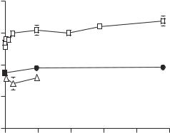
Figure 16.11. Changes in pulmonary arterial blood pressure in hypoxic pulmonary hypertension.
PULMONARY DISORDERS |
755 |
hypoxia is often associated with an increase in metabolic wastes, such as CO2, H+, and adenosine. These metabolites induce smooth muscle relaxation in the systemic arteries. However, the effect of these metabolites cannot overcome the constrictive effect of hypoxia in the pulmonary arteries.
Hypoxic pulmonary hypertension can induce rapid pathological changes in the pulmonary arteries. Primary changes include early bleb formation in the endothelial cells (Fig. 16.12), transient edematous swelling and disorganization of the medial elastic laminae (Chapter 16 opening Figure), and progressive hyperplasia of the pulmonary arterial smooth muscle cells and hypertrophy of the pulmonary arterial wall (Fig. 16.13). These are considered adaptive alterations in response to a rapid increase in the pulmonary arterial blood pressure and the stretching tensile stress in the vessel wall. In the presence of hypertension suppressors, such as nifedipine, these pathological changes were significantly inhibited in association with reduced pulmonary arterial blood pressure. These observations support the role of hypertension, but not hypoxia, in the induction of pulmonary arterial pathological changes. When hypoxia is removed, the pathological changes can be gradually recovered. However, the time required for recovery is significantly longer than the time required for the development of the pathological changes.
Rats and mice are often used to induce experimental hypoxic pulmonary hypertension. A hypoxic environment can be created by using a hypoxic chamber with a controlled flow of oxygen and nitrogen. It is necessary to install an oxygen sensor and a CO2 sensor to monitor the concentrations of oxygen and CO2, respectively. Animals can be placed in the chamber for a desired period. Pulmonary arterial blood pressure usually starts to increase within 5 min, continues to increase for about 5 days, and reaches a plateau after 5 days. The pulmonary arterial blood pressure can be monitored in living animals by implanting a catheter into the pulmonary arterial trunk or the right ventricle via the jugular vein and the right atrium.
Conventional Treatment of Pulmonary Hypertension [16.13, 16.14]. The principle of treating pulmonary hypertension is to induce arterial dilation and reduce the pulmonary arterial blood pressure. Vascular smooth muscle relaxants are often used to achieve such a goal. Common smooth muscle relaxants include direct smooth muscle relaxants (e.g., nitroprusside and nitroglycerine), β-adrenergic agonists (e.g., isoproterenol), α-adrenergic blockers (e.g., phentolamine and phenoxybenzamine), and calcium channel blockers (e.g., nifedipine). These agents are effective for the temporary relief of pulmonary hypertension. When pulmonary arterial embolism is the cause of pulmonary arterial hypertension, patients should be treated with anticoagulants.
Molecular Therapies for Pulmonary Hypertension. Primary pulmonary hypertension is a result of pulmonary arterial constriction due to hyperactivity of smooth muscle cells. Thus, the principle of treating pulmonary hypertension by molecular engineering is to relax smooth muscle cells, dilate pulmonary arteries, and reduce pulmonary arterial blood pressure. Because the genes responsible for the hyperactivity of smooth muscle cells remain poorly understood, there are few molecular engineering approaches available for removing the causative factors. Several genes encoding vasodilator proteins have been used in experimental models for the treatment of pulmonary hypertension. These include the nitric oxide synthase gene, the prostaglandin synthase gene, and the prepro-calcitonin gene-related peptide gene. These genes are briefly described as follows.