

Bioregenerative Engineering Principles and Applications - Shu Q. Liu
..pdfANATOMY AND PHYSIOLOGY OF THE URINARY SYSTEM |
847 |
fluid filtration from the glomerulus capillaries to Bowman’s capsule. The external membrane of the Bowman’s capsule, also called the parietal layer, is composed of epithelial cells, which are extended to the proximal tubule. The glomerulus is a capillary network with an afferent and an efferent arteriole. The afferent arteriole supplies blood to and the efferent arteriole drains blood from the glomerulus. The primary function of glomerulus is fluid filtration from the capillaries to Bowman’s capsule. There exist fenestrae in the capillary endothelial cells, allowing efficient fluid filtration.
Fluid generated in the renal corpuscle is processed through the renal tubular system, eventually forming urine. All renal tubules are composed of epithelial cells, which reside on a basement membrane. The first segment of the tubular system is the proximal convoluted tubule, which is located in the renal cortex. The proximal tubule is extended to the Henle’s loop, which is located in the renal medulla and consists of a descending and an ascending tubule. The ascending tubule is extended to the distal tubule, which is located in the renal cortex. The distal tubule is connected to the connecting tubule and then to cortical collecting tubules. A number of cortical collecting tubules join together to form a medullary collecting tubule, which merges into a larger collecting duct. The collecting duct is connected to the renal pelvis. The proximal, Henle loop, distal, and connecting tubules are associated with a rich network of capillaries, which are originated from the glomerular efferent arterioles.
The formation of urine is accomplished via several processes: glomerular filtration, renal tubular reabsorption, and secretion from peritubular capillaries. Fluid and substances from the blood, except for cells and proteins, can be freely filtered through the capillaries of the glomerulus. Major barriers for filtration are the fenestrated endothelial cells, the basement membrane, and the podocytes. Under physiological conditions, about 20% of bloodflow is filtered through the glomerular capillaries. Several factors determine the rate of glomerular filtration rate. These include the hydrostatic and colloid pressure within the glomerular capillaries, and the total surface area and conductivity of the glomerular capillaries. An increase in the hydrostatic pressure in the capillaries enhances filtration, whereas an increase in the hydrostatic pressure in Bowman’s capsule reduces filtration. An increase in colloid pressure (depending on the concentration of proteins) in the capillaries reduces filtration. An increase in the total area and conductivity of capillaries enhances filtration.
Filtered fluid and substances, collectively called filtrate, in Bowman’s capsule are not completely excreted from the kidney. Instead, they are reabsorbed and processed via coordinated work of the renal tubules and peritubular capillaries. Different substances are processed in different ways. For instance, certain types of substances, such as toxic chemicals and drugs, are filtered through the glomerular capillaries and completely excreted without reabsorption. Some substances, such as electrolytes (Na+, K+, Ca2+, Cl−, and HCO−), can be partially reabsorbed in the renal tubules, resulting in a decrease in the concentration of the substances in the excreted urine. Water can be partially reabsorbed. There are also substances that are completely reabsorbed after filtration. These include amino acids and glucose. Through various processing mechanisms, a large fraction of water and useful substances are reabsorbed, whereas waste and toxic substances are excreted via the formation of urine.
The reabsorption of water and electrolytes is regulated by hormones including the antidiuretic hormone (ADH) and proteins involved in the renin-angiotensin-aldosterone system. Antidiuretic hormone is produced in the posterior pituitary. The production of antidiuretic hormone is controlled by the osmolarity-sensitive cells located in the
848 URINARY REGENERATIVE ENGINEERING
supraoptic nuclei of the hypothalamus. These cells can sense changes in blood osmolarity. An increase in blood osmolarity induces the activation of the osmolarity-sensitive cells, which stimulate the ADH-producing cells to release ADH. A reduction in arterial blood pressure also stimulates the production and release of ADH. This hormone acts on the renal tubules, increasing water reabsorption. Such a process results in an increase in blood volume and a decrease in blood osmolarity.
Another mechanism that regulates water reabsorption involves the rennin– angiotensin–aldosterone system. As discussed in Chapter 15, cells in the renal juxtaglomerular apparatus produce and secret a proteolytic enzyme known as renin. A decrease in blood pressure and flow enhances renin secretion. Renin is an enzyme that converts angiotensinogen to angiotensin I. Another proteolytic enzyme, known as angiotensin converting enzyme (ACE), can covert angiotensin I to angiotensin II. Angiotensin II directly induces vascular smooth muscle contraction and stimulates the adrenal cortex to secret a steroid hormone called aldosterone. This hormone can act on the epithelial cells of the renal distal and collecting tubules, increasing Na+ transport and water reabsorption. Such a process results in an increase in blood volume.
Structure and Function of the Urinary Tract [20.1]
The urinary tract includes three parts: a pair of ureters (left and right), the urinary bladder, and the urethra. The left and right ureters are tubular structures originated from the left and right renal pelvis, respectively, and extended to the urinary bladder. The function of the ureters is to conduct urine from the kidneys to the urinary bladder. The urinary bladder is a muscular sac located in the lower abdominal cavity and anterior to the rectum. The function of the urinary bladder is for the storage of urine. The urethra is a muscular tube that removes urine from the urinary bladder.
The structure and cellular organization are similar for the ureters, urinary bladder, and urethra. There are four layers in the wall of these structures, including the epithelium, the lamina propria, muscular layer, and adventitia. The epithelium is composed of multilayered epithelial cells. The lamina propria is a connective tissue layer, consisting of fibroblasts and extracellular matrix. The muscular layer is thicker than other layers and is composed of smooth muscle cells. The adventitia is another connective tissue layer composed of similar components as the lamina propria.
DISORDERS OF THE URINARY SYSTEM
Acute Renal Failure
Pathogenesis, Pathology, and Clinical Features [20.2]. Acute renal failure is a disorder characterized by rapid deterioration of the renal function, resulting in the accumulation of blood urea and metabolic wastes. These metabolic wastes, when reaching a critical level, induce cell injury and death, resulting in the failure of the brain, heart, liver, and other organs. Pathogenic factors that induce acute renal failure include renal ischemia due to trauma and atherosclerosis, acute glomerulonephritis, toxicity of chemicals and substances, such as organic solvents, heavy metals, glycols, and radiographic contrast agents, and an increase in the blood concentration of myoglobin due to massive crush trauma, muscular ischemia, and massive infection. Renal ischemia causes glomerular and tubular
DISORDERS OF THE URINARY SYSTEM |
849 |
cell injury and death. Acute glomerulonephritis is a disorder characterized by rapid injury of glomerular capillaries and renal tubules, which results in a reduction in glomerular filtration, water and salt retention, proteinuria, and hematuria. Renal failure occurs in severe cases. Toxins impair the function of tubular epithelial cells. A sudden increase in myoglobin induces the obstruction of the renal tubules. All these changes cause a rapid reduction in the glomerular filtration and tubular processing capability, resulting in acute renal failure. When the causative factors are removed, acute renal failure is generally reversible, if there is no massive renal necrosis.
Acute renal failure is associated with a number of pathophysiological changes in the kidneys. Ischemia and toxins often result in three abnormalities: (1) reduction in the renal arterial perfusion pressure, leading to a decrease in the glomerular hydrostatic pressure and filtration; (2) reduction in the glomerular permeability and effective area, leading to a reduction in glomerular filtration; and (3) obstruction of renal tubules and reduction in tubular transport function, leading to a decrease in the capability of excreting toxic wastes, chemicals, and acidic substances. All these changes contribute to the accumulation of toxins and wastes in the blood. The accumulation of acidic substances results in a disorder known as metabolic acidosis. The accumulation of urea is referred to as uremia. In these cases, the toxic and acidic substances impair the function of the cells in almost all systems. The nerve cells and cardiomyocytes are especially sensitive to these substances. When the toxic and acidic substances are accumulated to a critical level, cells are injured and committed to death. Massive cell death ultimately results in the failure of tissues and organs.
The kidneys with acute renal failure exhibit a number of pathological abnormalities. Examinations by optical and electron microscopy often demonstrate disruption and necrosis of tubular epithelial cells, tubular obstruction, tubular dilation or collapse, interstitial edema, and leukocyte infiltration. In severe renal ischemia, large areas of renal infarction and tissue necrosis can be found. When recovered from acute renal failure, the kidneys usually regain normal structure. Acute renal failure may be associated with complications in the nervous, cardiovascular, and gastrointestinal systems. These complications are mostly due to cell injury or death resulting from uremia and acidosis. Nerve cell injury induces clinical symptoms including somnolence, disorientation, agitation, asterixis, and seizures. Cardiovascular complications induce arrhythmias, hypertension, circulatory congestion, and heart failure. Arrhythmias are due to electrolyte abnormalities and injury of the cardiac conductive system. Hypertension and circulatory congestion are induced by sodium and water retention resulting from renal dysfunction. Heart failure is a result of cardiac cell injury and death. In the gastrointestinal system, hemorrhage often occurs in association with symptoms such as nausea and vomiting.
Experimental Models of Acute Renal Failure [20.2]. Renal ischemia is a major cause of acute renal failure and is commonly used as a model of experimental renal failure. Mice and rats are often used for this model. To create renal ischemia, an animal can be anesthetized by peritoneal injection of sodium pentobarbital at a dose of 50 mg/kg body weight. The abdominal skin is sterilized, the abdominal cavity is opened, and the left or the right kidney is identified. The renal artery of the identified kidney can be isolated and tied off completely with a surgical suture for a period from 10 to 30 min. The severity of acute renal failure is dependent on the duration of ischemia. It is often desired to test a number of times (e.g., 10, 20, and 30 min) to determine an appropriate level of acute renal failure. The other kidney can be used as a control. The abdominal cavity is then closed and the animal is allowed to recover.
850 URINARY REGENERATIVE ENGINEERING
Acute renal failure can also be induced by administration of chemical toxins. A typical toxin is folic acid, which induces injury of the renal tubular epithelial cells and thus acute renal failure. Folic acid (250 mg/kg body weight in 150 mM sodium bicarbonate) can be introduced to the animal via a single intravenous injection. Animals with vehicle injection (150 mM sodium bicarbonate) can serve as controls. Assays and functional tests can be carried out at desired times following the injection.
Conventional Treatment [20.2]. Acute renal failure can be treated with several strategies, including (1) identification and treatment of causative diseases, such as renal ischemia, acute glomerulonephritis, and chemical toxicity; (2) establishment of urine output;
(3) control of the intake of proteins, water, and electrolytes; (4) control of the intake of any drugs and chemicals that are excreted from the kidneys; and (5) treatment of nervous and cardiovascular disorders, if any. When the causative factors are removed, acute renal failure can self-recover in most cases.
In severe cases when urea and acidic substances reach a critical level, renal dialysis ought to be conducted to remove the toxic substances. Renal dialysis is a process by which toxic substances are removed via diffusion from the blood to a dialysate across a semipermeable polymeric membrane based on the gradient of substance concentration. There are two types of dialysis: hemodialysis and peritoneal dialysis. Hemodialysis is a process by perfusing blood through a semipermeable polymeric membrane system. The semipermeable membrane is usually fabricated with cellophane, cellulose acetate, or polyacrylnitrile. A typical dialyzer is composed of two systems: blood and dialysate perfusion systems. The blood is separated from the dialysate by a dialysis membrane. There are two forms of dialyzer: flat plate and hollow fiber forms. In the plate form, semipermeable membranes are arranged to form narrow slits. Blood and dialysate are introduced to separated slit systems. In the hollow fiber form, blood is perfused through the lumen of the semipermeable hollow fibers, while dialysate flow is introduced to the outside the hollow fibers. The flow rate of blood and dialysate can be controlled based on the conditions of the patient. It is usually necessary to conduct 10–15-hr dialysis for patients with acute and chronic renal failure to bring down the toxic substances to an acceptable level. In hemodialysis, there are several common clinical problems, including blood coagulation, pulmonary embolism, and intimal hyperplasia in blood vessels for dialysis. These problems remain to be resolved.
Peritoneal dialysis is a process by which urea and toxic substances are removed by diffusion from the capillaries to the dialysate introduced into the peritoneal cavity. The peritoneal membrane on the surface of the abdominal organs are ideal semipermeable membranes and suitable for dialysis. Dialysate can be introduced into and removed from the peritoneal cavity via a catheter. A period of 20–70 hr dialysis is usually sufficient for most patients with acute and chronic renal failure. Compared to hemodialysis, peritoneal dialysis does not cause vascular complications and is relatively safe. However, peritoneal dialysis is not as efficient as hemodialysis.
When massive renal infarction occurs and cannot be rescued, it is necessary to carry out renal transplantation. Allogenic kidneys can be harvested and transplanted into the host patient by renal arterial and venous anastomosis. Since allogenic tissues cause acute immune rejection, it is necessary to administer immunity-suppressing agents.
Molecular Regenerative Engineering. Acute renal failure is a devastating disease with a rate of mortality rate of 20%. The primary cause of renal failure is the injury and death
DISORDERS OF THE URINARY SYSTEM |
851 |
of glomerular endothelial cells and tubular epithelial cells. In spite of tremendous efforts, few conventional approaches are available for the treatment of the disease. Given the fact that mitogenic molecules protect cells from injury and death, it is conceivable that the genes encoding these proteins can be used as therapeutic agents for the treatment of acute renal failure. There are several types of genes that can be potentially used for such a purpose, including growth factor, mitogenic signaling molecule, and cell death inhibitor genes. Representative genes for each group are discussed as follows.
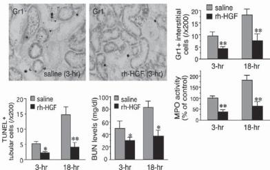
Growth Factor Genes [20.3]. Growth factors not only promote cell proliferation and differentiation, but also protect cells from injury and death. Thus, growth factors are potential therapeutic agents for the treatment of acute renal failure. Hepatocyte growth factor (HGF) is one of such agents. This growth factor is upregulated in the kidneys in response to renal injury, exerts renotropic, tubulogenic, antiapoptotic, and antifibrotic activities in glomerular endothelial cells and renal tubular epithelial cells (Fig. 20.2). The gene encoding hepatocyte growth factor can be transferred into the kidney to enhance the expression of HGF. In experimental models of renal injury induced by administration of nephrotoxic agents, such as cyclosporin A, the transfer of the HGF gene into the impaired kidney can induce significant upregulation of HGF expression. Upregulated HGF activates protein kinase B/Akt kinase and stimulates the expression of antiapoptotic factors such
A
B
Figure 20.2. Preventive effects of recombinant human hepatocyte growth factor (rh-HGF) on neutrophil infiltration, tubular apoptosis, and renal dysfunction in mice that underwent renal ischemia and reperfusion (I/R). (A) Changes in neutrophil accumulations in interstitial spaces of ischemic kidneys, treated with or without rh-HGF therapy (Gr1 staining, ×300). Renal tissues were collected at 3 and 18 hour after I/R challenge and then subjected to immunohistochemical procedures. Furthermore, granulocyte-specific myeloperoxidase (MPO) activities were measured in a part of samples used in immunohistochemistry. In this assay, the saline control level (3 h postischemia) was defined as 100%. (B) Decrease in tubular apoptosis levels and blood urea nitrogen (BUN) levels in the mouse model of renal I/R injury by rh-HGF treatment. Data are shown as means ± SD (n = 6). *P < 0.05; **P < 0.01 compared with the saline-injected control group. (Reprinted with permission from Mizuno S, Nakamura T: Am J Pathol 166:1895–1905, copyright 2005.)
852 URINARY REGENERATIVE ENGINEERING
as Bcl2. The expression of these factors is associated with a reduction in tubular epithelial cell injury and death, a decrease in interstitial leukocyte infiltration, and amelioration of renal impairment. Furthermore, the overexpression of HGF induces a decrease in the production of collagen, leading to a reduction in renal fibrosis. All these effects potentially improve the function of the kidneys.
Other growth factor genes such as the epidermal growth factor (EGF), fibroblast growth factor (FGF), platelet-derived growth factor (PDGF) genes can also be used for the treatment of acute renal failure (see Chapter 15 for the characteristics of these growth factors). It should be noted that growth factors can be directly used to treat acute renal failure. However, most proteins are rapidly degraded by the liver, and have a short half-life in the blood. It is a challenging task to maintain a sustained level of exogenously delivered protein. Thus, gene transfer is a more effective approach for the delivery of growth factors.
Genes Encoding Mitogenic Signaling Proteins [20.4]. Mitogenic signaling molecules represent a group of intracellular molecules that mediate the transduction of growthpromoting signals and the induction of cell proliferation and migration. Several signaling molecule genes have been tested in experimental models for the treatment of acute renal failure. Typical examples include protein kinase B, protein kinase C, mitogen-activated protein kinase (MAPK) genes. These genes encode proteins that mediate the transduction of growth factor-initiated signals and, thus, can be potentially used for the treatment of acute renal failure.
Genes Encoding Cell Death Inhibitors [20.4]. As described above, growth factor genes, such as platelet-derived growth factor, hepatocyte growth factor, and epidermal growth factor genes, can be used for the prevention of renal cell injury and death. In addition, there exist anti-apoptotic factors in the cells. A typical example is Bcl2, which inhibits the activity of apoptotic signaling molecules. Thus, the gene encoding Bcl2 can be used for the molecular treatment of acute renal failure.
Cell and Tissue Regenerative Engineering. Renal cell and tissue regenerative engineering is to restore the structure and function of disordered kidneys by inducing renal cell and tissue regeneration. There are several strategies for such a purpose, including: (1) kidney regeneration based on stem cells, (2) kidney regeneration based on embryonic renal tissues, and (3) construction of artificial kidneys based on adult renal tubular epithelial cells. The first two strategies have been tested in experimental models, and the third strategy has been tested in both experimental studies and clinical trials. These investigations have provided promising results for kidney regeneration and reconstruction.
Stem Cell-Based Kidney Regeneration [20.5]. As discussed on page 381, embryonic and fetal stem cells are multipotent cells that are capable of differentiating into specialized cell types. Under appropriate conditions, stem cells can differentiate into renal cells. A typical example is the regeneration of kidney-like structures by using stem cells from the embryonic metanephric mesenchyme and the ureteric bud, which are intermediate mesodermal tissues for kidney formation (see page 359). During the embryonic and fetal stage, the kidney is developed via reciprocal interactions between the metanephric mesenchyme and the ureteric bud. The metanephric mesenchyme forms within about 1 month in the intermediate mesoderm. Its formation stimulates the generation of branch-like ureteric
DISORDERS OF THE URINARY SYSTEM |
853 |
buds from an epithelial structure known as the nephric duct. The ureteric buds in turn induce mesenchyme cells to form epithelial nodules, which proliferate and differentiate into various renal cell types, including capsule cells, podocytes, and renal tubular cells, necessary for the constitution of the renal nephrons. At the same time, the ureteric buds give rise to the renal collecting ducts and the ureter. The newly generated nephrons and collecting ducts fuse with each other, forming the kidney.
In light of the fact that the metanephric mesenchyme and the ureteric bud are required for the development of the kidney, it is conceivable that stem cells from both structures are necessary for the regeneration of the kidney. In cell culture models, when stem cells from the metanephric mesenchyme and the ureteric bud are seeded in an extracellular matrix gel with mature renal epithelial cells, the stem cells from both embryonic tissues can proliferate and migrate. Interestingly, the stem cells can form branched and polarized tubules with internal lumens. A number of growth factors, including pleiotrophin, hepatocyte growth factor (HGF), fibroblast growth factor (FGF), and insulin-like growth factor (IGF)1, promote tubule formation and branching. In contrast, transforming growth factor (TGF)β inhibits branching morphogenesis. The presence of extracellular matrix is also required for the generation of the renal structures. These investigations suggest that the formation of the kidney is a complex process, requiring coordinated regulatory processes that involve multiple cells and mediators. Further investigations are needed to identify additional regulatory factors and clarify the regulatory mechanisms.
There are other types of stem cell that can be potentially used for kidney regeneration. The embryonic stem cells of the blastocyst are pluripotent stem cells that can differentiate into all types of specialized cells. Under appropriate conditions, embryonic stem cells can be induced to form renal glomerular endothelial and tubular epithelial cells. In addition, the adult bone marrow stem cells have the capability of differentiate in specialized cells. Experimental studies have shown that bone marrow stem cells can engraft to the kidney and differentiate into renal glomerular and tubular cells (Chapter 20 opening figure). These investigations have provided promising results, demonstrating the possibility of generating a functional kidney or a kidney-like structure.
Embryonic Tissue-Based Kidney Regeneration [20.5]. A kidney-like structure can be generated from the embryonic metanephros, a progenitor organ for the kidney. In experimental models, the embryonic metanephros can be collected and cultured in vitro for the generation of the kidney. Alternatively, the embryonic metanephric mesenchyme and the ureteric bud can be isolated from the metanephros and cultured together. The later model allows to understand the contributions of individual parts of the kidney progenitors. When the ureteric bud is placed in the proximity of the metanephric mesenchyme, the ureteric bud can form polarized branches with internal lumens and extend into the metanephric mesenchyme. At the same time, the metanephric mesenchyme can transform to epithelial structures and generate tubular nephrons. Interestingly, the mesenchyme tubular nephrons can form connections with the collecting tubules derived from the ureteric bud.
As for regeneration of the kidney based on stem cells, the formation of the renal structures from the embryonic metanephros are dependent on various growth factors and extracellular matrix. For example, pleiotrophin (see Table 20.1), fibroblast growth factor-1, and glial cell line-derived neurotrophic factor can induce branch formation of the ureteric bud in vitro. Fibroblast growth factor 2 enhances the survival of mesenchyme. In contrast, transforming growth factor β has been shown to inhibit the branch formation of the ureteric bud. Endostatin, a cleavage product of collagen XVIII present in the basement

854
TABLE 20.1. Characteristics of Selected Growth Regulatory Factors*
|
|
Amino |
Molecular |
|
|
Proteins |
Alternative Names |
Acids |
Weight (kDa) |
Expression |
Functions |
|
|
|
|
|
|
Pleiotrophin |
PTN, heparin-binding neurite |
168 |
19 |
Brain, pancreas, |
Stimulating cell proliferation and |
|
outgrowth promoting factor, |
|
|
kidney |
migration, inducing angiogenesis, and |
|
neurite growth-promoting |
|
|
|
enhancing tumor growth |
|
factor 1 (NEGF1), heparin- |
|
|
|
|
|
binding growth factor 8 |
|
|
|
|
|
(HBGF8), heparin-binding |
|
|
|
|
|
growth-associated molecule |
|
|
|
|
|
(HB-GAM), osteoblast-specific |
|
|
|
|
|
factor 1 (OSF1) |
|
|
|
|
Glial cell-derived |
Astrocyte-derived trophic factor 1, |
211 |
24 |
Nervous system, |
Existing as a homodimer, promoting |
neurotrophic |
glial cell-line-derived neurotrophic |
|
|
kidney, testis, |
survival and differentiation of |
factor |
factor |
|
|
lung |
dopaminergic neurons, protecting |
|
|
|
|
|
neurons from apoptosis, stimulating the |
|
|
|
|
|
proliferation of kidney cells, and |
Collagen XVIII α |
|
|
|
|
regulating kidney development |
COL18A1, type XVIII collagen |
1516 |
154 |
Heart, brain, liver, |
Generating endostatin, which serves as a |
|
chain |
|
|
|
kidney, overy, |
growth inhibitor and an antiangiogenic |
|
|
|
|
skeletal muscle, |
factor |
|
|
|
|
intestine, prostate |
|
|
|
|
|
gland |
|
|
|
|
|
|
|
*Based on bibliography 20.5.
DISORDERS OF THE URINARY SYSTEM |
855 |
membrane of the ureteric bud, also inhibits branch formation of the ureteric bud. These investigations show that kidney generation is a process regulated by a variety of signaling factors and extracellular matrix components. With an appropriate selection of the regulatory factors, it is possible to generate a kidney-like structure or “neokidney” based on embryonic kidney progenitor tissues. The kidney progenitor tissues can also be directly transplanted into an adult animal to generate a functional kidney-like structure.
Nuclear Transfer-Based Kidney Regeneration [20.6]. Nuclear transfer is a genetic technique that can be used to transfer adult or somatic cell nuclei into unfertilized oocytes and generate desired clones of tissues, organs, or animals with genetic characteristics of the donor somatic cells. Scientists have used such a technique to generate renal tissues for therapeutic purposes (Fig. 20.3). In experimental models, animal fetuses can be cloned by transferring adult cell nuclei into selected oocytes. At a selected stage, renal progenitor cells can be collected from the cloned fetus. The collected progenitor cells can be seeded in an extracellular matrix structure and implanted into a tissue of the cell nucleus donor. The implanted renal progenitor cells can differentiate into renal epithelial cells, which form tubule-like structures. Because half of the genome is from the donor, immune rejection response is reduced. These investigations have demonstrated the potential of the “cloning” technology for the regeneration of the kidney.
Adult Tubular Cell-Based Kidney Regeneration [20.7]. A cell-based artificial kidney can be constructed with two essential components: a dialyzer and a bioreactor with renal tubular epithelial cells (known as a renal tubule assist device). The dialyzer is similar to a conventional hemodialyzer, composed of semipermeable hollow fibers surrounded by an exterior chamber. The hollow fibers are for blood perfusion, and the exterior chamber is for dialysis filtrates. The bioreactor is also composed of semipermeable hollow fibers and an exterior chamber. The hollow fibers are for seeding and culturing renal tubular epithelial cells, and the exterior chamber is for blood perfusion. The dialyzer and the bioreactor are connected into a series. The dialyzer is connected to a selected vein from an animal model or a patient. Blood from the dialyzer is directed into the exterior chamber of the bioreactor, and the filtrate from the dialyzer is directed into the hollow fibers with renal tubular cells. Toxic substances and metabolic wastes can be dialyzed from the blood to the filtrate, whereas sodium, glucose, amino acids, and water are reabsorbed through the tubular epithelial cells in the bioreactor. Blood from the bioreactor is returned to host circulation and the waste-containing filtrate is discarded. Compared to conventional dialysis, the cell-based bioreactor not only serves as a blood filter but also provides necessary renal functions, including the reabsorption of nutrients and electrolytes, production of hormones, and synthesis of 1,25-dihydroxyvitamin D3 and glutathione. This type of artificial kidney has been used successfully in dog and pig models. In limited clinical trials, the renal tubule assist device can be functional for up to 24 h. These trials have provided fundamental information for the application of cell-based renal assist devices to renal failure.
Chronic Renal Failure
Pathogenesis, Pathology, and Clinical Features [20.8]. Chronic renal failure is a disorder characterized by a progressive reduction in the density of nephrons and deterioration