

Bioregenerative Engineering Principles and Applications - Shu Q. Liu
..pdfBONE AND CARTILAGE DISORDERS |
937 |
of IL-10 have been studied extensively. Here, IL-10 is used as an example to demonstrate how the antiinflammatory cytokines inhibit the activity of proinflammatory factors.
Interleukin-10 is a protein ( 18 kDa, 178 amino acids) present in the form of homodimer. IL10 is expressed in a umber of cell types, including monocytes, macrophages, T cells, B cells, natural killer cells, microglial cells, dendritic cells, eosinophils, and keratinocytes. The concentration of circulating IL10 is about 0.5 pg/mL in humans and other mammals. IL10 exerts its anti-inflammatory effect via interacting with the IL10 receptor. The IL10 receptor is a transmembrane glycoprotein complex composed of heterodimers α and β chains. The α chain is responsible for the binding of ligands, whereas the β chain transduces signals to the cytoplasmic signaling pathways. Each chain is composed of an extracellular, transmembrane, and cytoplasmic region. The binding of IL-10 to its receptor induces a number of antiinflammatory activities, including the inhibition of interferon-γ production and release from the T cells, suppression of chemokine secretion from neutrophils (e.g., MIP1α, MIP1β, and IL8), blockade of proinflammatory effects of IL1 and TNFα, reduction of inflammatory mediator generation in monocytes (e.g., IL8), and promotion of IL1Ra production. These activities results in the suppression of T-cell activation and immune responses, exerting beneficial effects for the treatment of rheumatoid arthritis. For therapeutic purposes, the genes encoding IL10 and IL10 receptor can be transferred into target cells in rheumatoid arthritis. Preliminary studies have demonstrated promising results for the treatment of arthritis by using IL10.
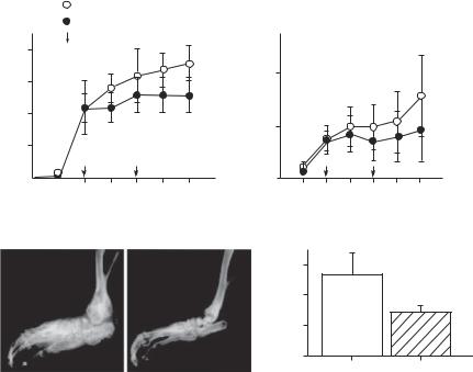
Dominant-Negative Mutant Ras Gene [22.19]. Ras is a protein that mediates the transduction of mitogenic factor signals. Acivated Ras stimulates cell proliferation and inflammatory reactions. Thus, Ras activation enhances the development of arthritis. The suppression of the Ras activity is an effective approach for the treatment of arthritis. One memthod for suppressing the Ras activity is to construct and transfer a dominant-negative mutant ras gene into the target cells. The dominant-negative mutant ras gene can be integrated into a gene-carrying vector such as a replication-deficient adenovirus vector. The vector can be delivered to the site of arthritis. Experimental investigations with such an approach have demonstrated that the delivered dominant-negative mutant ras gene can be expressed in target cells, resulting in a significant reduction in the level of inflammatory reactions in the joints (Fig. 22.12).
Osteoprotegerin [22.20]. Osteoprotegerin (OGP) is a protein of the cytokine tumor necrosis factor (TNF) receptor superfamily. This proetin serves as a decoy receptor for the osteoprotegerin ligand (OPGL) and can bind and inactivate OPGL. Osteoprotegerin ligand is a factor that stimulates the differentiation and proliferation of osteoclasts, which induce bone resorption and degeneration. Thus, osteoprotegerin can inhibit the activity of OPGL, suppress bone resorption, and enhance bone mineral deposition and bone formation. In the skeletal system, bone growth and resorption are controlled to a certain extent by the balance between OPGL and its decoy receptor osteoprotegerin. In transgenic animal models, the overexpression of osteoprotegerin is associated with eenhanced bone growth and reduced bone resorption. In contrast, the genetic disrutpion or knock out of the osteoprotegerin gene enhances bone resorption, resulting in osteoporosis-like alterations. Osteoprotegerin is present in the circulation and interstitial fluids of various tissues and organs. The direct delivery of osteoprotegerin or the transfer of the osteoprotegerin gene into the skeletal system prevents bone and cartilage resorption and destruction in inflammation and arthritis.


BONE AND CARTILAGE DISORDERS |
939 |
dividing or proliferating cells, nucleotides are taken up by cells for DNA synthesis. When nucleoside analogues (e.g., ganciclovir) are present, the viral thymidine kinase can covert the nucleoside analogues to nucleotides, which can be used for DNA synthesis. However, the incorporation of the nucleotide analogs results in the termination of DNA synthesis, because no more nucleotides can be added to the incorporated nucleotide analogues. By such a mechanism, the viral thymidine kinase gene, together with nucleoside analogues, can be used to suppress the proliferation of synoviocytes and reduce pathological changes in rheumatoid arthritis.
Bone and Cartilage Injury
Pathogenesis, Pathology, and Clinical Features [22.22]. Bone and cartilage injury occurs due to mechanical overload in sports and accidents. Various types of injury can be induced, depending on the magnitude and direction of the mechanical load. The most common injury is bone fracture. Such an injury is followed by several remodeling processes, including inflammatory, reparative, and modeling processes. The understanding of these processes is critical to the treatment of bone injury. Here, bone fracture is used as an example to discuss these remodeling processes.
Inflammatory Responses. Inflammation is a process that occurs in response to injury. Such a process is necessary for the self-healing of injured tissues or organs. While inflammation is common to all types of tissue, there are several unique features for bone injury. Bone injury induces transport and deposition of calcium and phosphorus, which do not occur in the inflammation of other tissues; also, since bone is subject to large mechanical loads, bone inflammation and recovery are influenced by mechanical forces.
Bone injury induces a series of inflammatory reactions. Blood vessel injury usually occurs with bone fracture, inducing hemorrhage and the formation of hematoma. At the same time, necrotic and injured cells can release cytokines and growth factors, which stimulate the infiltration of leukocytes and the migration of fibroblasts and other cell types to the injury site. Cytokines and growth factors also stimulate the proliferation of these cells and the production of extracellular matrix. All these reactions contribute to the formation of granulation tissue, which replaces the necrotic tissue and hematoma. Furthermore, angiogenesis occurs near the injury site, generating new blood vessels that provide oxygen and nutrients necessary for cell and tissue regeneration. Within about two weeks, calcium and phosphorus start to deposit to the injured tissue, resulting in the formation of immature membranous bone structure, which is referred to as a callus. A callus can be further mineralized to form a mature bone as described in the following section.
Reparative Reactions. Reparative reactions are initiated for the formation of mature bones based on calluses. Following the inflammatory phase, callus formation occurs at several locations. One is formed near the cortical surface of the bone at the injury site by the periosteum and adjacent skeletal muscle cells. In the medullary cavity at the injury site, the bone marrow cells can form a callus, which seals the fracture. Another type of callus forms between the two fracture-ends, serving to bridge the gap between the calluses at the ends. Additional calluses can be formed to join all separate calluses. Within about a month, a bone fracture can be filled with joined calluses.
940 BONE AND CARTILAGE REGENERATIVE ENGINEERING
Modeling Process. Modeling is a process by which the newly formed bone is further matured, organized, and aligned along the direction of the principal mechanical forces. The modeling process is thought to be regulated by mechanical stress. There are several events for the modeling phase. First, the newly generated bone structure is reshaped in response to the distribution of the mechanical stress. In regions with sufficient mechanical stress, the new bone is strengthened with additional mineralization, whereas in regions without sufficient mechanical stress new bone may be absorbed and degraded. Second, the medullary cavity and bone marrow are gradually restored. Third, the restoration of the natural form of bone structure (also referred to as bone reconstitution) is accomplished by coordinated bone resorption and regeneration. Bone resorption is induced by osteoclasts, whereas bone regeneration is induced by osteoblasts. Each type of cell may be activated in response to mechanical stress. A mechanical stress below a critical level may activate osteoclasts, initiating bone resorption. In contrast, a mechanical stress above a critical level may activate osteoblasts, resulting in bone regeneration. With such a stressregulated process, the reconstituted bone can be eventually shaped to the original natural form. Bone regeneration is a long-term process. The entire reconstitution process may take about several years.
Complications of Bone Injury. There are several complications that may occur during bone healing. The most common complications include fibrous union and nonunion. Fibrous union is a form of bone reconstitution with the establishment of a fibrous tissue bridge without mineralization between the fractured bones. A major cause for the formation of the fibrous tissue bridge is the lack of blood supply to the injured bone. A poor blood supply negatively influences the formation of calluses while promoting the formation of fibrous scar tissue, resulting in the formation of fibrous tissue bridges. Fibrous union often occurs in the injury of the distal pretibia and carpal navicular bone, which are associated with scarce blood vessels and insufficient blood circulation. Nonunion is a form of incomplete bone reconstitution, leaving a boneless gap between the ends of healed bone. Several factors, including bone loss, dislocation of fractured bone, infection, and severe soft tissue damage, may contribute to the bone nonunion. Such a consequence may occur in long-bone fracture.
Conventional Therapy [22.22]. The treatment of bone fracture and other types of bone injury is primarily dependent on the ability of the bone in self-healing and selfregeneration. Several approaches can be used to assist fractured bones in the healing processes. These include the realignment and restriction of dislocated bones to their natural positions by using external fixation devices, which ensures the anatomical reconstitution of the bones, stop of hemorrhage promptly, protection of injured bones from infection by administrating antibiotics, and treatment of bone non-union, if any, by bone transplantation or grafting. These approaches are usually effective for the treatment of bone fracture.
Molecular Regenerative Engineering [22.23]. The strategies for the molecular treatment of bone injury are to enhance bone regeneration and prevent bone nonunion or fibrous union. Although injured bones can be self-healed and conventional therapies are effective for most cases, bone reconstitution may take several months or longer. Furthermore, a fraction about 10% of bone fractures may experience delayed union or form nonunion structures. Bone regeneration can be greatly enhanced by using molecular regenerative
BONE AND CARTILAGE DISORDERS |
941 |
approaches. One effective approach is delivering bone formation-stimulating proteins or their genes into the cells responsible for bone regeneration. Typical factors that stimulate bone formation are bone morphogenetic proteins (BMPs), which can be used to enhance bone regeneration.
Bone morphogenetic protein 2, a member of the bone morphogenetic protein family, has been characterized and studied extensively in experimental bone injury models established in the rat, rabbit, and dog. The delivery of bone morphogenetic protein-2 or its gene to fractured bone enhances bone regeneration (Fig. 22.9). Clinical trials have demonstrated the effectiveness of locally delivered bone morphogenetic protein-2 in promoting bone regeneration. Bone morphogenetic protein 4 has also been used to enhance bone formation in experimental models of bone injury, demonstrating similar therapeutic effects as bone morphogenetic protein 2. The genes of these proteins can be used for gene therapy for improving bone regeneration. In addition to the bone morphogenetic protein genes, genes encoding angiogenic factors and vascular endothelial growth factor have been used for the promotion of bone regeneration. These genes can be delivered directly to the injury sites or indirectly delivered via the mediation of a matrix scaffold. Adenovirus vectors have been primarily used for mediating bone gene delivery.
Other growth factors, such as fibroblast growth factors (FGFs) and insulin-like growth factor (IGF), regulate the proliferation and differentiation of bone and cartilage cells (Fig. 22.13). These growth factors can be used to enhance the repair of injured bone and cartilage. In particular, fibroblast growth factors represent a family of potent growth factors for bone regeneration. The FGF family contains 23 known members. Among these members, FGF1 ( 18 kDa, 155 amino acids) and FGF2 ( 18 kDa, 155 amino acids) have been known to play an important role in the regulation of cell proliferation and differentiation. These growth factors are produced by many cell types, including the fibroblasts, endothelial cells, macrophages, hepatocytes, and keratinocytes. Both FGF1 and FGF2 can interact with FGF receptor tyrosine kinases and induce the proliferation of many cell
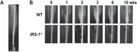
Figure 22.13. X-ray features of bone healing in WT and IRS-1−/− mice (IRS: insulin receptor substrate-1). (A) The fracture model used in this study. After exposing the right tibiae of 8-week-old mice, a transverse osteotomy was performed at the midshaft with a bone saw. The bone marrow cavity was then stabilized with an intramedullary nail. (B) Time course of the fracture healing in representative WT and IRS-1−/− mice. (Reprinted with permission from Shimoaka T et al: Impairment of bone healing by insulin receptor substrate-1 deficiency, J Biol Chem 279:15314–22, copyright 2004.)
942 BONE AND CARTILAGE REGENERATIVE ENGINEERING
types, including osteoblasts, chondrocytes, endothelial cells, and fibroblasts, thus enhacing bone and cartilage formation.
Cell Regenerative Engineering [22.24]. Bone regeneration involves the activation of osteoprogenitor cells, which differentiate to osteoblasts and other types of bone cells. It is thus conceivable to transplant osteoprogenitor cells or stem cells to promote bone formation. The bone marrow is known to contain osteoprogenitor cells. Bone marrow-derived cells have been used for the enhancement of bone regeneration in animal models as well as in humans. The transplantation of these cells to injured bones significantly facilitates bone formation and injury recovery. Other types of stem cells, such as embryonic, fetal, muscle-derived adult, and fat-derived adult stem cells, may also be used for bone regenerative engineering. These stem cells may be induced to differentiate to osteoblasts and other types of bone cells, when appropriate experimental conditions and extracellular environment are provided.
For the treatment of cartilage injury, several types of cells, including chondrocytes and osteochondrocytes, can be used for cell transplantation. The cells can be seeded in a scaffold constructed with an appropriate material (e.g., hyaluronan-based biopolymers or collagen matrix). A cell-seeded scaffold can be used to replace injured cartilage. In addition to chondrocytes, other cell types, such as embryonic stem cells and bone marrow stromal cells or mesenchymal stem cells, can be used for cartilage regeneration.
The type of matrix scaffolds may influence the differentiation of progenitor and stem cells. For instance, when bone marrow stem cells are cultured in a hyaluronan matrix with the supplement of transforming growth factor β1, the stem cells can differentiate into chondrocytes, forming a cartilage-like structure. When bone marrow stem cells are seeded in a porous calcium phosphate scaffold, the stem cells are transformed into osteoblasts, forming a bone-like structure. Bone morphogenetic proteins and growth factors can be used in cartilage constructs to facilitate cartilage formation. These approaches have been successfully used in experimental models for cartilage regeneration.
Progenitor and stem cells for bone and cartilage regeneration can be genetically transfected with genes encoding bone regeneration-promoting factors, such as the bone morphogenetic protein and vascular endothelial growth factor genes. Such cells may exhibit enhanced capability of differentiation and proliferation, thus facilitating bone regeneration and recovery. Alternatively, the bone formation-stimulating genes can be delivered by using gene carriers. Fibroblasts derived from soft connective tissue have been used as such gene carriers. Fibroblasts can be collected, cultured, and transfected with a selected gene in vitro, and used for cell transplantation in vivo. These approaches have been successfully used in experimental models. In human trials, mesenchymal stem cells have been used as gene carriers for bone-injury therapy. These trials have demonstrated encouraging results.
Tissue Regenerative Engineering [22.25]. While molecular and cellular therapies enhance bone regeneration, engineering manipulations at the tissue level is also important for the recovery from bone injury, especially in delayed bone union and nonunion. A major type of engineering manipulation is bone grafting or reconstruction with bone substitutes. Such a manipulation is necessary in the case of bone loss and destruction. There are several types of materials that can be used for such a purpose. These include autogenous cancellous bone specimens, metal prostheses, calcium phosphate ceramics, and polymeric materials. Among these materials, the autogenous cancellous bone is the gold standard material. The cancellous bone contains osteoprogenitor cells and osteoblasts, which play a critical role in bone repair and regeneration. However, in the case of large bone
BIBLIOGRAPHY 943
destruction, it is difficult to collect sufficient cancellous bone specimens. It is necessary to use other types of materials, such as synthetic materials and allogenic bones.
Synthetic materials have been used and tested for bone reconstruction. Osteoprogenitor or stem cells can be seeded in scaffolds of synthetic materials. A cell-containing scaffold can be tailored into a desired shape and used for bone grafting and reconstruction. However, synthetic materials cannon be integrated into the natural skeletal system. It is often difficult for cells and blood vessels to grow into the synthetic bone substitute. These limitations hinder the use of synthetic materials for bone reconstruction. Allogenic bone specimens are alternative materials for bone reconstruction. However, allogenic bones with living cells induce acute immune rejection reactions. It is necessary to administrate immune suppressive agents for patients with allogenic bone grafting. The removal of living cells from allogenic bone grafts can significantly reduce immune responses. Decellularized allogenic bone specimens can be used as bone substitutes for bone reconstruction.
BIBLIOGRAPHY
22.1. Anatomy and Physiology of the Bone and Cartilage
Li YC, Kong J, Wei M, Chen ZF, Liu SQ et al: 1,25-Dihydroxyvitamin D3 is a negative endocrine regulator of the renin-angiotensin system, J Clin Invest 110:229–38, 2002.
Suda T, Ueno Y, Fujii K, Shinki T: Vitamin D and bone, J Cell Biochem 88(2):259–66, 2003.
Guyton AC, Hall JE: Textbook of Medical Physiology, 11th ed, Saunders, Philadelphia, 2006.
McArdle WD, Katch FI, Katch VL: Essentials of Exercise Physiology, 3rd ed, Lippincott Williams & Wilkins, Baltimore, 2006.
Germann WJ, Stanfield CL (with contributors Niles MJ, Cannon JG), Principles of Human Physiology, 2nd ed, Pearson Benjamin Cummings, San Francisco, 2005.
Thibodeau GA, Patton KT: Anatomy & Physiology, 5th ed, Mosby, St. Louis, 2003.
Boron WF, Boulpaep EL: Medical Physiology: A Cellular and Molecular Approach, Saunders, Philadelphia, 2003.
Ganong WF: Review of Medical Physiology, 21st ed, McGraw-Hill, New York, 2003.
22.2. Pathogenesis, Pathology, and Clinical Features of Osteoporosis
Kanis JA, Melton III LJ, Christiansen C, Johnston CC, Khaltaev N: The diagnosis of osteoporosis, J Bone Miner Res 9:1137–41, 1994.
Downey PA, Siegel MI: Bone biology and the clinical implications for osteoporosis, Phys Ther 86(1):77–91, 2006.
Raisz LG: Pathogenesis of osteoporosis: Concepts, conflicts, and prospects, J Clin Invest 115(12):3318–25, 2005.
Rosen CJ, Brown SA: A rational approach to evidence gaps in the management of osteoporosis, Am J Med 118(11):1183–9, 2005.
Feng X: Regulatory roles and molecular signaling of TNF family members in osteoclasts, Gene 350(1):1–13, 2005.
Chien KR, Karsenty G: Longevity and lineages: Toward the integrative biology of degenerative diseases in heart, muscle, and bone. Cell 120(4):533–44, 2005.
Bassett JH, Williams GR: The molecular actions of thyroid hormone in bone, Trends Endocr Metab 14(8):356–64, 2003.
944 BONE AND CARTILAGE REGENERATIVE ENGINEERING
Harada S, Rodan GA: Control of osteoblast function and regulation of bone mass, Nature 423(6937):349–55, 2003.
Boyle WJ, Simonet WS, Lacey DL: Osteoclast differentiation and activation, Nature 423(6937):337– 42, 2003.
Schneider AS, Szanto PA: Pathology, 3rd ed, Lippincott Williams & Wilkins, Philadelphia, 2006.
Frazier MS, Drzymkowski JW: Essentials of Human Diseases and Conditions, 3rd ed, Elsevier Saunders, St Louis, 2004.
22.3. Vitamin D Receptor (VDR)
Morrison NA, Qi JC, Tokita A, Kelly P, Crofts L et al: Prediction of bone density from vitamin D receptor alleles, Nature 367:284–7, 1994.
Arai H, Miyamoto KI, Taketani Y, Yamamoto H, Iemori Y et al: A vitamin D receptor gene polymorphism in the translation initiation codon: Effect on protein activity and relation to bone mineral density in Japanese women, J Bone Miner Res 12:915–21, 1997.
Garnero P, Munoz F, Borel O, Sornay-Rendu E, Delmas PD: Vitamin D receptor gene polymorphisms are associated with the risk of fractures in postmenopausal women, independently of bone mineral density, J Clin Endocr Metab 90(8):4829–35, 2005.
Carling T, Rastad J, Akerstrom G, Westin G: Vitamin D receptor (VDR) and parathyroid hormone messenger ribonucleic acid levels correspond to polymorphic VDR alleles in human parathyroid tumors, J Clin Endocr Metab 83:2255–9, 1998.
Colin EM, Uitterlinden AG, Meurs JBJ, Bergink AP, van de Klift M et al: Interaction between vitamin D receptor genotype and estrogen receptor alpha genotype influences vertebral fracture risk, J Clin Endocr Metab 88:3777–84, 2003.
Ensrud KE, Stone K, Cauley JA, White C, Zmuda JM et al: Vitamin D receptor gene polymorphisms and the risk of fractures in older women, J Bone Miner Res 14:1637–45, 1999.
Ferrari S, Manen D, Bonjour JP, Slosman D, Rizzoli R: Bone mineral mass and calcium and phosphate metabolism in young men: Relationships with vitamin D receptor allelic polymorphisms,
J Clin Endocr Metab 84:2043–8, 1999.
Houston LA, Grant SFA, Reid DM, Ralston SH: Vitamin D receptor polymorphism, bone mineral density, and osteoporotic vertebral fracture: Studies in a UK population, Bone 18:249–52, 1996.
Hughes MR, Malloy PJ, Kieback DG, Kesterson RA, Pike JW et al: Point mutations in the human vitamin D receptor gene associated with hypocalcemic rickets, Science 242:1702–5, 1988.
Hustmyer FG, Peacock M, Hui S, Johnston CC, Christian J: Bone mineral density in relation to polymorphism at the vitamin D receptor gene locus, J Clin Invest 94:2130–4, 1994.
Kelly PJ, Hopper JL, Macaskill GT, Pocock NA, Sambrook PN et al: Genetic factors in bone turnover, J Clin Endocr Metab 72:808–13, 1991.
Lorentzon M, Lorentzon R, Nordstrom P: Vitamin D receptor gene polymorphism is associated with birth height, growth to adolescence, and adult stature in healthy Caucasian men: A crosssectional and longitudinal study, J Clin Endocr Metab 85:1666–71, 2000.
Malloy PJ, Eccleshall TR, Gross C, Van Maldergem L, Bouillon R et al: Hereditary vitamin D resistant rickets caused by a novel mutation in the vitamin D receptor that results in decreased affinity for hormone and cellular hyporesponsiveness, J Clin Invest 99:297–304, 1997.
Morrison NA, Qi JC, Tokita A, Kelly PJ, Crofts L et al: Prediction of bone density from vitamin D receptor alleles, Nature 367:284–7, 1994. Note: Erratum: Nature 387: 106 only, 1997.
BIBLIOGRAPHY 945
Riggs BL: Vitamin D-receptor genotypes and bone density, [editorial], New Engl J Med 337:125– 126, 1997.
Sainz J, Van Tornout JM, Loro L, Sayre J, Roe TF et al: Vitamin D-receptor gene polymorphisms and bone density in prepubertal American girls of Mexican descent, New Engl J Med 337:77–82, 1997.
Uitterlinden AG, Weel AEAM, Burger H, Fang Y, Van Duijn CM et al: Interaction between the vitamin D receptor gene and collagen type I-alpha-1 gene in susceptibility for fracture, J Bone Miner Res 16:379–85, 2001.
Yoshizawa T, Handa Y, Uematsu Y, Takeda S, Sekine K et al: Mice lacking the vitamin D receptor exhibit impaired bone formation, uterine hypoplasia and growth retardation after weaning, Nature Genet 16:391–6, 1997.
Human protein reference data base, Johns Hopkins University and the Institute of Bioinformatics, at http://www.hprd.org/protein.
22.4. Type I Collagen Gene
Aitchison K, Ogilvie D, Honeyman M, Thompson E, Sykes B: Homozygous osteogenesis imperfecta unlinked to collagen I genes, Hum Genet 78:233–6, 1988.
Bateman JF, Chan D, Walker ID, Rogers JG, Cole WG: Lethal perinatal osteogenesis imperfecta due to the substitution of arginine for glycine at residue 391 of the alpha-1(I) chain of type I collagen, J Biol Chem 262:7021–7, 1987.
Bateman JF, Lamande SR, Dahl HHM, Chan D, Cole WG: Substitution of arginine for glycine 664 in the collagen alpha-1(I) chain in lethal perinatal osteogenesis imperfecta, J Biol Chem 263:11627–30, 1988.
Boedtker H, Fuller F, Tate V: The structure of collagen genes, Int Rev Connect Tissue Res 10:1–63, 1983.
Bonadio J, Ramirez F, Barr M: An intron mutation in the human alpha-1(I) collagen gene alters the efficiency of pre-mRNA splicing and is associated with osteogenesis imperfecta type II, J Biol Chem 265:2262–8, 1990.
Cabral WA, Mertts MV, Makareeva E, Colige A, Tekin M et al: Type I collagen triplet duplication mutation in lethal osteogenesis imperfecta shifts register of alpha chains throughout the helix and disrupts incorporation of mutant helices into fibrils and extracellular matrix, J Biol Chem 278:10006–12, 2003.
Chamberlain JR, Schwarze U, Wang PR, Hirata RK, Hankenson KD et al: Gene targeting in stem cells from individuals with osteogenesis imperfecta, Science 303:1198–1201, 2004.
Chu ML, de Wet W, Bernard M, Ramirez F: Fine structural analysis of the human pro-alpha-1(I) collagen gene: Promoter structure, AluI repeats, and polymorphic transcripts, J Biol Chem 260:2315–20, 1985.
Constantinou CD, Nielsen KB, Prockop DJ: A lethal variant of osteogenesis imperfecta has a single base mutation that substitutes cysteine for glycine 904 of the alpha-1(I) chain of type I procollagen: The asymptomatic mother has an unidentified mutation producing an overmodified and unstable type I procollagen, J Clin Invest 83:574–84, 1989.
Dalgleish R: The human type I collagen mutation database, Nucleic Acids Res 25:181–7, 1997. Di Lullo GA, Sweeney SM, Korkko J, Ala-Kokko L, San Antonio JD: Mapping the ligand-binding
sites and disease-associated mutations on the most abundant protein in the human, type I collagen, J Biol Chem 277:4223–31, 2002.
Grant SFA, Reid DM, Blake G, Herd R, Fogelman I et al: Reduced bone density and osteoporosis associated with a polymorphic Sp1 binding site in the collagen type I-alpha 1 gene, Nature Genet 14:203–5, 1996.
Long JR, Liu PY, Lu Y, Xiong DH, Zhao LJ et al: Association between COL1A1 gene polymorphisms and bone size in Caucasians, Eur J Hum Genet 12:383–8, 2004.