

Bioregenerative Engineering Principles and Applications - Shu Q. Liu
..pdf976 OCULAR REGENERATIVE ENGINEERING
3 mm × 3 mm
|
|
Oral mucosal tissue |
Oral surgery |
|
|
|
|
|
|
|
|
Suspended
cells
3T3 feeder cells treated with mitomycin C
Culture at 37°C for two weeks
Reduce temperature to 20°c
Autologous oral epithelial-cell sheet
Transplantation without sutures
Figure 23.5. Transplantation of autologous tissue-engineered epithelial cell sheets fabricated from oral mucosal epithelium to injured cornea. Oral mucosal tissue (3 × 3 mm) was removed from a patient’s cheek. Isolated epithelial cells are seeded onto temperature-responsive cell culture inserts. After 2 weeks at 37ºC, these cells grow to form multilayered sheets of epithelial cells. The viable cell sheet was harvested with intact cell-to-cell junctions and extracellular matrix in a transplantable form simply by reducing the temperature of the culture to 20ºC for 30 min. The cell sheet is then transplanted directly to the diseased eye without sutures. (Reprinted with permission from Nishida K et al: Corneal reconstruction with tissue-engineered cell sheets composed of autologous oral mucosal epithelium, New Engl J Med 351:1187–96, copyright 2004 Massachusetts Medical Society. All rights reserved.)
OCULAR DISORDERS |
977 |
Preoperative |
Postoperative |
Patient 1
Patient 2
Patient 3
Patient 4
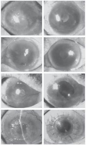
Figure 23.6. Eyes of patients before and after transplantation of sheets of tissue-engineered autologous epithelial cells. These photographs were taken just before transplantation of the cell sheets and postoperatively at 13, 14, or 15 months. (Reprinted with permission from Nishida K et al: Corneal reconstruction with tissue-engineered cell sheets composed of autologous oral mucosal epithelium, New Engl J Med 351:1187–96, copyright 2004 Massachusetts Medical Society. All rights reserved.)
addition to epithelial progenitor cells and mature epithelial cells, embryonic stem cells can also be used for corneal reconstruction. An example is shown in Fig. 23.7.
Corneal Reconstruction [23.7]. When severe opacity exists in the cornea, it is necessary to conduct complete corneal reconstruction. Although allogenic corneal transplantation is an option, there is a shortage of the supply of allogenic corneas. Thus, it is necessary to construct artificial corneal substitutes. Extracellular matrix components, including collagen and proteoglycans, are potential constituents for the construction of artificial corneas. Collagen type I and a type of glycosaminoglycan called chondroitin sulfate can be blended together and molded into a cornea-like structure in vitro. The presence of chondroitin

OCULAR DISORDERS |
979 |
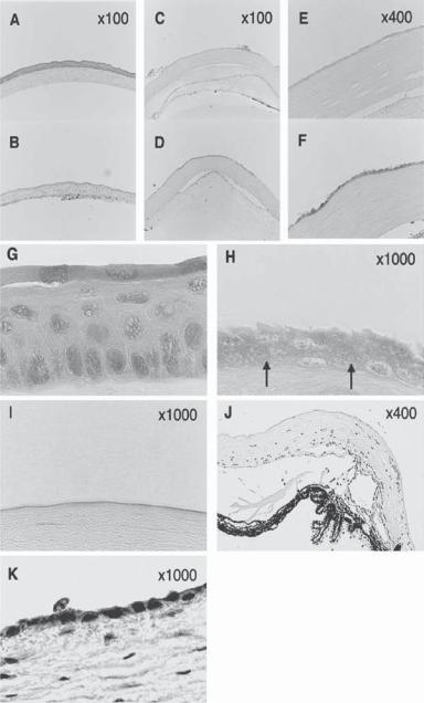
Figure 23.7. Histologic analysis of injured cornea, with or without transplantation of the embryonic stem (ES)-cell-derived epithelial progenitor cells. The ES-cell-derived epithelial progenitor cells (day 8 culture) were transplanted to n-heptanol-injured cornea of mice. (A) Normal mouse cornea.
(B). n-Heptanol-injured cornea without transplantation. (C–F). Mouse eyes were injured with n- heptanol. At 1 h (C, E) and 12 h (D, F) after transplantation, the eyes were enucleated. Cryostat sections were fixed with 20% formaldehyde in methanol, stained with H&E, and compared with those of normal cornea. (G) Higher magnification of the normal corneal epithelium shown in (A).
(H) Higher magnification of another preparation of the ES-cell-derived epithelial progenitor cells at 12 h after transplantation. Arrows: the basal or wing-cell–like transplanted cells. (I) Higher magnification of n-heptanol-injured cornea without transplantation 12 hours after the injury. No corneal epithelial cells were observed. (J) Limbus of n-heptanol-injured cornea without transplantation 24 h after the injury. Migration of the host-originated progenitor cells onto the corneal surface was not observed. (K) Immunostaining for E-cadherin of the corneal epithelial cells 12 h after transplantation of ES-cell-derived graft cells. E-cadherin-positive epithelial cells are stained red. (Reprinted with permission from Homma R et al: Induction of epithelial progenitors in vitro from mouse embryonic stem cells and application for reconstruction of damaged cornea in mice, Invest Ophthalmol Vis Sci 45:4320–6, copyright 2004.)
type of corneal scaffold is suitable for the growth and expansion of corneal cells and can be potentially used for corneal reconstruction. However, matrix structures generated by aldehyde-induced cross-linking are not natural and may exert side effects on the ocular system.
Another approach for the construction of corneal substitutes is to integrate extracellular matrix components into a synthetic polymer material, forming a natural and synthetic copolymer material. Scientists have used hydrated collagen and a polymeric material called poly(N-isopropylacrylamide–coacrylic acid–coacryloxysuccinimide) to fabricate a copolymer stromal scaffold. A cornea-like structure can be generated by casting a corneal mold with mixed collagen gel and synthetic polymer. Corneal cells can be seeded and grown on the matrix scaffold to establish a functional corneal substitute. Such a matrix scaffold can also be used for nerve innervation. In addition to collagen and glycosaminoglycans, other biological matrix molecules, such as fibrin and fibronectin, have been used for the construction of corneal substitutes. These investigations have demonstrated that corneal substitutes generated with these materials can be potentially used for the reconstruction of malfunctioned cornea.
Glaucoma
Pathogenesis, Pathology, and Clinical Features [23.8]. Glaucoma is a disorder characterized by an increase in the pressure of the intraocular aqueous humor. Under physiological conditions, the aqueous humor maintains a narrow range of pressure about 15 mm Hg. When the outflow of the aqueous humor is obstructed, the intraocular pressure increases, resulting in glaucoma. Such a disorder is often induced by the abnormality and occlusion of the trabecular meshwork and the canal of Schlemm. Glaucoma is diagnosed when the aqueous humor pressure is increased over 22 mm Hg. Increased aqueous humor pressure in the anterior compartment is transmitted to the posterior compartment via the vitreous humor, resulting in the compression and impairment of the retina and optic nerves. These alterations exert harmful effects on the retinal neurons, eventually leading to blindness. In the United States, glaucoma is the second leading cause of blindness.
980 OCULAR REGENERATIVE ENGINEERING
Based of on pathogenic mechanisms, glaucoma can be classified into two types: primary glaucoma due to the obstruction of the trabecular meshwork and secondary glaucoma as a complication of other disorders such as leukemia, rheumatoid arthritis (collagen disorder), infectious diseases (rubella and onchocerciasis), amyloidosis, cancer metastases, asthma, emphysema, renal disorders, administration of corticosteroids, chemical toxicity, Marfan’s syndrome, and ocular trauma. The pathogenic mechanisms of the primary obstruction of the trabecular meshwork are not fully understood. Glaucoma may be associated with ocular pain and corneal edema. The diagnosis of glaucoma relies on the measurement of intraocular pressure and visual field tests. In severe cases, partial or complete visual loss may occur.
Conventional Treatment of Glaucoma [23.8]. Strategies for glaucoma treatment are to reduce the resistance of the trabecular meshwork to the outflow of the aqueous humor and reduce the intraocular pressure. There are two conventional approaches that can be used to achieve these goals: administration of pharmacological agents and conduction of laser trabeculoplasty. Several types of agents, including cholinergic agonists and β-adrenergic antagonists, have been used to reduce the resistance of the trabecular meshwork. Cholinergic agonists, such as pilocarpine and carbachol, stimulate the contraction of the circumferential smooth muscle cells of the iris. This action shrinks the pupil, decreases the thickness of the iris, and increases the diameter of the canal of Schlemm, thus reducing the resistance to the outflow of the aqueous humor. A treatment with β-adrenergic antagonists, such as timolol maleate, reduces the formation of aqueous humor, thus lowering the intraocular pressure. When these drugs are ineffective, it is necessary to carry out trabeculoplasty, a laser-based surgical procedure that widens the trabecular meshwork and reduces resistance to the outflow of the aqueous humor. Another surgical intervention is to create a fistula from the anterior chamber to the subconjunctival gap, a procedure known as filtration surgery. This approach facilitates the outflow of the aqueous humor.
Molecular Regenerative Engineering. There are several strategies for the molecular treatment of glaucoma. These include facilitation of the aqueous humor outflow through the trabecular meshwork, prevention of scar formation and occlusion of surgically created aqueous humor fistula, and protection of retinal neurons from glaucoma-induced injury and death. A number of genes can be used for these purposes. These genes are discussed as follows.
Facilitation of Aqueous Humor Outflow through the Trabecular Meshwork [23.9]. Primary glaucoma is induced by increased resistance of the trabecular meshwork to the outflow of the aqueous humor. Extracellular matrix components, including proteoglycans, in the trabecular meshwork contribute to the resistance. It has been thought that an increase in the production and a decrease in the degradation of proteoglycans may play a role in the induction of primary glaucoma. Extracellular matrix components are degraded by a class of proteinases, known as matrix metalloproteinases (MMPs). Stromelysins (Table 23.4) are a group of matrix metalloproteinases that degrade proteoglycans, several types of collagen, and other matrix components. This group of proteinases includes three known members: stromelysin 1, 2, and 3. Stromelysin 1 and 2, also known as matrix metalloproteinase 3 and 10, respectively, degrade proteoglycans, collagen types III, IV, V, and IX, fibronectin, and laminin. Stromelysin 3, known as matrix metalloproteinase 11, degrades primarily fibronectin and laminin. The transfer of stromelysin genes into the cells of the
|
|
|
|
OCULAR DISORDERS |
981 |
||
TABLE 23.4. Characteristics of Stromelysin Isoforms* |
|
|
|
|
|||
|
|
|
|
|
|
|
|
|
|
|
Molecular |
|
|
|
|
|
|
Amino |
Weight |
|
|
|
|
Proteins |
Alternative Names |
Acids |
(kDa) |
Locus |
Expression |
|
Functions |
|
|
|
|
|
|
|
|
Stromelysin 1 |
Matrix |
477 |
54 |
11q22.3 |
Skin, |
Degrading |
|
|
metalloproteinase-3 |
|
|
|
connective |
|
fibronectin, |
|
(MMP 3), transin, |
|
|
|
tissue, |
|
laminin, |
|
progelatinase |
|
|
|
heart |
|
collagens III, |
|
|
|
|
|
|
|
IV, IX, and |
|
|
|
|
|
|
|
X, and |
|
|
|
|
|
|
|
proteoglycans |
Stromelysin 2 |
Matrix |
476 |
54 |
11q22.3q23 |
Heart, lung, |
Similar to |
|
|
metalloproteinase-10, |
|
|
|
liver, |
|
those of |
|
MMP10 |
|
|
|
kidney |
|
stromelysin 1 |
Stromelysin 3 |
Matrix |
488 |
55 |
22q11.2 |
Skin, |
Degrading |
|
|
metalloproteinase-11 |
|
|
|
connective |
|
fibronectin |
|
(MMP11) |
|
|
|
tissue |
|
and laminin |
|
|
|
|
|
|
|
|
*Based on bibliography 23.9.
trabecular meshwork may upregulate the expression of these proteinases, enhance the degradation of extracellular matrix, and reduce the resistance of the trabecular meshwork. Experimental investigations have provided promising results for the use of these proteinases. Thus, the genes encoding stromelysins are candidate genes for the molecular therapy of human primary glaucoma. Another potential gene is the interleukin-1 gene. Interleukin- 1 is known to stimulate the expression of trabecular matrix metalloproteinases. The overexpression of the interleukin-1 gene in the cells of the trabecular meshwork has been shown to enhance the outflow of the aqueous humor in experimental models. For gene transfer into the eye, electroporation has been proven an effective method (Fig. 23.8).
Prevention of the Occlusion of Surgically Created Aqueous Humor Fistula [23.10]. Surgical construction of a fistula through the trabecular meshwork is an effective approach for the treatment of glaucoma. However, surgical trauma induces inflammatory reactions, cell proliferation, extracellular matrix production, and scar formation, ultimately leading to the occlusion and failure of the fistula. To resolve such a problem, it is necessary to apply anti-inflammatory and anti-proliferative agents to the fistula. While pharmacological agents have been used for such a purpose, these agents can be degraded rapidly and it is necessary to conduct daily multiple deliveries. The delivery of therapeutic genes to the surgical site can provide long-term effects.
A large number of proteins have been known to participate in the regulation of inflammatory and proliferative activities. While some proteins, such as growth factors, cell cycle regulatory proteins, and mitogenic protein kinases, enhance inflammatory and proliferative activities, others exert an opposite effect. A typical example of negative regulators is the p21 WAF-1/Cip-1 protein (Table 23.5). This protein induces cell cycle arrest, thus suppressing cell proliferation and production of extracellular matrix. The gene encoding p21 WAF-1/Cip-1 has been tested extensively in experimental glaucoma. The transfer of the p21 WAF-1/Cip-1 gene results in a reduction in inflammatory reactions, cell proliferation, and scar formation in the surgical site of ocular fistula. Genes that encode matrix metalloproteinases can also be used to prevent fistula occlusion. As discussed above,
982 |
OCULAR REGENERATIVE ENGINEERING |
|||
|
|
|
|
|
|
|
(A) |
|
(B) |
|
|
|
|
|
(C) |
|
(D) |
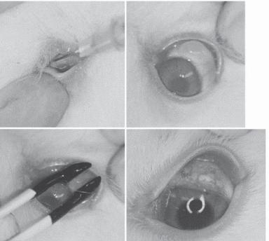
Figure 23.8. Photographic demonstration of surgical procedures of electroporation of MMP-3 cDNA into rabbit conjunctiva followed by trabeculectomy: (A) Injection of 0.1 mL of PBS containing CMV/MMP-3 vector (0.5 mg DNA/mL) into superior conjunctiva with 26 G needle; (B) bleb formation; (C) electroporation-mediated transfection using cup-shaped electrodes; (D) 7 days after trabeculectomy, which was performed 3 days after electroporation. (Reprinted from Mamiya K et al: Effects of matrix metalloproteinase-3 gene transfer by electroporation in glaucoma filter surgery, Exp Eye Res 79:405–10, copyright 2004, with permission from Elsevier.)
matrix metalloproteinases degrade extracellular matrix, thus reducing the rate of occlusion of surgically established ocular fistula. The stromelysin genes have been used for such a purpose in experimental models.
Protection of Retinal Neurons from Glaucoma-Induced Injury and Death [23.11]. A glaucoma-associated increase in the intraocular pressure, when reaching a certain level, often induces injury and apoptosis of the retinal neurons, a common cause for blindness. A strategy to prevent retinal neuron injury and death is to deliver genes encoding survival factors or antiapoptotic factors to the retina. Most growth factors are known to promote cell survival and their genes can be used for such a purpose. An example of cell survival factors is the brain-derived neurotrophic factor (BDNF). The transfer of the gene encoding this factor into the retina can effectively protect the retinal neurons from apoptosis and prolong the survival of these cells. In addition, genes encode ciliary neurotrophic factor (CNTF) and glial cell line-derived neurotrophic factor (GDNF) can also be used for such a purpose. These genes have been tested in experimental models, demonstrating promising results. Characteristics of several growth factors are listed in Table 23.6.

|
|
|
|
OCULAR DISORDERS |
983 |
|
TABLE 23.5. Characteristics of p21* |
|
|
|
|
||
|
|
|
|
|
|
|
|
|
Amino |
Molecular |
|
|
|
Proteins |
Alternative Names |
Acids |
Weight (kDa) |
Expression |
Functions |
|
|
|
|
|
|
|
|
P21 WAF1/ |
p21, CDK-interaction |
164 |
18 |
Heart, eye, |
Binding to and |
|
Cip-1 |
protein 1 (CIP1), |
|
|
bone |
inhibiting |
|
|
wildtype |
|
|
marrow, |
the activity |
|
|
p53-activated |
|
|
mammary |
of cyclin- |
|
|
fragment 1 |
|
|
gland, |
CDK2 or |
|
|
(WAF1), |
|
|
kidney |
cyclin- |
|
|
melanoma- |
|
|
|
CDK4 |
|
|
differentiation- |
|
|
|
and inducing |
|
|
associated |
|
|
|
cell cycle |
|
|
protein 6 |
|
|
|
arrest at G1 |
|
|
(MDA6), DNA |
|
|
|
and G2 |
|
|
synthesis |
|
|
|
|
|
|
inhibitor, |
|
|
|
|
|
cyclindependent kinase inhibitor 1
*Based on bibliography 23.10.
Cataract
Pathogenesis, Pathology, and Clinical Features [23.12]. A major lens disorder is cataract, which is characterized by progressive opacification of the lens. Lens opacity may be found in the center or the peripheral region of the lens. Pathogenic factors that cause cataract include infectious diseases (such as rubella, herpes simplex, and syphilis), ocular trauma, exposure to radiation, chemical toxicity, diabetes-related metabolic disorders, and aging (senile cataract). Senile cataract occurs much earlier in diabetics than in the general population. Long-term administration of corticosteroids enhances the progression of cataract. Cataract is often associated with visual impairment, such as image blurriness and duplication, alterations in color perception, and reduction in visual acuity.
Conventional Treatment of Cataract [23.12]. When cataract is a secondary disorder due to other diseases, such as infectious diseases, metabolic disorders, and diabetes, it is necessary to treat these causative diseases. The alleviation of these diseases prevents or slows down the progression of cataract. When the transparency of the lens is significantly reduced and the visual acuity is severely impaired, surgical removal of the lens is an effective approach for the treatment of cataract.
Molecular Regenerative Engineering [23.13]. The development of cataract is related to cell proliferation in the lens, often induced by diabetes-induced pathogenic alterations or inflammatory reactions in infectious diseases. An important strategy in molecular therapy for cataract is to deliver genes that encode anti-inflammatory and antiproliferative or proapoptotic proteins. There are two approaches that have been used for the treatment of cataract: transferring genes encoding proteins that activate cell mitosis-inhibiting or proapoptotic mechanisms and delivering antisense oligonucleotides or siRNA that inhibit the translation of mitogenic mRNAs.

984
TABLE 23.6. Characteristics of Selected Growth Factors*
|
|
Amino |
Molecular |
Gene |
|
|
Proteins |
Alternative Names |
Acids |
Weight (kDa) |
Locus |
Expression |
Functions |
|
|
|
|
|
|
|
Brain-derived |
BDNF |
247 |
28 |
11p13 |
Central nervous system |
Regulating the survival of |
neurotrophic |
|
|
|
|
(cortex, retina, and spinal |
neurons and stimulating |
factor |
|
|
|
|
cord), fetal testis |
embryonic development |
Ciliary neurotrophic |
CNTF |
200 |
23 |
11q12.2 |
Brain |
Promoting neurotransmitter |
factor |
|
|
|
|
|
synthesis and neurite |
|
|
|
|
|
|
outgrowth and regulating the |
|
|
|
|
|
|
survival of neurons and |
|
|
|
|
|
|
oligodendrocytes |
Glial cell line-derived |
Astrocyte-derived |
211 |
24 |
5p13.1-p12 |
Nervous system, kidney |
Promoting the survival and |
neurotrophic |
trophic factor 1, and |
|
|
|
|
differentiation of |
factor |
glial-cell-derived |
|
|
|
|
dopaminergic neurons, and |
|
neurotrophic factor |
|
|
|
|
preventing the apoptosis of |
|
|
|
|
|
|
motor neurons |
|
|
|
|
|
|
|
*Based on bibliography 23.11.
OCULAR DISORDERS |
985 |
For the first approach, the herpes simplex virus-thymidine kinase (HSV-tk) gene is a typical example. This gene encodes a protein kinase that catalyzes the phosphorylation of deoxynucleosides. When a deoxynucleoside analogue such as ganciclovir (an analog of 2′-deoxyguanosine) is present, the analogue can be phosphorylated into a deoxynucleotide, which can be incorporated into the genome of newly formed cells during mitosis. Unlike the natural types of deoxynucleotides, the incorporation of the deoxynucleotide analogues results in the termination of DNA synthesis, an effective process suppressing cell proliferation. Thus the herpes simplex virus-thymidine kinase gene and ganciclovir can be codelivered to the lens cells for the inhibition of cell proliferation. Such an approach prevents or reduces the progression of cataract.
For the second approach, an example is the use of the antisense oligonucleotides specific to the mRNAs of cell cycle regulatory proteins, such as cyclin G1. The selected genes can be injected into the anterior compartment of the eye. Experimental investigations have demonstrated that the transfer of antisense cyclin G1 oligonucleotides into the human lens epithelial cells in vitro induces downregulation of the cyclin G1 gene, an increase in cell apoptosis, and a decrease in cell proliferation. These observations suggest that antisense oligonucleotides to cell cycle regulators or mitogenic factors may be potentially used for the treatment of cataract.
Retinopathy
Pathogenesis, Pathology, and Clinical Features [23.14]. Retinopathy is a retinal disorder characterized by retinal arterial stenosis and occlusion, hemorrhage, edema, neuronal injury and apoptosis, reduction in visual acuity, and blindness. Retinopathy occurs due to several diseases, including atherosclerosis in the retinal arteries, systemic hypertension, and diabetes. Atherosclerosis induces stenosis and occlusion of the retinal arteries, leading to injury and death of retinal neurons. Systemic hypertension can induce pathological alterations in the retinal structures, including arteriolar reduction, hemorrhage, retinal edema, and focal ischemia. These alterations influence the function of the retinal neurons, reducing visual acuity. Long-term hypertension may cause severe retinal damage and blindness. Diabetes often induces wall thickening of the retinal arterioles, microaneurysms, hemorrhage, and neovascularization or angiogenesis. These changes result in the injury and death of retinal neurons and thus reduce visual acuity. The ultimate consequence of retinopathy is blindness. Retinopathy is the most common cause of visual loss in the elders.
Another major type of retinal disorder is inherited retinal degeneration. This disorder is characterized by progressive apoptosis of retinal neurons, ultimately leading to blindness. Retinitis pigmentosa is a common form of retinal degeneration. The pathogenic mechanisms of this disorder remain poorly understood. Molecular research has demonstrated that the mutation of several genes may contribute to the development of retinal degeneration. Potential genes include the retinal cyclic GMP phosphodiesterase (PDE) gene, the peripherin 2 gene, and the retinal pigmented epithelium (RPE) 65 gene. In transgenic animal models, the mutation of these genes is associated with retinal degeneration and visual impairment. The retinal cyclic GMP phosphodiesterase gene encodes a protein that regulates phototransduction in the retinal neurons. This protein is composed of catalytic α and β subunits and two inhibitory γ subunits. The mutation of the γ gene results in the loss of the catalytic activity of the α and β units, in association with retinal degeneration similar to that found in human retinitis pigmentosa. The mutation of other subunits also induces retinal degenerative changes. Peripherin 2 is a membrane