

Bioregenerative Engineering Principles and Applications - Shu Q. Liu
..pdf966 OCULAR REGENERATIVE ENGINEERING
muscles and the shape change of the lens control the focal distance of the eye. The ciliary body is connected to the iris, which contains smooth muscle cells and surrounds the pupil, the central opening of the eye. The iris is composed of circumferentially and radially aligned smooth muscle cells. The circumferential smooth muscle cells are controlled by parasympathetic nerves. The contraction of these muscles reduces the size of the pupil, resulting in a decrease in the amount of light entering the eye. The radial smooth muscle cells are controlled by sympathetic nerves. Their contraction induces the dilation of the pupil, increasing the amount of light entering the eye.
The nervous tunica is also called the retina, which is the internal layer of the eye. The retina is composed of a surface epithelial layer, known as the pigmented retina, and a neuronal layer, known as the sensory retina. The sensory retina consists of a large number of rod and cone photoreceptor neurons and relay neurons. The retina covers the internal surface of the eye except for the front area with the lens and the ciliary body. In the posterior center, there are two spots that contain highly concentrated photoreceptor neurons: the macula lutea and fovea centralis. These structures are capable of identifying fine objects and images. Near these structures, there is another spot, known as the optic disc, a location where blood vessels enter and the nerve fiber bundles from photoreceptor neurons leave the eye. The optic disc is not able to sense light because of the lack of photoreceptors.
The sensory retina is composed of three layers: photoreceptor neurons, bipolar neurons, and ganglionic neurons. Between these layers, there exist various types of association neurons. The photoreceptor layer contains rod and cone neurons. The rod neurons can sense light of low intensity and are insensitive to colors. These cells contain a protein complex, known as rhodopsin, which is responsible for the sensation of dim light. In addition, rhodopsin participates in the regulation of light adaptation. When a person is suddenly exposed to bright light, rhodopsin is rapidly degraded, reducing light-initiated stimulatory signaling activities. In contrast, when a person is suddenly exposed to dim light, the production of rhodopsin increases, enhancing the sensitivity of the retina to dim light.
The cone neurons are responsible for the sensation of colors and ordinary light. Color identification by these cells requires the presence of a critical level of bright light. Below such a critical level, these cells lose the capability of color identification. Cone cells contain protein complexes called iodopsins. These complexes are composed of three types of opsin proteins for the sensation of red, blue, and green colors. Each type is only sensitive to a narrow spectrum of light corresponding to a specific color. The distribution of the cone neurons differs from that of the rod neurons. The fovea centralis is primarily composed of cone neurons with almost no rod neurons. Thus this structure is for the sensation of color and bright light and, especially, for accurate identification of images. In contrast, the rod neurons are spread over the remaining retina, a distribution essential for the sensation of dim light.
The bipolar and ganglionic neurons in other retinal layers play critical roles in the transmission of optic signals from the rod and cone neurons to the central visual centers. The bipolar neurons synapse with the rod and cone neurons at one side and synapse with the ganglionic neurons at the other side. In these layers, there are several types of association neurons, including horizontal neurons, amacrine neurons, and interplexiform neurons. These neurons synapse with the photoreceptor, bipolar, and ganglionic neurons, and relay, integrate, and modify signals from the photoreceptor neurons. Nerve fibers from the ganglionic neurons converge to the optic disc, where they form the optic nerve, exit the eye,
OCULAR DISORDERS |
967 |
and enter the central visual centers of the brain, including the superior colliculi, lateral geniculate nuclei of thalamus, and visual cortex.
Enclosed within the eyeball are two compartments: the anterior and posterior compartments. The anterior compartment is the chamber between the cornea and the lens, and is filled with aqueous humor, a fluid that is produced by the ciliary processes, released into the anterior compartment, and returned to the vein through the trabecular meshwork and the canal of Schlemm. The aqueous humor circulates constantly with a stable hydrostatic pressure and supplies oxygen and nutrients to the cornea and lens. The obstruction of the trabecular meshwork and the canal of Schlemm results in an increase in the intraocular pressure, a disorder known as glaucoma (see page 979). The posterior compartment is the chamber surrounded by the retina and filled with vitreous humor, a transparent gel-like structure. The vitreous humor plays a critical role in the maintenance of the ocular shape and transmission of light.
The lens is an avascular, transparent, biconvex structure that is composed of two types of epithelial cells, including the cuboidal and fiber-like epithelial cells. The cuboidal epithelial cells are found on the anterior surface, whereas the fiber-like epithelial cells are found in the remaining body of the lens. The fiber-like cells are specially differentiated cells that do not contain nuclei and cellular organelles. Instead, these cells contain a special type of protein known as crystalline. The presence of crystalline renders the lens highly transparent. The crystalline-containing cells are enclosed by an elastic layer of tissue, which is connected to the ciliary body via the suspensory ligaments.
The ocular accessory structures include the eyelid, conjunctiva, lacrimal apparatus, and eye muscles. The eyelid is composed of several layers, including the skin, areolar connective tissue, skeletal muscles, tarsal plate, and palpebral conjunctiva. The function of the eyelid is to protect the eye from injury. The conjunctiva is a fibrous membrane that covers the internal surface of the eyelid (the palpebral conjunctiva) and the anterior surface of the eyes (the bulbar conjunctiva). The palpebral conjunctiva directly interacts with the cornea. Because of the presence of fluids, the friction between the conjunctiva and cornea is small. The lacrimal apparatus is composed of the lacrimal gland, lacrimal canaliculi, lacrimal sac, and nasolacrimal duct. The lacrimal gland produces tears, which are released to the external surface of the eye. The production and release of tears are controlled by the parasympathetic nerves. Tears serve as a lubricant for the interaction of the eyeball with the eyelid. Excessive tears enter the lacrimal canaliculi through two openings called punctas, flow into the nasal cavity via the lacrimal sac and nasolacrimal duct. Each eyeball is associated with six skeletal muscle bundles. These muscles are anchored to the external surface of the sclera and control the movement of the eyeball.
OCULAR DISORDERS
Corneal Injury
Pathogenesis, Pathology, and Clinical Features [23.2]. The cornea is a structure exposed to the exterior environment and is subject to various hazards, such as mechanical injury, chemical corrosion, radiation, and infection by bacteria and viruses. Corneal injury due to mechanical trauma and chemical corrosion is commonly seen. Corneal injury often induces inflammatory reactions, followed by fibrosis and scar formation in the cornea, reducing light transmission. Since the cornea is a collagen-rich structure, disorders with collagen degradation may affect the function of the cornea. Metabolic disorders can also
968 OCULAR REGENERATIVE ENGINEERING
induce dysfunction of the cornea. For instance, hypercalcemia is associated with calcium precipitation underneath the cornea epithelial cells. Cystinosis can cause the formation of cystine crystals in the cornea. Hypercholesterolemia induces cholesterol deposition in the cornea. All these disorders influence light transmission through the cornea and induce visual impairment.
Conventional Treatment of Cornea Injury [23.2]. There are two strategies for the treatment of corneal disorders: removing the factors that cause corneal abnormalities and conducting corneal transplantation. When causative factors can be identified, these factors should be removed, if possible, to reduce or stop the progression of corneal abnormalities. For example, ocular bacterial infection should be controlled by local administration of antibiotics. When hypercalcemia is identified as a causative disorder, the blood calcium concentration should be reduced to the normal level. When severe corneal scars and opacity develop, the cornea can be replaced with an allogenic corneal specimen, a procedure known as corneal transplantation.
Molecular Regenerative Engineering. Molecular engineering approaches can be applied to corneal disorders. Corneal disorders often involve molecular activities, such as activation of pro-inflammatory factors, upregulation of proliferative genes, and production of extracellular matrix. Thus, molecular strategies for the treatment of corneal disorders are to suppress inflammation and selectively inhibit the proliferation of certain cell types such as fibroblasts. Selected genes can be prepared and used for the treatment of ocular disorders. For example, corneal haze and cloudiness after mechanical injury are due to excessive inflammatory reactions, including leukocyte infiltration, cell proliferation, and extracellular matrix deposition. Genes encoding anti-inflammatory and antiproliferative proteins can be used to suppress inflammatory reactions and fibrous changes. In addition, dominant negative genes for proinflammatory and mitogenic factors can also be used for treating corneal inflammation. Another example is the molecular treatment of primary glaucoma. This disorder is induced by the obstruction of the trabecular meshwork by excessive production of extracellular matrix. Genes encoding matrix metalloproteinases, which degrade extracellular matrix components, can be used for the treatment of glaucoma.
Given the anatomical features of the ocular system, several approaches can be used for gene delivery. For the molecular treatment of the corneal epithelial disorders, a topical gene delivery is effective. For disorders of the iris, ciliary body, and trabecular meshwork, gene injection into the anterior compartment is required. For retinal disorders, it is necessary to conduct intravitreal gene injection. As for other organs and tissues, various methods can be used to mediate gene delivery to the ocular system, depending on the anatomical features of and cell types in the target tissue. For instance, electroporation is an effective method for gene delivery to the corneal epithelial cells, but may not be a suitable method for gene delivery to the intraocular structures. Genetically modified adenoviruses and retroviruses are often used for mediating gene delivery into ocular tissues, including the cornea, trabecular meshwork, and retina. These mediating methods have been shown to be more effective than other mediating methods, such as saltand liposome-mediated delivery, for the ocular system.
Molecular engineering approaches have been developed and used for treating several corneal disorders, including immune rejection of corneal transplants, corneal inflammation and haze, and corneal complications due to metabolic disorders such as mucopolysaccharidosis. These approaches are discussed in the following sections.
OCULAR DISORDERS |
969 |
Molecular Therapies for Corneal Immune Rejection [23.3]. Allogenic corneal transplantation is an effective approach for the treatment of corneal dysfunction. However, the presence of functional epithelial cells, which are essential for successful corneal transplantation, often causes immune reactions, resulting in acute rejection. It is often necessary to administrate immune suppressor agents to patients with corneal transplantation. However, these immune suppressor agents induce side effects by inhibiting the activity of the entire immune system. Furthermore, it is required to conduct daily agent deliveries. Molecular engineering approaches can be used to overcome these problems. Genes that encode immune suppressor cytokines and antisense oligonucleotides for immune activator genes can serve as immune suppressor agents. Given the anatomical features of the cornea, it is relatively easier to deliver genes to the cornea than to the internal structures. Three approaches may be used for corneal gene delivery: application of genes to the exterior surface of the cornea, gene injection to the anterior compartment, and augmentation of gene delivery by electroporation (see page 444 for these methods). Typical genes for corneal disorders include the CD152 and interleukin (IL)10 genes, which have been used for the treatment of corneal transplant immune rejection.
The CD152 gene (CTLA-4) encodes a membrane protein in the T lymphocytes. The CD152 protein exerts an inhibitory effect on T lymphocyte-related immune reactions. When allogenic tissues are transplanted to the host, the allogenic antigens activate antigenpresenting cells (APCs), which in turn interact with the T lymphocytes, leading to activation of the T lymphocytes and initiating immune reactions. In particular, a cell membrane protein known as CD80 can interact with another membrane protein CD86 to form complexes. The CD80 and CD86 complexes on the APC surface interact with CD28 (see Table 23.1) on the T lymphocyte surface, eliciting co-stimulating signals for the activation of the T lymphocytes. The CD152 complexes on the T lymphocyte surface, when present, can bind to CD80 and CD86 in antigen-presenting cells, suppressing the activity of these cells as well as the T lymphocytes. The overexpression of the CD152 gene by gene transfer has been shown to induce the arrest of T lymphocyte division, reduce immune responses, prevent corneal immune rejection, and prolong the survival of transplanted allogenic cornea. The CD152 gene can be conjugated with an Ig gene, forming a recombinant gene complex, which can facilitate gene delivery and expression.
Another gene used for the treatment of corneal transplant immune rejection is the interleukin-10 gene (see page 634 for characteristics of IL10). This gene encodes a cytokine that suppresses the activity of T lymphocytes. Experimental investigations have demonstrated that the interleukin-10 gene can be effectively transferred into more than 70% of the epithelial cells of the cornea with a virus-mediated gene transfer approach. The transferred gene can be expressed for about 3 weeks. Such an approach has been shown to reduce immune responses in transplanted allogenic cornea and prolong corneal survival.
Molecular Therapies for Corneal Inflammation and Fibrosis [23.4]. Corneal inflammation is induced by trauma and therapeutic keratectomy. Inflammation often results in epithelial cell proliferation, extracellular matrix production, and fibrosis. Thus, the principle of molecular engineering therapy for corneal inflammation is to introduce genes that encode antiproliferative proteins. A gene encoding the dominant negative cyclin G1 (Table 23.2) protein has been constructed and used to treat corneal inflammation. Cyclin G1 plays a critical role in stimulating the progression of the cell division cycle. The

970
TABLE 23.1. Characteristics of Selected Molecules that Regulate Immune Responses*
|
|
Amino |
Molecular |
|
|
Proteins |
Alternative Names |
Acids |
Weight (kDa) |
Expression |
Functions |
|
|
|
|
|
|
CD28 |
Antigen CD28, T-cell antigen CD28 |
220 |
25 |
T cell, B cell |
Regulating CD4-positive T-cell survival |
|
|
|
|
|
and proliferation, inducing |
|
|
|
|
|
interleukin-2 production from T cells, |
|
|
|
|
|
and promoting the development of |
|
|
|
|
|
T-helper type-2 (Th2) cells |
CD80 |
CD28 antigen ligand 1 (CD28LG1), |
288 |
33 |
B cell, dendritic cell, monocyte, |
Interacting with CD28 on T cells and |
|
B lymphocyte activation antigen |
|
|
mast cell, nervous system, |
regulating T-cell proliferation and |
|
B7-1, activation B7-1 antigen, |
|
|
blood vessels |
activation |
|
B7-1 antigen |
|
|
|
|
CD86 |
CD86 antigen, CD28 antigen |
329 |
38 |
T cell, B cell, monocytes, |
A member of the immunoglobulin |
|
ligand 2 (CD28LG2), B |
|
|
macrophages, dendritic cells, |
superfamily expressed by antigen- |
|
lymphocyte activation antigen |
|
|
vascular endothelial cells, |
presenting cells, binding to CD28 on |
|
B7-2, Activation B7-2 antigen, |
|
|
smooth muscle cells, bone |
T cells to regulate T-cell proliferation, |
|
CTLA4 counterreceptor B7.2, |
|
|
marrow, intestinal epithelial |
survival, and activation, and binding |
|
LAB7-2, B70 |
|
|
cells |
to cytotoxic T-lymphocyte-associated |
|
|
|
|
|
protein 4 on T cells to negatively |
|
|
|
|
|
regulate T-cell activation and |
|
|
|
|
|
diminish T-cell-mediated immune |
|
|
|
|
|
responses |
CD152 |
Cytotoxic T lymphocyte associated |
223 |
25 |
T cell |
A member of the immunoglobulin |
|
4, cytotoxic T lymphocyte |
|
|
|
superfamily that inhibits the activity of |
|
antigen 4 (CTLA4) |
|
|
|
T cells |
|
|
|
|
|
|
*Based on bibliography 23.3.

|
|
|
|
OCULAR DISORDERS |
971 |
|
TABLE 23.2. Characteristics of Cyclin G1* |
|
|
|
|||
|
|
|
|
|
|
|
|
Alternative |
Amino |
Molecular |
|
|
|
Proteins |
Names |
Acids |
Weight (kDa) |
Expression |
Functions |
|
|
|
|
|
|
|
|
Cyclin G1 |
Cyclin G, |
295 |
34 |
Lymphocytes, lung, |
Regulating cell |
|
|
CCNG |
|
|
kidney, intestine, |
division |
|
|
|
|
|
spleen, thymus, |
|
|
testis, ovary, prostate gland, skeletal muscle
*Based on bibliography 23.4.
negative dominant cyclin G1 protein competes with the wild-type or natural form of cyclin G1 for substrate binding, but does not activate the substrate. Thus, the introduction of this dominant negative gene to the corneal epithelial cells suppresses the activity of the natural cyclin G1, induces the arrest of the cell division cycle, and reduces cell proliferation and matrix production. These processes are associated with a decrease in inflammatory reactions and suppression of extracellular matrix production and haze development. Experimental investigations have demonstrated promising results for the transfer of the dominant negative cyclin G1 gene into the cornea with laser keratectomy-induced injury. Dominant negative genes constructed for other types of mitogenic signaling factors, such as growth factor receptors, protein tyrosine kinases, and cell cycle regulators, can also be used to inhibit corneal inflammatory reactions.
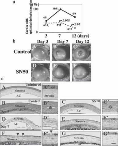
Pharmacological inhibitors can be used to suppress the activities of inflammatory factors. One example is the use of the nuclear factor κB inhibitor SN50. Nuclear factor κB is a transcription factor that stimulate the expression of inflammatory genes. The suppression of the activity of nuclear factor κB may reduce inflammatory reactions in corneal injury. Experimental investigations have demonstrated that the topical application of SN50 can facilitate the healing process of alkali-induced corneal injury (Fig. 23.2).
Molecular Therapies for Corneal Complications Due to Mucopolysaccharidosis Type VII (MPSVII) [23.5]. Mucopolysaccharidosis type Vll is an hereditary disorder due to the deficiency of the enzyme β-glucuronidase (GUSB) (Table 23.3), which breaks down proteoglycans. This disorder is associated with the lysosomal accumulation of undegraded glycosominogycans (GAGs), resulting in corneal abnormalities and opacity. The transfer of the β-glucuronidase gene into the corneal epithelial cells induces the over-expression of β-glucuronidase, prevents the accumulation of undegraded glycosominogycans, and reduces corneal opacity.
Cellular and Tissue Engineering. Cellular and tissue engineering approaches have been established and used to treat ocular disorders in experimental models and preliminary clinical trials. As for other organ and tissue systems, a successful replacement of a malfunctioned ocular tissue requires the construction of a functional cellular and tissue structure and the integration of the constructed structure into the ocular system. To date, cellular and tissue engineering approaches have been used to repair and reconstruct disordered cornea in experimental models and clinical trials. However, the application of cellular and tissue approaches to other ocular tissues has been limited because of difficulties in the construction and assembly of functional ocular structures, such as the retina and lens.
972 OCULAR REGENERATIVE ENGINEERING
Figure 23.2. Healing of alkali-burned mouse cornea treated with topical SN50, an inhibitor of NFκB. (a) Percentage of corneas with epithelial defect (including ulceration) at each healing interval. The incidence of epithelial defect/ulceration is significantly higher in the control group than in the SN50-treated group at days 7 and 12 as judged by the χ2 test. (b) Macroscopic observation shows similar initial resurfacing in both control (A) and SN50-treated groups (D) at day 3 after alkali burning. Recurrence of the epithelial defect with stromal opacification is observed more frequently in the control group at days 7 (B) and 12 (C) as compared with SN50-treated group (E, F). (c) Histology of burned corneas stained with H&E. (A) An uninjured cornea. Stratified epithelium and stroma are seen. There is no histological difference between central corneas in the control (B) and SN50-treated group (C) at day 3. The epithelium shows a large defect and many inflammatory cells are observed. At day 7 the burned cornea in the control (D) shows more stromal inflammation, and a large epithelial defect as compared with the SN50-treated corneas that has been resurfaced with a thin epithelium (E). At day 12 the control cornea still shows marked inflammation and hypercellularity in the stroma (F), whereas the treated cornea exhibits a wellregenerated epithelium with a less stromal inflammation (G). Regenerated epithelium in control exhibits conjunctiva-like appearance with goblet cells (arrowheads). A′–G′ are high-magnification pictures of the central area of the healing corneas in A–G, respectively. Scale bar: 100 μm (A–G), 25 μm (A′–G′). (Reprinted with permission from Saika S et al: Am J Pathol 166:1393–1403, copyrights 2005.)

|
|
|
|
OCULAR DISORDERS |
973 |
|
TABLE 23.3. Characteristics of b-Glucuronidase* |
|
|
|
|||
|
|
|
|
|
|
|
|
Alternative |
Amino |
Molecular |
|
|
|
Proteins |
Names |
Acids |
Weight (kDa) |
Expression |
Functions |
|
|
|
|
|
|
|
|
β-Glucuronidase |
βG1 |
651 |
75 |
Retinal |
An enzyme that |
|
|
|
|
|
pigmented |
degrades |
|
|
|
|
|
epithelial |
proteoglycans |
|
cells,
leukocyte, liver, kidney, spleen, placenta, intestine, pancreas
*Based on bibliography 23.5.
Corneal dysfunction requires corneal replacement by transplantation. Allogenic cornea is often used for such a purpose. However, there two major problems for allogenic cornea transplantation: immune rejection and shortage of cornea donors. In particular, immune responses induce injury and death of corneal epithelial and endothelial cells, leading to opacification of the transplanted cornea. Several cellular and tissue engineering strategies have been developed and used to overcome these problems, including: (1) corneal surface reconstruction with epithelial stem cells or autogenous epithelial cells, which can be applied to native or transplanted corneas with injured epithelial cells and (2) corneal reconstruction with extracellular matrix and polymeric materials.
Corneal Surface Reconstruction [23.6]. The cornea is covered at the external surface with an epithelial layer. The injury or denudation of this layer induces inflammatory reactions and fibrosis, resulting in alterations in the optical properties of the cornea and visual acuity. In such a case, it is necessary to reconstruct the corneal surface. Corneal surface reconstruction can be accomplished by using an epithelial cell layer constructed in vitro. Several types of cells can be used to construct an epithelial cell layer: limbal epithelial stem cells, adult epithelial cells, and stem cells. Epithelial stem cells are present in the limbal region or the junction of the cornea and conjunctiva. These cells can differentiate into corneal epithelial cells in corneal injury. The deficiency of limbal stem cells can cause ocular surface disorders, leading to blindness. In corneal injury, limbal stem cells can be collected and used for enhancing corneal cell regeneration by cell transplantation.
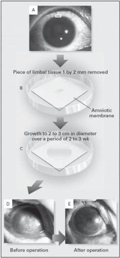
Limbal stem cells can be collected from several sources, including the cadaver eyes, the conjunctival limbal tissue from the donors, and autogenous limbal tissue. Harvested limbal epithelial stem cells by biopsy can be cultured and expanded in vitro on a suitable carrier membrane, such as a polymeric or natural matrix membrane, forming a transplantable epithelial membrane structure. The cultured epithelial cells can produce extracellular matrix, which serve as a basal meshwork for the formation of a stable epithelial membrane structure. The epithelial membrane is readily adhesive and can be used for the construction of a corneal epithelium-like structure (Fig. 23.3). Clinical investigations have shown that this approach can be used to effectively prevent corneal inflammatory reactions and improve visual acuity (Fig. 23.4).
Natural biological membranes can also be used to serve as epithelial cell carriers. A typical example is the human amniotic membrane, which can be used as a basal membrane
974 OCULAR REGENERATIVE ENGINEERING
Figure 23.3. Transplantation of autologous limbal epithelial cells cultured on amniotic membrane. Limbal tissue (1 × 2 mm) was removed by lamellar keratectomy from the superior limbus of the healthy contralateral eye (panel A). The explanted tissue was placed on amniotic membrane in a 35-mm dish containing 1.5 mL of culture medium (panel B). After 2–3 weeks, the epithelial cells had grown and spread to form a circular sheet of cells with a diameter of 2–3 cm (panel C). The cultured limbal epithelial cells with amniotic membrane were then transplanted to the diseased eye (panels D and E). (Reprinted with permission from Tsai RJ, Li LM, Chen JK: New Engl J Med 343:86–93, copyright 2000 Massachusetts Medical Society. All rights reserved.)
for culturing epithelial cells and constructing corneal epithelial layers. The constructed epithelial layer can be directly applied to the exterior surface of the cornea. Since the epithelial cell layer is usually thin and self-adhesive, it is not necessary to fast the cell layer with suture stitches or adhesives. Compared to synthetic biomaterials, a biological membrane is compatible with cells and provides a suitable substrate for the formation of an epithelial cell layer.
OCULAR DISORDERS |
975 |
(A) |
|
(B) |
|
(C) |
(D) |
|
(E) |
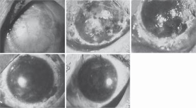
Figure 23.4. Serial photographs of the eye of patient 6 before and after transplantation initial examination. Panel A reveals corneal opacity with central corneal erosion and neovascular growth extending into the entire cornea for 4–6 mm before operation. Lamellar keratectomy was performed to remove the entire opacified limbal and corneal area to a thickness of 1/3rd of the corneal layer (panel B). Limbal epithelial cells with the amniotic membrane substrate were transplanted onto the denuded limbal and corneal area. Photographs were taken 1 day (panel B), 7 days (panel C), 30 days (panel D), and 450 days (panel E) after the operation. (Reprinted with permission from Tsai RJ, Li LM, Chen JK: New Engl J Med 343:86–93, copyright 2000 Massachusetts Medical Society. All rights reserved.)
Adult epithelial cells can proliferate and can be used for constructing the corneal external surface. Ideally, autogenous corneal epithelial cells should be used for corneal reconstruction. However, in patients with corneal disorders, corneal epithelial cells are usually not available. Given the fact that most epithelial cell types on the exterior surface of the body exhibit certain common phenotypes, it is conceivable that epithelial cells from other exterior tissues may be used for constructing a corneal epithelial structure. A candidate epithelial cell type is the oral mucosal epithelial cells. These cells can be easily identified, harvested, manipulated, and expanded in culture. A major advantage of using the oral mucosal epithelial cells is that cells can be harvested from the host patients, thus avoiding immune rejection responses.
Prepared oral mucosal epithelial cells can be seeded on a membrane for the construction of an epithelial layer. Researchers have developed a temperature-sensitive synthetic polymer material, which can be used to construct membranous cell carriers (Fig. 23.5). Epithelial cells or stem cells can be seeded on the carrier for expansion and formation of an epithelial membrane. A reduction in temperature causes shrinkage of the carrier polymeric material, inducing the separation of the cells from the carrier membrane. Since this approach does not require the use of proteinases for cell separation from the membrane carrier, epithelial cells remain intact and functional.
Epithelial membranes constructed with autogenous oral epithelial cells have been shown to express corneal epithelial markers, such as keratin-3 (Chapter 23 opening figure), and have been applied to patients with complete denudation of the corneal epithelium with severe impairment of visual acuity. These investigations have demonstrated that reepithelialization of the corneal surface occurs within one week, resulting in the restoration of the corneal transparency and significant improvement of visual acuity (Fig. 23.6). In