

Bioregenerative Engineering Principles and Applications - Shu Q. Liu
..pdf1006 OCULAR REGENERATIVE ENGINEERING
Leonard DG, Gorham JD, Cole P, Greene LA, Ziff EB: A nerve growth factor-regulated messenger RNA encodes a new intermediate filament protein, J Cell Biol 106:181–93, 1988.
Moncla A, Landon F, Mattei MG, Portier MM: Chromosomal localisation of the mouse and human peripherin genes, Genet Res 59:125–9, 1992.
Thompson MA, Ziff EB: Structure of the gene encoding peripherin, an NGF-regulated neuronalspecific type III intermediate filament protein, Neuron 2:1043–53, 1989.
Bennett J, Tanabe T, Sun D, Zeng Y, Kjeldbye H et al: Photoreceptor cell rescue in retinal degeneration (rd) mice by in vivo gene therapy, Nat Med 2:649–54, 1996.
Bennett J, Zeng Y, Bajwa R, Klatt L, Li Y et al: Adenovirus-mediated delivery of rhodopsin-pro- moted bcl-2 results in a delay in photoreceptor cell death in the rd/rd mouse, Gene Ther 5:1156–64, 1998.
Tsang SH, Gouras P, Yamashita CK, Kjeldbye H, Fisher J et al: Retinal degeneration in mice lacking the gamma subunit of the rod cGMP phosphodiesterase, Science 272:1026–9, 1996.
Human protein reference data base, Johns Hopkins University and the Institute of Bioinformatics, at http://www.hprd.org/protein.

1008 SKIN REGENERATIVE ENGINEERING
ANATOMY AND PHYSIOLOGY OF THE SKIN [24.1]
The skin is composed of two layers: the epidermis and dermis. The epidermis is a layer of stratified squamous epithelial cells. There are several types of cell in the epidermal layer: keratinocytes, melanocytes, and sensory cells. Keratinocytes are the largest group of epithelial cells, which produce and release keratin, a protein forming the surface keratin layer of the epidermis. The keratin layer protects the epidermis from mechanical injury and chemical corrosion. Melanocytes are pigmented epithelial cells that determine the color of the skin. Sensory cells are specialized nerve cells. The endings of these cells are responsible for sensing mechanical contacting and temperature changes. The epidermis resides on a matrix layer known as the basement membrane, which separates the epidermis from the dermis.
The dermis is a layer of connective tissue and serves as the base for the epidermis, providing structural support and mechanical strength to the skin. The dermis is composed of several cell types, including the fibroblasts, adipocytes, and macrophages. Fibroblasts are responsible for the generation of extracellular matrix components such as collagen, elastin, and proteoglycans. Adipocytes are cells for the metabolism and storage of lipids. Macrophages are originated from the bloodborne monocytes, which are transformed to macrophages when migrating from the blood to tissue. These cells are responsible for the destruction of bacteria and clearance of cell debris. All cell types in the dermal layer reside in a meshwork of extracellular matrix, composed of collagen fibers, elastic fibers, and ground substances. In the dermal tissue, collagen fibers are constructed primarily with collagen type I. Elastic fibers consist of elastin, microfibrils, and microfibril-associ- ated proteins. Ground substances are composed of proteoglycans (see Chapter 4 for details). These matrix components are responsible for the organization, integrity, stability, elasticity, and strength of the dermis. The dermis is also composed of several other structures, including blood vessels (arteries, capillaries, and veins), lymphatic vessels, nerve endings, hair follicles, and sweat and sebaceous glands.
The dermis resides on a deeper layer of connective tissue, known as subcutaneous tissue. This layer is composed of several cell types, including fibroblasts, adipocytes, and macrophages, and extracellular matrix. Large blood vessels (arteries and veins) and nerve bundles are surrounded by the subcutaneous tissue. A large fraction of adipocytes in the body is found in this type of tissue. The subcutaneous tissue provides a connection between the skin and internal structures, such as bones and skeletal muscles.
There are several functions for the skin, including protection, regulation of temperature, and sensation. Given the anatomical location and histological structure, the skin serves to protect the internal tissues and organs from injury induced by environmental factors, including chemical toxins, corrosive agents (acids and bases), mechanical forces (stretching, compressing, and shearing), microorganisms (bacteria and viruses), and radiations (UV and X-ray). The skin contains various types of sensory nerve endings, which can sense temperature changes, pressure, mechanical contacting, and chemical corrosion. These sensory structures are critical to the protection of the body from dangerous environmental factors. The skin consists of a rich network of blood vessels, which play an important role in the regulation of body temperature. An increase in the body temperature induces arteriolar dilation, leading to an increase in blood flow to the body surface and thus facilitating heat loss. A decrease in the body temperature exerts an opposite effect. The excretion of sweat is another mechanism that facilitates the loss of body heat and reduces body temperature. The skin participates in the synthesis of vitamin D, a hormone

SKIN DISORDERS |
1009 |
that regulates the metabolism of calcium and phosphate (stimulating the absorption of calcium and phosphate in the intestines, and increasing the level of blood calcium and phosphate). Vitamin D is synthesized from a cholesterol molecule, 7-dehydrocholesterol, which is converted into cholecalciferol under the stimulation of ultraviolet light. Cholecalciferol is released into the blood and converted into vitamin D in the liver via hydroxylation. In addition, the skin is an important structure that protects the body from water loss.
SKIN DISORDERS
Skin Injury
Pathogenesis, Pathology, and Clinical Features [24.2]. The skin directly interacts with the external environment and is often subject to hazard factors, such as heat, chemical toxins, radiation, electricity, and mechanical impacts. These factors induce various types of skin injury ranging from reversible inflammation to the destruction of the skin. The severity of skin injury is dependent on the skin layers involved and is classified into three degrees. First-degree skin injury is defined as injury limited to the epidermis, characterized by the presence of redness, an increase in local temperature, itching, and pain in the injured area. Examples of first-degree injury include scalds and sunburn. Second-degree injury involves the epidermis and dermis, and is characterized by severe local pain, swelling, and the formation of blisters containing fluids or blood, which arise from tissue cleavage within the epidermis (intraepidermal vesication) or the separation of the epidermis from the dermis (subepidermal vesication). Sweat glands and hair follicles are often damaged in the second degree of injury. Third-degree of skin injury involves the epidermis, dermis, and subcutaneous tissue, characterized by the destruction of all skin layers, the exposure of the subcutaneous tissue or other internal tissues, such as the skeletal muscles, tendons, ligaments, and bones, in association with massive swelling. Since sensory nerve endings are destroyed, third degree skin injury may not be associated with severe pain. The first and second degrees of skin injury can be self-healed within 1 to 3 weeks without the formation of scars. The recovery of the third degree of skin injury is often associated with scar formation. In severe cases with the exposure of skeletal muscles, tendons, ligaments, and bones, distortion and malfunction of the internal tissues may occur.
The impact of skin injury on the function of the skin and internal tissues is dependent on the area and degree of the injury. A small area of skin injury, referred to as minor injury, may not significantly influence the function, except that scars may form when the subcutaneous tissue is involved. A large area of third-degree skin injury, referred to as major injury, can cause pathological changes in not only the skin but also internal tissues and organs. Major changes in the skin include loss of the protective function and loss of body fluids. The loss of the protective function renders the skin vulnerable to bacterial and viral infection. Patients with fireor chemical-induced injury are often associated with bacterial infection, the most common cause of death in these patients. Because of the destruction of the skin, water loss by evaporation is greatly enhanced in the area of injury, resulting in a reduction in the volume of blood and interstitial fluids. In addition, scar formation may result in distortion of the subcutaneous and internal tissues. When scars form over a joint, the flexibility of the joint will be reduced, influencing the mobility of the joint and extremities.

1010 SKIN REGENERATIVE ENGINEERING
The pathological effects of skin injury are not limited to the skin. A large area of third-degree skin injury can result in malfunction of the internal tissues, organs, and systems. When internal tissues such as skeletal muscles and bones are injured, the functions of these tissues are impaired. Severe skin injury may also involves the vascular, lymphatic, and hormonal systems. Injured cells can release inflammatory mediators, such as histamine and prostaglandins. These mediators act on capillary endothelial cells and increase the permeability of capillaries, resulting in the transport of fluids from the blood to the interstitial tissue and a decrease in the volume of circulating blood. In severe cases, the deficit of blood volume may affect the performance of the heart and leads to oxygen deficiency in peripheral tissues and organs. In the case of bacterial infection, toxins released by the bacteria, together with inflammatory mediators released by infected and injured cells, may induce cell malfunction in the brain, heart, kidney, and liver, resulting in the impairment and failure of these organs.
Conventional Treatment of Skin Injury [24.2]. For the first and second degree of skin injury, injured skin can usually be self-cured. However, the third degree of skin injury cannot be self-cured and may result in serious clinical consequences. There are several strategies for the treatment of third-degree skin injury. These include the restoration of fluid and electrolyte balance, protection of the skin from bacterial infection, coverage of injured skin to prevent water loss and bacterial infection, and prevention of scar formation. Water loss and increased capillary permeability result in a reduction in the volume of circulating blood and imbalance of electrolytes. Thus, patients with severe skin injury should be treated to restore the water and electrolyte balance. Bacterial infection occurs in almost all cases of sever skin injury. Antibiotics should be used to prevent bacterial infection. The most important treatment for severe injury is skin transplantation. In principle, all areas of third-degree skin injury should be covered with autogenous skin specimens collected from intact areas of the patient. This is a critical treatment for the prevention of water loss and bacterial infection. In most cases of third-degree skin injury, however, it is difficult to collect sufficient skin specimens. Artificial skin substitutes can be constructed and used for the treatment of severe skin injury. When scar formation influences the function of the joints and extremities, the scar should be removed and replaced with intact skin specimens or skin substitutes.
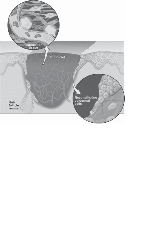
Skin Regenerative Engineering. Skin regenerative engineering is to develop cell-based functional skin substitutes that can be used for the coverage of injured skin or replacement of the lost skin. Skin regenerative approaches are established based on the natural healing processes of skin wounds. In response to skin injury, the wound site is rapidly plugged with fibrin clots. Injured epithelial cells and fibroblasts at the wound site may release cytokines and growth factors, which attract leukocytes and fibroblasts to the wound site, inducing inflammatory reactions and formation of granulation tissue. The vascular endothelial cells are activated in response to angiogenic factors to induce the formation of new blood vessels. The epithelial cells at the edge of the wound are stimulated to proliferate and migrate over the wound area to form a new epidermal layer. The fibroblasts within the wound produce and release extracellular matrix, which contributes to fibrosis and scar formation (Fig. 24.1). When the skin wound only involves the epidermis and a small area of the dermis (firstand second-degree of wounds), the wound can be completely selfhealed. However, when the wound involves the deep subcutaneous tissue (third-degree wounds), the wound cannot be self-healed, often resulting in permanent scars and distor-
SKIN DISORDERS |
1011 |
Figure 24.1. Schematic representation of the key players in the healing of a skin wound. The defect is temporarily plugged with a fibrin clot, which is infiltrated by inflammatory cells, fibroblasts, and a dense capillary plexus of new granulation tissue. An epidermal covering is reconstituted from the edges of the wound and from the cut remnants of hair follicles. At the migrating keratinocyte leading edge, cells bore a passageway enabling them to crawl beyond the cut basal lamina and over provisional matrix and healthy dermis, Cell division occurs back from the leading edge. Monocytes emigrate from wound capillaries into the granulation tissue, which contracts by means of smooth- muscle-like myofibroblasts that tug on one another and the surrounding collagen matrix. (Reprinted with permission from Martin P: Wound healing—aiming for perfect skin regeneration, Science 276:75–81, copyright 1997 AAAS.)
tion of the skin and involved joints. For third-degree of skin wounds, it is necessary to cover the wounds with skin substitutes and reduce inflammatory reactions, which facilitate the healing process of skin wounds. Skin regenerative engineering approaches are established for these purposes. A skin substitute can be generated by seeding and growing stem cells or epidermal cells on extracellular matrix or synthetic scaffolds. Several factors should be taken into account for the construction of skin substitutes. These include cell types, matrix types and forms, appropriate growth stimulators, and a suitable growth system. These factors are discussed as follows.
Cell Types for Constructing Skin Substitutes [24.3]. Several cell types can be used for the construction of skin substitutes. These include embryonic stem cells, multipotent fetal

1014 SKIN REGENERATIVE ENGINEERING
CD34 and AC133-2 (an isoform of CD133), have also been identified as potential markers for epidermal stem cells. Antibodies for these proteins can be used for the identification of epidermal stem cells. However, mature epidermal cells and follicle cells may express some of these proteins in certain developmental stages. These marker proteins should be used in caution. The use of multiple protein markers may help to identify epidermal stem cells.
Matrix Scaffolds for Constructing Skin Substitutes [24.4]. Epidermal cells reside on the dermis, a soft connective tissue containing fibroblasts and extracellular matrix that is constituted with collagen fibers, elastic fibers, and proteoglycans. These matrix components not only provide a structural basis for the integrity and mechanical strength of the skin, but also play critical roles in the regulation of cell attachment, proliferation, migration, regeneration, and wound repair. Thus, extracellular matrix is an essential component for skin regeneration and reconstruction. Collagen matrix is commonly used as a substrate for culturing epidermal cells and constructing skin substitutes. Artificially constituted collagen gel and natural collagen matrix collected from the host or allogenic patients can be used for such a purpose. Collagen stimulates cell adhesion, proliferation, and migration, thus enhancing skin regeneration. Other matrix components, such as fibronectin, proteoglycans, and fibrin, can also be used or added to the collagen matrix to construct matrix scaffolds for skin regeneration and reconstruction.
In addition to natural extracellular matrix components, synthetic polymers, such as polyethylene glycol and polyglycolide, can be used for skin regeneration and reconstruction. These polymers are often used as substrates for seeding and culturing cells or for delivering biological substances that are required for cell growth and differentiation. In particular, the use of biodegradable polymers, such as polyglycolide and poly(glycolide- L-lactide), may provide suitable conditions for the growth of epidermal cells. Biological substances can be easily incorporated into the polymer matrix and delivered to the target cells. The polymer matrix can serve as a substrate for cell attachment, growth, migration, and pattern formation. By controlling the degradation of the polymeric matrix, seeded cells can gradually form a natural structure with cell-synthesized extracellular matrix, which replaces the polymeric matrix and provide a permanent substrate for grown cells.
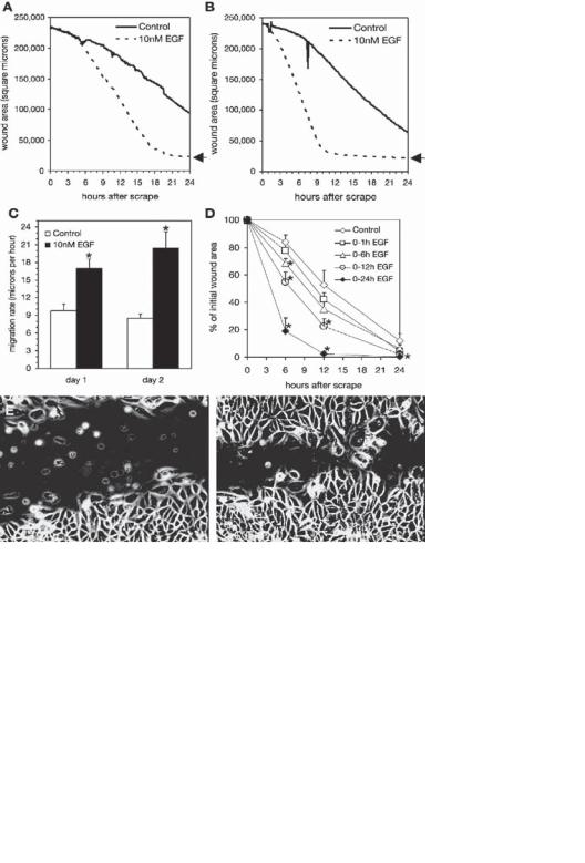
Growth Factors for Stimulating the Growth of Epidermal Cells [24.5]. Epidermal cells express a number of growth factors and cytokines, including epidermal growth factor (EGF), keratinocyte growth factor (KGF), fibroblast growth factor (FGF), platelet-derived growth factor (PDGF), hepatocyte growth factor (HGF), vascular endothelial growth factor (VEGF), insulin-like growth factor (IGF), macrophage colony-stimulating factor (M-CSF), granulocyte-macrophage colony-stimulating factor (GM-CSF), and interleukin- 1,3,6,8,10,18. These growth factors and cytokines play important roles in regulating the proliferation and differentiation of the epidermal cells. Figure 24.4 shows the effectiveness of EGF in improving the proliferation and migration of epithelial cells.
Although the exact mechanisms of cell differentiation remain poorly understood, timedependent dynamic activation of selected growth factors and cytokines is critical to the differentiation of epidermal stem cells. Thus, these factors can be applied to cultured epidermal stem cells to stimulate cell differentiation and growth and facilitate skin regeneration. Alternatively, the genes that encode these factors can be used and transferred into epidermal stem cells to enhance the expression of these factors and facilitate cell differentiation and growth. (See Table 24.1 for further details.)
SKIN DISORDERS |
1015 |
E |
|
F |
Figure 24.4. Epidermal growth factor (EGF) enhances serum-dependent closure of CV-1 cell scrape wounds: (A) parallel serum-starved CV-1 cell cultures were scrape wounded on the microscope stage, and images were collected at 6-min intervals for 24 h. The area of the scrape wound was measured for each image and is plotted for cells cultured in 10% calf-serum-containing medium (control) or in 10% calf-serum-containing medium supplemented with 10 nM EGF. (B) At the end of the experiment shown in panel A, the medium in both cultures was replaced with 10% calf serum and the assay was repeated. No EGF was present on day 2 of the experiment. The arrows to the right of the plots in A and B indicate the point of complete wound closure. (C) the sustained maximum migration rates were calculated using linear regression (means ± SE). *Significant increase (P < 0.05) in the monolayer migration rate compared with time-matched controls. (D) Cells cultured in calf-serum-containing medium were treated with EGF during the indicated intervals on day 1, and wound closure on day 2 was measured using a static assay. Values are means ± SE from triplicate determinations in 4 independent experiments. *Significant reduction (P < 0.05) in wound area compared with the time-matched control treated with calf serum only. The morphology of wound closure after 15 h is shown for cells cultured in serum (E) or in serum plus 10 nM EGF (F). See Video 3 for an example of time lapse. (Reprinted with permission from Kurten RC et al: Coordinating epidermal growth factor-induced motility promotes efficient wound closure, Am J Physiol Cell Physiol 288:C109–21, copyright 2005.)