

Bioregenerative Engineering Principles and Applications - Shu Q. Liu
..pdf886 SKELETAL MUSCLE REGENERATIVE ENGINEERING
glycoproteins (see Table 21.1 chapter for dystroglycan, a major dystrophin-associated glycoprotein) and integrin α7β1. These two molecules coordinately regulate muscular cell attachment to extracellular matrix. In the case of dystrophin deficiency, cell attachment mediated by the dystrophin-associated glycoproteins is impaired. The overexpression of the integrin α7β1 gene can partially rescue the functional loss of dystrophin (Fig. 21.5). Thus, the transfer of the integrin α7β1 gene into target muscular cells is a potential approach for the treatment of muscular dystrophy.
Characteristics of several therapeutic molecules for muscular dystrophy are presented in Table 21.2.
ADAM12. ADAM (A disintegrin and metalloprotease or meltrin)12 is a molecule that possesses integrin-binding and metalloproteinase activities. This molecule is expressed in skeletal muscle cells during development and regeneration, and plays a critical role in the regulation of muscular formation and morphogenesis. Furthermore, ADAM12 enhances cell attachment to extracellular matrix through interacting with syndecans and promotes cell spreading via binding to β1 integrin-containing complexes. In the mdx mouse model of muscular dystrophy, the overexpression of the ADAM12 gene results in a reduction in pathological changes found in muscular dystrophy and enhancement of muscular cell regeneration. Such effects may be related to the function of ADAM12 in regulating cell adhesion via interacting with integrins and syndecans.
CALPASTATIN. Calpastatin is a protein that inhibits the activity of calpain, a calciumdependent protease that induces autoproteolysis and cell death. Calpain may participate in the regulation of cell degeneration in muscular dystrophy. In the transgenic mdx mouse model of muscular dystrophy, the transfer of the calpastatin gene into the target skeletal muscle cells results in the suppression of the activity of calpain in association with a reduction in muscle cell death and degeneration.
NITRIC OXIDE SYNTHASE. Nitric oxide synthase is an enzyme that catalyzes the formation of nitric oxide from L-arginine. Nitric oxide has been shown to exert an inhibitory effect on inflammatory reactions in various systems. Skeletal muscle cells express nitric oxide synthase, which is localized to the cell membrane. In muscular dystrophy, the expression of the nitric oxide synthase gene is impaired and translocation of nitric oxide synthase occurs, resulting in a reduction in the production of nitric oxide. These changes are associated with profound inflammatory reactions in the skeletal muscles, which are thought to contribute to the development of muscular dystrophy. The overexpression of the nitric oxide synthase gene in the skeletal muscle cells of the mdx mouse muscular dystrophy model induces an increase in the level of nitric oxide as well as a reduction in inflammatory reactions and muscle cell death (Fig. 21.6).
INSULIN-LIKE GROWTH FACTOR. Cell degeneration is a critical process that leads to muscular dystrophy. One treatment strategy for muscular dystrophy is to enhance cell regeneration. Insulin-like growth factor is a molecule that stimulates such a process. Experimental investigations have shown that the overexpression of the insulin-like growth factor gene in the mouse mdx muscular dystrophy model results in a reduction in muscular cell death and improvement of muscular cell survival and regeneration. The insulin-like growth
DISORDERS OF THE SKELETAL MUSCLE SYSTEM |
887 |
Wild type
mdx
T mdx
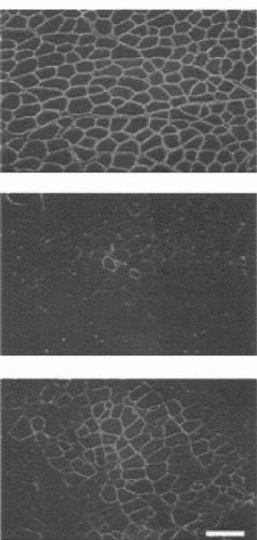
Figure 21.5. Systemic delivery of microdystrophin to dystrophic mice. (A) Antidystrophin immunofluorescence microscopy of tibialis anterior muscles from treated mdx mice (Tmdx) administered 1 × 10¹² vector genomes of rAAV6–CK6–microdystrophin and 10 μg VEGF, compared with wildtype and untreated mdx mice, a model of muscular dystrophy. Dystrophin expression is increased in the muscles of treated compared with untreated mdx mice, but remains mosaic compared with wildtype mice. Scale bars: 100 mm. (Reprinted by permission from Macmillan Publishers Ltd.: Gregorevic P et al: Systemic delivery of genes to striated muscles using adeno-associated viral vectors, Nature Med 10:828–34, copyright 2004.)

888
TABLE 21.2. Characteristics of Selected Therapeutic Proteins for Muscular Dystrophy*
|
|
Amino |
Molecular |
|
|
Proteins |
Alternative Names |
Acids |
Weight (kDa) |
Expression |
Functions |
|
|
|
|
|
|
Integrin α7 |
ITGA7 |
1137 |
124 |
Heart, skeletal muscle, nervous |
Joining with integrin β1 to form |
|
|
|
|
system, lung, intestine, ovary, |
an integrin complex, which |
|
|
|
|
prostate gland |
is a major integrin complex |
|
|
|
|
|
expressed in differentiated |
|
|
|
|
|
muscular cells (note that all |
|
|
|
|
|
integrins are heterodimeric |
|
|
|
|
|
integral membrane proteins |
|
|
|
|
|
composed of an α chain and |
|
|
|
|
|
a β chain), binding to the |
|
|
|
|
|
extracellular matrix protein |
|
|
|
|
|
laminin-1, and regulating cell |
|
|
|
|
|
attachment to extracellular |
|
|
|
|
|
matrix |
Integrin β1 ITGB1, fibronectin receptor β |
825 |
92 |
Heart, nervous system, skeletal |
Joining with an integrin α subunit |
|
|
subunit (FNRB), fibronectin |
|
|
muscle, lymphocytes, liver, bone, |
to form integrin complexes, |
|
receptor β subunit-like, very |
|
|
cartilage, skin |
regulating cell adhesion to |
|
late activation protein β |
|
|
|
extracellular matrix, and |
|
polypeptide (VLA β) |
|
|
|
regulating various cellular |
activities, including embryogenesis, cell proliferation and migration, immune responses, and metastasis of tumor cells

890 SKELETAL MUSCLE REGENERATIVE ENGINEERING
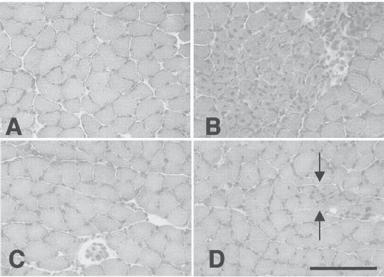
Figure 21.6. Influence of NOS expression on the morphology of skeletal muscle cells: (A) C57 control muscle showing fibers of uniform diameter, no central nucleation, and no clusters of inflammatory cells between adjacent fibers; (B) mdx muscle showing a typical focus of muscle pathology characterized by fiber populations of variable diameters and central nucleation (note that transgenic mdx mice are null mutants for dystrophin). Dark staining nuclei of inflammatory cells appear between adjacent fibers; (C) NOS Tg/mdx muscle showing typical histology, where fiber diameter is more uniform than age-matched mdx muscle, and there is little inflammation or central nucleation (note that NOS Tg/mdx is a transgenic mouse model with deficient dystrophin but with the expression of the NOS transgene); (D) NOS Tg/mdx muscle showing an example of the relatively small lesions that appear in NOS Tg/mdx muscle (between arrows) where there are small clusters of small-diameter, central-nucleated fibers. All micrographs are at the same magnification. Scale bar: 250 μm. (Reprinted with permission from Wehling M et al: Clonal isolation of muscle-derived cells capable of enhancing muscle regeneration and bone healing, J Cell Biol 155:123–32, copyright 2001.)
factor gene can serve as a candidate gene for the molecular treatment of muscular dystrophy.
MYOSTATIN. Myostatin is a protein that negatively regulates the development and regeneration of skeletal muscles. In animal models with myostatin gene mutation, hyperplasia and hypertrophy occur in the skeletal muscle system. The overexpression of the myostatin gene results in the degeneration of the skeletal muscle cells. Thus, it is conceivable that the suppression of the activity of the myostatin gene is beneficial for the treatment of muscular dystrophy. Such a goal can be achieved by delivering antisense oligonucleotides or small interfering RNA specific to the myostatin mRNA to compromise the translation of the myosin protein. In addition, local delivery of antimyostatin antibody and myostatin inhibitors can achieve the same goal.
DISORDERS OF THE SKELETAL MUSCLE SYSTEM |
891 |
MINIAGRIN. Miniagrin is a fragment of a large protein called agrin, which is known to induce the aggregation of acetylcholine receptors and other postsynaptic proteins in muscular cells and regulate the formation of the neuromuscular junction. Agrin also interacts with laminin and dystroglycan, enhancing cell adhesion and survival. There are two important domains within the agrin protein: the N-terminal domain (responsible for binding to laminin) and the C-terminal domain (responsible for binding to dystroglycan). A miniagrin gene has been constructed with the N- and C-terminal domains. The delivery of the constructed miniagrin gene into target skeletal muscle cells in a laminin-2-deficient model (associated with impairment of cell adhesion and muscle weakness) results in upregulation of laminin and crosslink of laminin with dystroglycan. These activities are associated with enhanced cell adhesion, reduced muscle degeneration and dystrophic symptoms, and improved muscular contractility.
Cellular Regenerative Engineering for Muscular Dystrophy. Cell transplantation is a potential approach for the treatment of muscular dystrophy. There are two potential cell types, including muscular progenitor cells and stem cells, which can be used for cell transplantation. Cell transplantation may elicit two possible therapeutic effects: (1) transplanted cells can differentiate into muscular cells, replacing cells with muscular dystrophy; and (2) transplanted cells can serve as carriers for the delivery of therapeutic genes such as the dystrophin gene and “booster” genes.
Muscular Progenitor Cells [21.12]. The skeletal muscle system contains muscular progenitor cells, also known as skeletal myoblasts, myogenic cells, and satellite cells, which are capable of differentiating to mature muscular cells. Experimental investigations have identified muscular progenitor cells based on stem celland progenitor cell-specific markers. The muscular progenitor cells express a cell surface molecule specific for stemlike cells, known as stem cell antigen1 (Sca1). In addition, these cells may coexpress other cell surface markers, including CD34, myf5, and m-cadherin. The muscular progenitor cells can be identified by immunochemical labeling of specific surface markers in conjunction with a cell sorting approach, such as magnetic bead-assisted cell sorting (tagging iron beads with a specific antibody and enriching antibody-labeled cells by magnetization) and fluorescence-activated cell sorting or FACS (labeling cells with a fluorescent antibody and enriching antibody-labeled cells by fluorescence-based cell sorting). Once muscular progenitor cells are identified and enriched, the cells can be used for transplantation into target muscular cells. In the mouse mdx muscular dystrophy model, the transplanted muscular progenitor cells are capable of differentiating into mature muscular cells, replacing dystrophic cells, and reducing pathological changes and symptoms of muscular dystrophy (Fig. 21.7). Furthermore, the muscular progenitor cells can be transfected with therapeutic genes for muscular dystrophy, such as the dystrophin gene and “booster” genes, and used as gene delivery carriers. When the progenitor cells are transplanted into target muscular cells, the proteins produced by the transplanted cells can elicit a therapeutic effect on the host dystrophic cells.
Stem Cells [21.13]. As discussed on Chapter 9, there are several types of stem cells, including embryonic, fetal, and adult stem cells. These stem cell types can be potentially
892 SKELETAL MUSCLE REGENERATIVE ENGINEERING
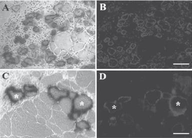
Figure 21.7. In vivo differentiation of mc13 cells into myogenic lineage after intramuscular (IM) and intravenous (IV) injection (note that mc13 cells are muscle-derived stem cells and are capable of differentiating into myogenic and osteogenic lineage in vitro and in vivo). The mc13 cells were stably transfected with a plasmid DNA construct encoding LacZ, dystrophin, and neomycin resistance genes and injected intramuscularly into hind limbs of mdx mice. After 7 days, and hind-limb musculature was isolated for histology. Many LacZ-positive myofibers (A) were found at the injected site that colocalized with dystrophin-positive myofibers (B). Some LacZ (C,*) and dystrophin positive myofibers (D,*) were also found in the hind limb muscle of mdx mice after IV injection of mc13. Scale bars: 100 μm (A,B); 50 μm (C,D). (Reprinted with permission from Lee JY et al: J Cell Biol 150:1085–100, copyright 2000.)
used for the treatment of muscular dystrophy. A desired type of stem cells can be identified, enriched, and transplanted into target muscular cells. These procedures are technically similar to those described above for the transplantation of muscular progenitor cells. In experimental investigations, multipotent embryonic stem cells have been used for the treatment of muscular dystrophy in the mouse mdx dystrophindeficient model. These investigations have shown that embryonic stem cell transplantation is an effective approach for the prevention of pathological changes and the relief of the symptoms of muscular dystrophy. In addition, extensive investigations have been conducted to demonstrate the possibility of using adult stem cells for the treatment of muscular dystrophy. Bone marrow stem cells have been identified, enriched, and delivered into the circulation of the mdx mouse with dystrophin deficiency. The delivered cells can integrate into the skeletal muscle system and express dystrophin, improving the function of the dystrophic muscular cells. Other types of adult stem cells, such as the liver adipocytes, have also been used for transplantation into dystrophic muscular cells. Such an approach results in beneficial effects for the treatment of dystrophin deficiency-induced muscular dystrophy.
BIBLIOGRAPHY 893
Potential Limitations [21.14]. There are several potential problems for the cellular treatment of muscular dystrophy. First, in patients with dystrophin deficiency and muscular dystrophy, the muscular progenitor cells or other types of stem cells are unlikely capable of expressing dystrophin. Thus, therapeutic cells can only be collected from an allogenic source or a donor individual. The transplantation of living allogenic cells induces acute immune rejection responses, resulting in rapid death of the transplanted cells. Second, it is impossible to deliver therapeutic cells to all dystrophic muscular cells over the entire body. The effects of cellular therapy are often limited to a small area around the cell delivery site. Although cells can be delivered through the blood circulation, the rate of cell integration into the skeletal muscle system is very low. Further investigations are needed to resolve these problems.
BIBLIOGRAPHY
21.1. Anatomy and Physiology
Guyton AC, Hall JE: Textbook of Medical Physiology, 11th ed, Saunders, Philadelphia, 2006.
McArdle WD, Katch FI, Katch VL: Essentials of Exercise Physiology, 3rd ed, Lippincott Williams & Wilkins, Baltimore, 2006.
Germann WJ, Stanfield CL (with contributors Niles MJ, Cannon JG), Principles of Human Physiology, 2nd ed, Pearson Benjamin Cummings, San Francisco, 2005.
Thibodeau GA, Patton KT: Anatomy & Physiology, 5th ed, Mosby, St Louis, 2003.
Boron WF, Boulpaep EL: Medical physiology: A cellular and molecular approach, Saunders, Philadelphia, 2003.
Ganong WF: Review of Medical Physiology, 21st ed, McGraw-Hill, New York, 2003.
21.2. Pathogenesis, Pathology, and Clinical Features of Muscular Dystrophies
Koenig M, Hoffman EP, Bertelson CJ, Monaco AP, Feener C et al: Complete cloning of the Duchenne muscular dystrophy (DMD) cDNA and preliminary genomic organization of the DMD gene in normal and affected individuals, Cell 50:509–17, 1987.
Hoffman EP, Brown RH Jr, Kunkel LM: Dystrophin: the protein product of the Duchenne muscular dystrophy locus, Cell 51:919–28, 1987.
Cohn RD, Campbell KP: Molecular basis of muscular dystrophies, Muscle Nerve 23:1456–71, 2000.
Bornemann A, Anderson LV: Diagnostic protein expression in human muscle biopsies, Brain Pathol 10:193–214, 2000.
Watkins SC, Hoffman EP, Slayter HS, Kunkel LM: Immunoelectron microscopic localization of dystrophin in myofibres, Nature 333:863–6, 1988.
Ohlendieck K, Campbell KP: Dystrophin constitutes 5% of membrane cytoskeleton in skeletal muscle, FEBS Lett 283:230–4, 1991.
Hoffman EP, Brown RH Jr, Kunkel LM: Dystrophin: The protein product of the Duchenne muscular dystrophy locus, Cell 51:919–28, 1987.
Koenig M, Monaco AP, Kunkel LM: The complete sequence of dystrophin predicts a rod-shaped cytoskeletal protein, Cell 53:219–26, 1988.
Ohlendieck K, Campbell KP: Dystrophin-associated proteins are greatly reduced in skeletal muscle from mdx mice, J Cell Biol 115:1685–94, 1991.
BIBLIOGRAPHY 895
Chelly J, Kaplan JC, Maire P, Gautron S, Kahn A: Transcription of the dystrophin gene in human muscle and non-muscle tissues, Nature 333:858–60, 1988.
Bies RD: X-linked dilated cardiomyopathy, New Engl J Med 330:368–9, 1994.
Bogdanovich S, Krag TOB, Barton ER, Morris LD, Whittemore LA et al: Functional improvement of dystrophic muscle by myostatin blockade, Nature 420:418–21, 2002.
Cox GA, Cole NM, Matsumura K, Phelps SF, Hauschka SD et al: Overexpression of dystrophin in transgenic mdx mice eliminates dystrophic symptoms without toxicity, Nature 364:725–9, 1993.
England SB, Nicholson LVB, Johnson MA, Forrest SM, Love DR et al: Very mild muscular dystrophy associated with the deletion of 46% dystrophin, Nature 343:180–2, 1990.
Forrest SM, Cross GS, Speer A, Gardner-Medwin D, Burn J et al: Preferential deletion of exons in Duchenne and Becker muscular dystrophies, Nature 329:638–40, 1987.
Goyenvalle A, Vulin A, Fougerousse F, Leturcq F, Kaplan JC et al: Rescue of dystrophic muscle through U7 snRNA-mediated exon skipping, Science 306:1796–9, 2004.
Gussoni E, Soneoka Y, Strickland CD, Buzney EA, Khan MK et al: Dystrophin expression in the mdx mouse restored by stem cell transplantation, Nature 401:390–4, 1999.
Harper SQ, Hauser MA, DelloRusso C, Duan D, Crawford RW et al: Modular flexibility of dystrophin: Implications for gene therapy of Duchenne muscular dystrophy, Nature Med 8:253–61, 2002.
Hoffman EP, Knudson CM, Campbell KP, Kunkel LM: Subcellular fractionation of dystrophin to the triads of skeletal muscle, Nature 330:754–8, 1987.
Hoffman EP, Monaco AP, Feener CC, Kunkel LM: Conservation of the Duchenne muscular dystrophy gene in mice and humans, Science 238:347–50, 1987.
Kronqvist P, Kawaguchi N, Albrechtsen R, Xu X, Daa Schroder H et al: ADAM12 alleviates the skeletal muscle pathology in mdx dystrophic mice, Am J Pathol 161:1535–40, 2002.
Kunkel LM: Analysis of deletions in DNA from patients with Becker and Duchenne muscular dystrophy, Nature 322:73–7, 1986.
Lee CC, Pearlman JA, Chamberlain JS, Caskey CT: Expression of recombinant dystrophin and its localization to the cell membrane, Nature 349:334–6, 1991.
Lu QL, Mann CJ, Lou F, Bou-Gharios G, Morris GE et al: Functional amounts of dystrophin produced by skipping the mutated exon in the mdx dystrophic mouse, Nature Med 9:1009–14, 2003.
Monaco AP, Neve RL, Colletti-Feener C, Bertelson CJ, Kurnit DM et al: Isolation of candidate cDNAs for portions of the Duchenne muscular dystrophy gene, Nature 323:646–50, 1986.
Muntoni F, Cau M, Ganau A, Congiu R, Arvedi G, Mateddu A et al: Deletion of the dystrophin muscle-promoter region associated with X-linked dilated cardiomyopathy, New Engl J Med 329:921–5, 1993.
Ray PN, Belfall B, Duff C, Logan C, Kean V et al: Cloning of the breakpoint of an X;21 translocation associated with Duchenne muscular dystrophy, Nature 318:672–5, 1985.
Scott MO, Sylvester JE, Heiman-Patterson T, Shi YJ, Fieles W et al: Duchenne muscular dystrophy gene expression in normal and diseased human muscle, Science 239:1418–20, 1988.
Sicinski P, Geng Y, Ryder-Cook AS, Barnard EA, Darlison MG et al: The molecular basis of muscular dystrophy in the mdx mouse: a point mutation, Science 244:1578–80, 1989.
Tinsley JM, Potter AC, Phelps SR, Fisher R, Trickett JI et al: Amelioration of the dystrophic phenotype of mdx mice using a truncated utrophin transgene, Nature 384:349–53, 1996.
Toyo-Oka T, Kawada T, Nakata J, Xie H, Urabe M et al: Translocation and cleavage of myocardial dystrophin as a common pathway to advanced heart failure: A scheme for the progression of cardiac dysfunction, Proc Natl Acad Sci USA 101:7381–5, 2004.