

Bioregenerative Engineering Principles and Applications - Shu Q. Liu
..pdf916 BONE AND CARTILAGE REGENERATIVE ENGINEERING
and homeostasis. Vitamin D is synthesized in the epidermal cells under the stimulation of ultraviolet radiation from the sunlight. Such a stimulation can induce a photobiochemical process, by which 7-dehydrocholesterol undergoes a conformational change, resulting in the formation of previtamin D3. Previtamin D3 can be spontaneously converted to vitamin D3 under the stimulation of the body temperature. Vitamin D3 is transported from the epidermis to the blood via the mediation of vitamin D-binding proteins. In the liver, vitamin D3 is further converted to 25-hydroxyvitamin D [25(OH)D], a major form of vitamin D that exists in the blood. The blood concentration of 25-hydroxyvitamin D varies considerably under physiological conditions, ranging from 5 to 80 ng/mL. However, 25-hydroxyvitamin D is not active. This form of vitamin D is metabolized in the kidney and converted to an active form known as 1,25-hydroxyvitamin D [1,25(OH)2D] under the action of 25(OH)D-1α hydroxylase. 1,25-hydroxyvitamin D is an active form of vitamin D.
The level of 1,25-hydroxyvitamin D is controlled by a number of factors, including the level of sunlight exposure, the intensity of ultraviolet light, aging, the calcium concentration, and the level of parathyroid hormone. An increase in exposure to sunlight enhances vitamin D3 synthesis and promotes the formation of 1,25-hydroxyvitamin D. Aging is associated with a progressive reduction in the rate of vitamin D3 synthesis. A decrease in the blood concentration of calcium stimulates the conversion of 25-hydroxyvitamin D to 1,25-hydroxyvitamin D. Such a calcium change also stimulates the secretion of parathyroid hormone, which acts on renal tubule cells and enhances the formation of 1,25hydroxyvitamin D. There exist many forms of vitamin D metabolites in the blood and extracellular matrix. These forms are mostly products of vitamin D degradation and possess vitamin D activity. However, the vitamin D metabolites are not as active as 1,25hydroxyvitamin D.
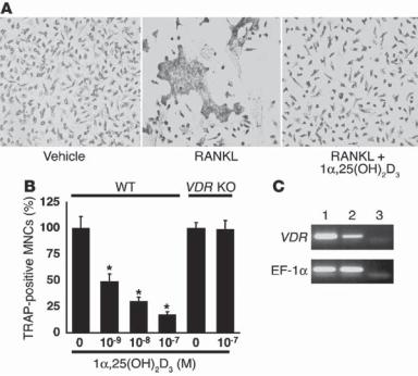
There are a number of functions for 1,25-hydroxyvitamin D. These include the regulation of calcium and phosphorus absorption in the epithelial cells of the small intestine, the regulation of bone resorption, and the control of cell proliferation and differentiation. It is well known that 1,25-hydroxyvitamin D stimulates the absorption of calcium and phosphorus in the epithelial cells of the small intestine, resulting in an elevation in the blood concentration of these ions. The influence of vitamin D on bone resorption is dependent on the concentration of vitamin D. At a high concentration in the extracellular space, 1,25-hydroxyvitamin D enhances bone resorption. This is possibly due to the stimulatory effect of vitamin D on the bone-resorption activity of the parathyroid hormone. Such an effect results in an increase in the concentration of calcium and phosphorus in the extracellular space and blood. However, at a low level, 1,25-hydroxyvitamin D enhances bone mineralization and matrix formation. A possible mechanism for this phenomenon is that a low level of vitamin D may reduce the activity of the parathyroid hormone. 1,25hydroxyvitamin D has also been hsown to inhibit the development and activation of osteoclasts (Fig. 22.7), thus reducing bone resorption.
Experimental investigations have demonstrated that 1,25-hydroxyvitamin D exerts inhibitory effects on the proliferation of normal and cancer cells. For instance, 1,25hydroxyvitamin D inhibits the generation and secretion of renin in the renal arteries, resulting in a decrease in the level and activity of angiotensin II. Since angiotensin II enhances the proliferation of vascular smooth muscle cells, 1,25-hydroxyvitamin D suppresses vascular mitogenic activities via mediating the function of angiotensin II. Vitamin D is also known to promote cell differentiation A treatment with 1,25-hydroxyvitamin D can induce the differentiation of monocytes to osteoclast-like cells.
ANATOMY AND PHYSIOLOGY OF THE CARTILAGE |
917 |
Figure 22.7. 1α-25(OH)2D3 inhibits osteoclast development through VDR by acting directly on osteoclast precursor cells in bone marrow. (A) Osteoclast precursor cells were isolated from the bone marrow of wildtype C57BL/6J and VDR Knockout mice as M-CSF–dependent adherent cells, and were further treated with RANKL (40 ng/mL) in the absence or presence of 10−7 M 1α25(OH)2D3 for 3 days. Note that the development of TRAP-positive multinucleate osteoclasts induced by RANKL (receptor activator of NFκB ligand) was markedly inhibited by cotreatment with 1α-25(OH)2D3. (B) The inhibitory effect of 1α-25(OH)2D3 on the formation of TRAP-positive multinucleate cells (MNCs) was dose-dependent and was not seen in marrow cultures derived from VDR knockout mice, even at the highest dose of 10−7 M. Data are expressed as a percentage of vehicle-treated cultures. *P < 0.05 versus vehicle group, n = 6. (C) Expression of VDRs in the intestine (lane 1) and osteoclast precursor cells (lane 2) as detected by RT-PCR. EF-1α mRNA served as control for PCR. Lane 3 contained water as a negative control. (Reprinted with permission from Takasu H et al: c-Fos protein as a target of antiosteoclastogenic action of vitamin D, and synthesis of new analogs, J Clin Invest 116:528–35, copyright 2006.)
Mammalian cells express a nuclear receptor for 1,25-hydroxyvitamin D. The interaction of 1,25-hydroxyvitamin D with this receptor induces the phosphorylation and activation of the receptor. The complex of 1,25-hydroxyvitamin D and activated nuclear receptor serves as a transcription factor and can translocate to the cell nucleus, initiating the transcription of specific target genes. The gene products in turn regulate related physiological activities.
ANATOMY AND PHYSIOLOGY OF THE CARTILAGE [22.1]
Cartilage is a connective tissue that is mostly associated with the bone except for several organs such as the large airways and ears. Cartilage is composed of two structures: peri-
918 BONE AND CARTILAGE REGENERATIVE ENGINEERING
chondrium and internal cartilage. The perichondrium contains an external and internal connective tissue layer. The external layer is composed of fibroblasts, extracellular matrix (collagen fibers, elastic fibers, and proteoglycans), blood vessels, and nerves. The internal layer is composed of cells known as chondroblasts. These cells can generate cartilage matrix components, including collagen and proteoglycans. Chondroblasts can selfreproduce and are responsible for cartilage growth. The internal cartilage is composed of chondrocytes and cartilage matrix. Chondrocytes are developed from chondroblasts and are surrounded by cartilage matrix. The cartilage matrix is an amorphous structure, which contains scattered chondrocytes. It should be noted that a cartilage layer can be found on the terminal surface of the bone at a joint. This type of cartilage, called articular cartilage, does not contain perichondrium.
There are several functions for the cartilage. It provides support to adjacent bones, serves as a structural framework for several organs, including the trachea, nose, and ear, and constitutes the surface structure of the joint, ensuring smooth interaction between bone surfaces. In addition, cartilage plays a critical role in mediating bone growth. In long bones such as the femur and tibia, there exists a cartilage plate between the end and the body of the bone. This plate is responsible for the growth of the bone, which determines the height of the entire body. When bone growth is ceased, the cartilage growth plate is ossified into a thin bone matrix structure known as the epiphyseal line.
BONE AND CARTILAGE DISORDERS
Osteoporosis
Pathogenesis, Pathology, and Clinical Features [22.2]. Osteoporosis is a bone disorder characterized by increased degeneration or resorption of the bone matrix in the entire skeletal system, resulting in a progressive reduction in the mass of the bone, the thickness of the compact bone, and the size and number of the trabeculae of the cancellous bone. Osteoporosis often results in bone deterioration and fracture. Osteoporosis is a consequence of imbalance between bone formation and resorption. In the healthy population, the rate of bone formation is dynamically balanced with the rate of bone resorption. When the rate of bone resorption exceeds that of bone formation, the bone mass reduces and osteoporosis occurs. In such a case, the skeletal system is no longer able to resist the physiological level of mechanical loads. A slight increase in the mechanical load may result in bone fracture. In the normal population, bone resorption starts to exceed bone formation at the age of 40–50. The rate of bone resorption increases with aging. Osteoporosis is a common disorder in the population over the age of 60.
Based on etiological factors, osteoporosis is classified into several types, including idiopathic, postmenopausal, glucocorticoid, thyrotoxicotic, and inherited osteoporosis. Idiopathic osteoporosis is defined as osteoporosis without identified etiological factors. This disorder is often found in young people, especially premenopausal women. The disorder is associated with a decrease in blood calcium and phosphorus. Idiopathic osteoporosis can be self-cured spontaneously within several years. Postmenopausal osteoporosis is found in women with reduced production and release of estrogen, a hormone that suppresses bone resorption. A decrease in the level of estrogen in postmenopausal women induces increased bone resorption and osteoporosis.
BONE AND CARTILAGE DISORDERS |
919 |
An excessive increase in glucocorticoids, as seen in Cushing’s syndrome, can reduce bone formation and simultaneously enhance bone resorption, often resulting in osteoporosis. Glucocorticoids have been shown to enhance the effect of parathyroid hormone, which activates the osteoclasts and mobilizes calcium from the bone matrix. Furthermore, glucocorticoids inhibit calcium absorption in the intestine. All these effects contribute to the development of osteoporosis.
Thyrotoxicity is a condition with increased secretion of the thyroid hormone, which is also referred to as hyperthyroidism. Increased thyroid hormone mobilizes calcium and phosphorus from the bone, promotes bone resorption, and enhances the excretion of calcium and phosphorus via urine and feces. These changes can lead to osteoporosis if hyperthyroidism prolongs. In postmenopausal women, hyperthyroidism may induce more severe osteoporosis than the general population.
Osteogenesis imperfecta is an inherited form of osteoporosis and is characterized by heterogeneous reduction in the mass of bone matrix. There are two types of disorder: autosomal dominant and autosomal recessive osteogenesis imperfecta. The autosomal dominant type of imperfecta is associated with relatively mild bone resorption and functional defects. In contrast, the autosomal recessive subtype is often found within a short period after birth and is associated with a severe reduction in the bone mass. Patients may experience recurrent fracture of long bones such as femurs and tibias. Pathological examinations usually demonstrate reduced synthesis of type I collagen and altered organization of collagen matrix.
Conventional Treatment [22.2]. Osteoporosis is a group of disorders induced by different etiological factors. One of the strategies for the treatment of osteoporosis is to eliminate or alleviate the primary etiological factors, if known. For instance, postmenopausal osteoporosis is caused by a reduction in the level of estrogen. Administration of estrogen is the primary choice of method for the treatment of postmenopausal osteoporosis. In osteoporosis induced by increased level of glucocorticoids and thyroid hormone, a primary approach is to treat the original diseases that enhance the production and secretion of these hormones. Since calcium is a major component of the bone matrix, calcium administration is often necessary. An increase in the level of extracellular calcium reduces bone resorption. In addition, fluorides can be incorporated into the bone matrix, enhancing the crystal formation and strength of the bone. Thus fluorides have been used to treat osteoporosis.
Molecular Engineering Therapy. Osteoporosis is induced by progressive degeneration of the bone matrix. Molecular regenerative approaches can be used to protect the bone from degeneration and promote bone matrix formation. There are a number of genes encoding proteins that are known to regulate bone matrix formation and mineralization, including vitamin D receptor, estrogen receptor alpha, type I collagen, transforming growth factor, and interleukin-6. The mutation of these genes and/or disorders in regulating the expression of these genes may predispose to bone degeneration, leading to osteoporosis. Thus, the genetic manipulation of these genes may potentially enhance bone formation and prevent bone degeneration. Here the application of these genes to osteoporosis is briefly discussed.
Vitamin D Receptor (VDR) [22.3]. The vitamin D receptor (Table 22.1) interacts with vitamin D and participates in the regulation of calcium metabolism and bone formation.
920 |
BONE AND CARTILAGE REGENERATIVE ENGINEERING |
|
||||
TABLE 22.1. Characteristics of Vitamin D Receptor* |
|
|
||||
|
|
|
|
|
|
|
|
|
|
Amino |
Molecular |
|
|
Proteins |
|
Alternative Names |
Acids |
Weight (kDa) |
Expression |
Functions |
|
|
|
|
|
|
|
Vitamin D |
VDR, 1,25- |
427 |
48 |
Ubiquitous |
Serving as a |
|
receptor |
dihydroxyvitamin |
|
|
|
trans-acting |
|
|
|
D3 receptor |
|
|
|
transcriptional |
|
|
|
|
|
|
regulatory |
|
|
|
|
|
|
factor, which |
|
|
|
|
|
|
is similar to |
|
|
|
|
|
|
steroid and |
|
|
|
|
|
|
thyroid |
|
|
|
|
|
|
hormone |
|
|
|
|
|
|
receptors in |
|
|
|
|
|
|
structure, |
|
|
|
|
|
|
regulating |
|
|
|
|
|
|
genes involved |
|
|
|
|
|
|
in mineral |
|
|
|
|
|
|
(calcium) |
|
|
|
|
|
|
metabolism |
|
|
|
|
|
||
*Based on bibliograhpy 22.3. |
|
|
|
|
||
TABLE 22.2. Characteristics of Selected Collagen Molecules* |
|
|||||
|
|
|
|
|
|
|
|
|
|
Amino |
Molecular |
|
|
Proteins |
|
Alternative Names |
Acids |
Weight (kDa) |
Expression |
Functions |
|
|
|
|
|
|
|
Collagen |
COL1A1, |
1464 |
139 |
Bone, skin, |
Constituting |
|
type I α1 |
collagen |
|
|
tendon |
collagen type 1 |
|
|
|
α1(I) chain |
|
|
|
together with |
|
|
|
|
|
|
collagen type I |
|
|
|
|
|
|
α2 |
Collagen |
COL1A2, |
1366 |
129 |
Bone, skin, |
Constituting |
|
type I α2 |
collagen I α2 |
|
|
tendon |
collagen type 1 |
|
|
|
polypeptide |
|
|
|
together with |
|
|
|
|
|
|
collagen type I |
|
|
|
|
|
|
α1 |
|
|
|
|
|
|
|
*Based on bibliograhpy 22.4.
At a relatively low level, vitamin D can activate the vitamin D receptor, enhancing calcium deposition and bone mineralization. Furthermore, vitamin D inhibits the activity of osteoclasts, suppressing bone resorption. The mutation of the vitamin D receptor gene induces disorder of calcium metabolism, influencing the mineralization process of the bone matrix. When the mutation of the vitamin D receptor is an identified cause for osteoporosis, the transfer of the wildtype vitamin D receptor gene may be considered for the treatment of the disorder.
Type I Collagen Gene [22.4]. Type I collagen is a major type of matrix molecule in the bone. This collagen is composed of collagen type I α1 and collagen type I α2 chains (both are listed in Table 22.2). The genes encoding these chains are COLIA1 and COLIA2,
BONE AND CARTILAGE DISORDERS |
921 |
respectively. The mutation of these genes may influence the formation of the collagen matrix. For instance, there exists a polymorphism in the introns of the COLIA1 gene. This polymorphism induces alterations in the regulation of gene transcription, resulting in a shift in the ratio of the alpha1 to alpha2 collagen chains. These abnormalities affect the formation of type I collagen as well as the collagen matrix in the bone. Since the integrity of the collagen matrix is critical to the mineralization of the bone matrix, any disorder in the formation of collagen matrix may contribute to bone degeneration and the development of osteoporosis. When disordered formation of collagen matrix is the cause of osteoporosis, the transfer of the wildtype type I collagen gene should be considered for the treatment of the disorder. Furthermore, when gene mutation is the cause of bone disorders, targeted correction of mutant genes by transfecting cells with viral vectors may be an effective approach for the treatment of bone disorders.
Estrogen Receptor [22.5]. This receptor interacts with the hormone estrogen and enhances bone formation. Postmenopausal women are often associated with osteoporosis because of estrogen reduction or deficiency. Estrogen is known to suppress the activity of cytokines, including tumor necrosis factor α, interleukin (IL)1, IL6, IL11, macrophagecolony stimulating factor, and prostaglandin E, via interaction with the estrogen receptor. The loss of estrogen and/or estrogen receptor is often associated with activation of these cytokines, which stimulate the proliferation of osteoclasts. Activated osteoclasts induce bone resorption. Mutation of the estrogen receptor gene may also lead to enhanced bone resorption. In the case of osteoporosis due to the malfunction of the estrogen receptor gene, the transfer of a functional gene into the skeletal system may help to enhance bone formation.
Characteristics of several estrogen receptors are listed in Table 22.3.
Calcitonin [22.6]. Calcitonin (Table 22.4) is a peptide hormone that is synthesized by the parafollicular cells of the thyroid. Its function is to inhibit the activity of osteoclasts, resulting in the suppression of bone resorption and a reduction in serum calcium. Calcitonin exerts an effect opposite to that of the parathyroid hormone. The protein form of calcitonin can be delivered to disordered bones for the treatment of osteoporosis. Clinical studies have provided promising results for the use of calcitonin. Alternatively, the gene of calcitonin can be cloned, amplified, and used for therapeutic purposes. As for other therapeutic genes, the delivery of the calcitonin gene can prolong the effectiveness of the hormone compared with direct delivery of the calcitonin protein.
Osteoprotegerin (OPG) and Osteoprotegerin Ligand (OPGL) [22.7]. Osteoprotegerin is a secreted soluble receptor protein that belongs to the tumor necrosis factor receptor superfamily and exists in the extracellular matrix. Osteoprotegerin is produced by the osteoblasts and is capable of suppressing the activity of osteoclasts and thus inhibiting bone resorption. Osteoprotegerin can bind to a ligand known as osteoprotegerin ligand (OPGL), which is a member of the tumor necrosis factor cytokine family and is also known as tumor necrosis factor ligand superfamily member 11 (TNFSF11). Osteoprotegerin ligand is a protein that can interact with a cell membrane receptor in hematopoietic cells to induce the differentiation of hematopoietic stem cells to osteoclasts. The binding of osteoprotegerin, which serves as a decoy receptor in the extracellular matrix, to osteoprotegerin ligand inhibits the interaction of the osteoprotegerin ligand to cell membrane receptor and thus suppresses the formation of osteoclasts from hematopoietic stem cells.

922
TABLE 22.3. Characteristics of Selected Estrogen Receptors*
|
|
Amino |
Molecular |
|
|
|
|
Proteins |
Alternative Names |
Acids |
Weight (kDa) |
|
Expression |
|
Functions |
|
|
|
|
|
|
|
|
Estrogen |
Estrogen receptor 1 (ESR1), |
595 |
66 |
Uterus, ovary, breast, bones, skeletal |
|
Serving as a nuclear receptor and |
|
receptor α |
estrogen receptor (ER) |
|
|
|
muscle, blood vessels, lung, skin |
|
transcription factor, regulating |
|
|
|
|
|
|
|
the development of the sex |
|
|
|
|
|
|
|
organs, and regulating cell |
|
ER β, estrogen receptor 2 |
|
|
|
|
|
proliferation |
Estrogen |
530 |
59 |
Uterus, ovary, breast, prostate gland, |
|
Similar to functions of estrogen |
||
receptor β |
(ESR2), ESRB, ESR β |
|
|
|
bones, skeletal muscle, blood vessels, |
receptor α |
|
|
|
|
|
|
brain, lung, skin |
|
|
|
|
|
|
|
|
|
|
*Based on bibliograhpy 22.5. |
|
|
|
|
|
|
|
TABLE 22.4. Characteristics of Calcitonin* |
|
|
|
|
|
|
|
|
|
|
|
|
|
|
|
|
|
Amino |
Molecular |
|
|
|
|
Proteins |
Alternative Names |
Acids |
Weight (kDa) |
Expression |
|
Functions |
|
|
|
|
|
|
|
|
|
Calcitonin |
CT, α calcitonin, katacalcin |
141 |
15 |
|
Nervous system, bone, |
Suppressing bone resorption and |
|
|
|
|
|
|
thyroid gland |
|
reducing the level of serum calcium |
|
|
|
|
|
|
|
|
*Based on bibliograhpy 22.6.
BONE AND CARTILAGE DISORDERS |
923 |
Since osteoclasts are responsible for bone resorption, the activation of osteoprotegerin leads to a reduction in bone resorption. Animals without the osteoprotegerin gene usually develop osteoporosis-like disorders. The application of osteoprotegerin to osteoporotic bones has been shown to prevent bone degeneration. In animal models with oophorectomy (surgical removal of the ovary), the administration of osteoprotegerin reduces estrogen deficiency-induced bone degeneration. The transfer of the osteoprotegerin gene into osteoporotic bones is a potential approach for the treatment of osteoporosis.
Properties of osteoprotegerin and its ligand are listed in Table 22.5.
Integrin-Binding Proteins [22.8]. The integrin complex αvβ3 is expressed in osteoclasts, plays a role in regulating the adhesion and proliferation of osteoclasts, and thus enhances osteoclast-mediated bone resorption. A family of integrin-binding proteins, known as disintegrins, can bind to the αvβ3 integrin and inhibit the activity of the integrin, resulting in the suppression of osteoclast activation and bone resorption. Disintegrins are small proteins found in the venom of snakes. The disintegrin family includes several members, which are echistatin, kistrin, albolabrin, bitistatin, elegantin, flavoridin, halysin, and triflavin. These proteins can specifically bind to β1 and β3 integrins and block the interaction of integrins with extracellular matrix components. Disintegrins also inhibit the activity of parathyroid hormone-induced bone resorption. Thus, the proteins or genes of disintegrins can be potentially used for the treatment of osteoporosis.
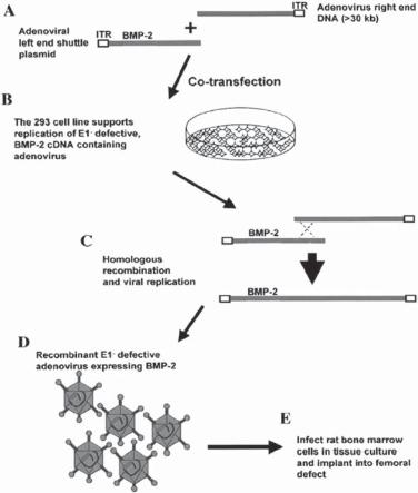
Growth Factors [22.9]. Several growth factors, including insulin-like growth factor (IGFβ1), fibroblast growth factor (FGF), the bone morphogenetic proteins (BMPs), and transforming growth factor-beta (TGFβ), are known to stimulate osteoblast proliferation and promote bone formation. Mutation of the genes encoding these growth factors is associated with an increased incidence of osteoporosis. A typical example is the association of the mutation of the transforming growth factor β gene (e.g., C509→T polymorphism) with osteoporosis. Local delivery of these growth factors enhances bone regeneration and recovery from osteoporosis. Alternatively, the genes of these growth factors can be used and transferred into disordered bones. As shown in experimental investigations, the overexpression of the bone morphogenetic protein gene in osteoblasts by gene transfer enhances bone formation. The construction of the bone morphogenetic protein gene vector is shown in Fig. 22.8. The effectiveness of the bone morphogenetic protein gene in bone formation is shown in Fig. 22.9.
Cell Therapy for Bone Regeneration [22.10]. Osteoporosis is a disorder induced by reduced osteogenesis due to impaired function of the osteoblasts, which regulate calcium deposition and bone formation. A potential approach for improving the function of osteoblasts is to transplant stem or progenitor cells to target bone tissue and replace malfunctioned osteoblasts. Candidate stem and progenitor cell types include embryonic, fetal, and adult stem and progenitor cells. It is important to point out the osteoporosis is a disorder that involves the entire skeletal system. Thus a systematic approach, such as intravenous delivery of osteogenic stem or progenitor cells, is required for the treatment of the disorder. To achieve a therapeutic goal, it is necessary to carry out several steps: (1) identify and collect a stem or progenitor cell type; (2) expand the cells in vitro; (3) genetically manipulate the cells (e.g., transfection of the cells with desired genes to enhance selected functions), if necessary; and (4) deliver expanded cells to the venous system.

924
TABLE 22.5. Characteristics of Osteoprotegerin and Osteoprotegerin Ligand*
|
|
Amino |
Molecular |
|
|
Proteins |
Alternative Names |
Acids |
Weight (kDa) |
Expression |
Functions |
|
|
|
|
|
|
Osteoprotegerin |
OPG, osteoclastogenesis |
401 |
46 |
Thyroid gland, kidney |
Inhibiting the formation and |
|
inhibitory factor (OCIF), |
|
|
|
activation of osteoclasts, |
|
tumor necrosis factor |
|
|
|
suppressing bone resorption, |
|
receptor superfamily |
|
|
|
and regulating lymph node |
|
member 11B (TNFRSF11B) |
|
|
|
organogenesis and vascular |
|
|
|
|
|
calcification |
Osteoprotegerin ligand |
OPGL, tumor necrosis factor |
317 |
35 |
T-cell, dendritic cell, thymus, |
Stimulating osteoclast |
|
ligand superfamily, |
|
|
lymph node |
differentiation and activation, |
|
member 11 (TNFSF11), |
|
|
|
inducing bone resorption, |
|
receptor activator of NKκB |
|
|
|
serving as a dentritic cell |
|
ligand (RANKL), |
|
|
|
survival factor, and regulating |
|
TNF-related activation- |
|
|
|
T-cell-dependent immune |
|
induced cytokine, |
|
|
|
responses |
|
osteoclast differentiation |
|
|
|
|
|
factor, ODF |
|
|
|
|
|
|
|
|
|
|
*Based on bibliograhpy 22.7. |
|
|
|
|
|
BONE AND CARTILAGE DISORDERS |
925 |
Figure 22.8. Diagram showing the construction of recombinant adenovirus containing rhBMP-2 cDNA. (A) Adenovirus E1 genes were deleted and replaced by BMP-2 cDNA on a plasmid (shuttle plasmid) containing the left inverted terminal repeat (ITR) required for viral replication. This BMP-2 shuttle plasmid and the large adenoviral right genome ( 30 kb) were transfected into a human embryonic kidney fibroblast cell line, referred to as 293 cells. (B) The 293 cells contain integrated adenoviral E1 genes and express E1 proteins (key growth-regulatory proteins of the adenovirus) constitutively. Thus, the E1-defective adenovirus (the E1 genes have been deleted) can be propagated only in the 293 cells. (C) The cotransfected BMP-2 shuttle plasmid DNA and the adenoviral right end DNA can undergo recombination through the shared homologous viral sequence in vivo in the 293 cells. The resultant BMP-2-expressing E1-defective adenovirus will be able to replicate and form plaques on the 293 cells. (D) BMP-2 recombinant adenoviral clones are further purified and expanded from individual plaques, and their DNA structure is confirmed. (E) The purified BMP-2 recombinant adenovirus then can be used to infect the rat-bone-marrow cells that have been grown in tissue culture. (Reprinted with permission from Lieberman JR et al: The effect of regional gene therapy with bone morphogenetic protein-2-producing bone-marrow cells on the repair of segmental femoral defects in rats, J Bone Joint Surg 81:905–17, copyright 1999.)