

Biotechnology for Biomedical Engineers - Martin L. Yarmush et al
.pdf6-12 Biotechnology for Biomedical Engineers
Morat,J.P.1882.Sur l’innervation motrice de l’estomac.Lyon.Med., 40:289–296.
Morat, J.P.1893. Sur quelques particularites de l’innervation motrice de l’estomac et de l’intestin. Arch. de Physiol Norm. et Path., 5:142–153.
Nelson,T.S. and Becker, J.C. 1968. Simulation of the electrical and mechanical gradient of the small intestine. Am. J. Physiol., 214:749–757.
Ormsbee, H.S., Telford, G.L., and Mason, G.R. 1979. Required neural involvement in control of canine migrating motor complex. Am. J. Physiol., 237:E451-E456.
Pffafenbach,B.,Adamek,R.J.,Kuhn,K.,andWegener,M.1995,Electrogastrography in healthy subjects. Evaluation of normal values: influence of age and gender. Dig. Dis. Sci., 40:1445–1450.
Pick,T.P. and Howden, R. 1977. Gray’s Anatomy, Bounty Books, NewYork.
Puestow, C.B. 1933. Studies on the origins of the automaticity of the intestine:The action of certain drugs on isolated intestinal transplants. Am. J. Physiol., 106:682–688.
Qiao, W., Sun, H.H., Chey, W.Y., and Lee, K.Y. 1998. Continuous wavelet analysis as an aid in the representation and interpretation of electrogastrographic signals.Ann.Biomed.Eng., 26:1072–1081.
Rees,W.D.W.,Malagelada,J.R.,Miller,L.J.,and Go,V.L.W.1982.Human interdigestive and postprandial gastrointestinal motor and gastrointestinal hormone patterns. Dig. Dis. Sci., 27:321–329.
Reinke, D.A., Rosenbaum,A.H., and Bennett, D.R. 1967. Patterns of dog gastrointestinal contractile activity monitored in vivo with extraluminal force transducers. Am. J. Dig. Dis., 12:113–141.
Robertson-Dunn,B.and Linkens,D.A.1974.A mathematical model of the slow wave electrical activity of the human small intestine. Med. Biol Eng., 12:750–758.
Ruckebusch,Y. and Fioramonti, S. 1975. Electrical spiking activity and propulsion in small intestine in fed and fasted states. Gastroenterology, 68:1500–1508.
Ruckebusch,Y. and Bueno, L. 1976.The effects of feeding on the motility of the stomach and small intestine in the pig. Br. J. Nutr., 35:397–405.
Ruckebusch, Y. and Bueno, L. 1977. Migrating myoelectrical complex of the small intestine.
Gastroenterology, 73:1309–1314.
Sanders,K.M.1996.A case for interstitial cells of Cajal as pacemakers and mediators of neurotransmission in the gastrointestinal tract. Gastroenterology, 111(2):492–515.
Shearin, N.L., Bowes, K.L. and Kingma, Y.J. 1978. In vitro electrical activity in canine colon. Gut, 20:780–786.
Sarna, S.K., Daniel, E.E., and Kingma,Y.J. 1971. Simulation of slow wave electrical activity of small intestine. Am. J. Physiol., 221:166–175.
Sarna, S.K., Daniel, E.E., and Kingma,Y.J. 1972. Simulation of the electrical control activity of the stomach by an array of relaxation oscillators. Am. J. Dig. Dis., 17:299–310.
Sarna, S.K. and Daniel, E.E. 1973. Electrical stimulation of gastric electrical control activity. Am. J. Physiol., 225:125–131.
Sarna, S.K. 1975a. Models of smooth muscle electrical activity. In Methods in Pharmacology, E.E. Daniel and D.M. Paton, Eds., Plenum Press, NewYork, 519–540.
Sarna, S.K. 1975b. Gastrointestinal electrical activity: terminology. Gastroenterology, 68:1631–1635. Sarna, S.K. and Daniel, E.E. 1975c. electrical stimulation of small intestinal electrical control activity.
Gastroenterology, 69:660–667.
Sarna, S.K., Bardakjian, B.L., Waterfall, W.E., and Lind, J.F. 1980. Human colonic electrical control activity (ECA). Gastroenterology, 78:1526–1536.
Sarna, S.K., Stoddard, C., Belbeck L, and McWade D. 1981. Intrinsic nervous control of migrating myoelectric complexes. Am. J. Physiol., 241:G16-G23.
Sarna,S.,Condon,R.E.,and Cowles,V.1983.Enteric mechanisms of initiation of migrating myoelectric complexes in dogs. Gastroenterology, 84:814–822.
Sarna, S.K. 1991. Physiology and pathophysiology of colonic motor activity. Dig. Dis. Sci., 6:827–862. Sarr M.G. and Kelly, K.A. 1981. Myoelectric activity of the autotransplanted canine jejunoileum.
Gastroenterology, 81:303–310.
The Gastrointestinal System |
6-13 |
Serio,R.,Barajas-Lopez,C.,Daniel,E.E.,Berezin,L,and Huizinga,J.D.1991. Pacemaker activity in the colon: Role of interstitial cells of Cajal and smooth muscle cells. Am.J. Physiol., 260:G636-G645.
Stanciu, C. and Bennett, J.R. 1975.The general pattern of gastroduodenal motility: 24 hour recordings in normal subjects. Rev. Med. Chir. Soc. Med. Nat. Iasi., 79:31–36.
Skinner, F.K. and Bardakjian, B.L. 1991.A barrier kinetic mapping unit.Application to ionic transport in gastric smooth muscle. Gastrointest. J. Motil., 3:213–224.
Stern, R.M. and Koch, K.L. 1985. Electrogastrography: Methodology,Validation, and Applications. Praeger Publishers, NewYork.
Summers, R.W., Anuras, S., and Green, J. 1982. Jejunal motility patterns in normal subjects and symptomatic patients with partial mechanical obstruction or pseudo-obstruction.In Motility of the Digestive Tract, M.Weinbeck, Ed., Raven Press, NewYork, 467–470.
Suzuki, N., Prosser, C.L., and Dahms,V., 1986. Boundary cells between longitudinal and circular layers: Essential for electrical slow waves in cat intestine. Am. J. Physiol., 280:G287-G294.
Szurszewski, J.H. 1969. A migrating electric complex of the canine small intestine. Am. J. Physiol., 217:1757–1763.
Szurszewski, J.H. 1987. Electrical basis for gastrointestinal motility. In Physiology of the Gastrointestinal Tract, L.R. Johnson, Ed., Raven Press, NewYork, chap. 12.
Thompson, D.G.,Wingate, D.L., Archer, L, Benson, M.J., Green,W.J., and Hardy, R.J. 1980. Normal patterns of human upper small bowel motor activity recorded by prolonged radiotelemetry. Gut, 21:500–506.
Vanasin, B., Ustach, T.J., and Schuster, M.M. 1974. Electrical and motor activity of human and dog colon in vitro. Johns Hopkins Med. J., 134:201–210.
Vantrappen, G., Janssens, J.J., Hellemans, J. and Ghoos,Y. 1977.The interdigestive motor complex of normal subjects and patients with bacterial overgrowth of the small intestine.J.Clin.Invest., 59:1158– 1166.
Wang, Z., He, Z., and Chen, J.D.Z. 1999. Filter banks and neural network-based feature extraction and automatic classification of electrogastrogram. Ann. Biomed. Eng., 27:88–95.
Weisbrodt,N.W.,Copeland,E.M.,Moore,E.P.,Kearly,K.W.,and Johnson,L.R.1975.Effect of vagotomy on electrical activity of the small intestine of the dog. Am. J. Physiol., 228:650–654.
Wheelon, H., andThomas, J.E. 1921.Rhythmicity of the pyloric sphincter. Am.J. Physiol, 54:460–473. Wheelon, H., and Thomas, J.E. 1922. Observations on the motility of the duodenum and the relation
of duodenal activity to that of the pars pylorica. Am. J. Physiol., 59:72–96.

|
|
|
7 |
|
|
|
Respiratory System |
||
|
|
|
|
|
|
7.1 |
Respiration Anatomy |
7-1 |
|
Arthur T.Johnson |
|
Lungs • Conducting Airways • Alveoli • Pulmonary |
|
|
|
Circulation • Respiratory Muscles |
|
||
University of Maryland |
|
|
||
7.2 |
LungVolumes and Gas Exchange |
7-5 |
||
Christopher G.Lausted |
||||
7.3 |
Perfusion of the Lung |
7-6 |
||
University of Maryland |
7.4 |
Gas Partial Pressures |
7-7 |
|
Joseph D.Bronzino |
7.5 |
Pulmonary Mechanics |
7-10 |
|
Trinity College/The Biomedical |
7.6 |
Respiratory Control |
7-11 |
|
Engineering Alliance and |
7.7 |
The Pulmonary Function Laboratory |
7-12 |
|
Consortium (BEACON) |
|
Spirometry • Body Plethysmography • Diffusing Capacity |
|
|
As functioning units, the lung and heart are usually considered a single complex organ, but because these organs contain essentially two compartments—one for blood and one for air—they are usually separated in terms of the tests conducted to evaluate heart or pulmonary function.This chapter focuses on some of the physiologic concepts responsible for normal function and specific measures of the lung’s ability to supply tissue cells with enough oxygen while removing excess carbon dioxide.
7.1 Respiration Anatomy
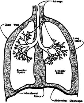
The respiratory system consists of the lungs, conducting airways, pulmonary vasculature, respiratory muscles, and surrounding tissues and structures (Fig. 7.1). Each plays an important role in influencing respiratory responses.
Lungs
There are two lungs in the human chest; the right lung is composed of three incomplete divisions called lobes, and the left lung has two, leaving room for the heart.The right lung accounts for 55% of total gas volume and the left lung for 45%. Lung tissue is spongy because of the very small (200 to 300 × 10–6 m diameter in normal lungs at rest) gas-filled cavities called alveoli, which are the ultimate structures for gas exchange.There are 250 million to 350 million alveoli in the adult lung, with a total alveolar surface area of 50 to 100 m2 depending on the degree of lung inflation [Johnson, 1991].
Conducting Airways
Air is transported from the atmosphere to the alveoli beginning with the oral and nasal cavities, through the pharynx (in the throat),past the glottal opening, and into the trachea or windpipe. Conduction of air begins at the larynx, or voice box, at the entrance to the trachea, which is a fibromuscular tube 10 to 12 cm in length and 1.4 to 2.0 cm in diameter [Kline, 1976]. At a location called the carina, the trachea terminates and divides into the left and right bronchi. Each bronchus has a discontinuous cartilaginous support in its wall.Muscle fibers capable of controlling airway diameter are incorporated into the walls of
0-8493-1811-4/03/$0.00+$1.50 |
|
© 2003 by CRC Press LLC |
7-1 |
7-2 |
Biotechnology for Biomedical Engineers |
FIGURE 7.1 Schematic representation of the respiratory system.
the bronchi,as well as in those of air passages closer to the alveoli.Smooth muscle is present throughout the respiratory bronchiolus and alveolar ducts but is absent in the last alveolar duct,which terminates in one to several alveoli.The alveolar walls are shared by other alveoli and are composed of highly pliable and collapsible squamous epithelium cells.
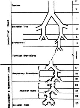
The bronchi subdivide into subbronchi, which further subdivide into bronchioli, which further subdivide, and so on, until finally reaching the alveolar level. Table 7.1 provides a description and dimensions of the airways of adult humans.A model of the geometric arrangement of these air passages is presented in Fig. 7.2. It will be noted that each airway is considered to branch into two subairways. In the adult human there are considered to be 23 such branchings, or generations, beginning at the trachea and ending in the alveoli.
Movement of gases in the respiratory airways occurs mainly by bulk flow (convection) throughout the region from the mouth to the nose to the fifteenth generation.Beyond the fifteenth generation,gas diffusion is relatively more important.With the low gas velocities that occur in diffusion,dimensions of the space over which diffusion occurs (alveolar space) must be small for adequate oxygen delivery into the walls; smaller alveoli are more efficient in the transfer of gas than are larger ones.Thus animals with high levels of oxygen consumption are found to have smaller-diameter alveoli compared with animals with low levels of oxygen consumption.
Alveoli
Alveoli are the structures through which gases diffuse to and from the body.To ensure gas exchange occurs efficiently, alveolar walls are extremely thin. For example, the total tissue thickness between the inside of the alveolus to pulmonary capillary blood plasma is only about 0.4×10⁻⁶ m. Consequently, the principal barrier to diffusion occurs at the plasma and red blood cell level, not at the alveolar membrane [Ruch and Patton, 1966].
Molecular diffusion within the alveolar volume is responsible for mixing of the enclosed gas.Due to small alveolar dimensions,complete mixing probably occurs in less than 10 ms,fast enough that alveolar mixing time does not limit gaseous diffusion to or from the blood [Astrand and Rodahl, 1970].

Respiratory System |
7-3 |
TABLE 7.1 Classification and Approximate Dimensions of Airways of Adult Human Lung (Inflated to about 3/ 4 of TLC)*
airwaysineachgenerationisbasedonregulardichotomousbranching. |
tolastgenerationineachgroup. |
Thenumberof |
Numbersrefer |
* |
† |
Source: Used with permission from Staub [1963] and Weibel [1963]; adapted by Comroe [1965].
7-4 |
Biotechnology for Biomedical Engineers |
FIGURE 7.2 General architecture of conductive and transitory airways.(Used with permission fromWeibel,1963.) In the conductive zone, air is conducted to and from the lungs while in the respiration zone, gas exchange occurs.
Of particular importance to proper alveolar operation is a thin surface coating of surfactant.Without this material, large alveoli would tend to enlarge and small alveoli would collapse. It is the present view that surfactant acts like a detergent, changing the stress-strain relationship of the alveolar wall and thereby stabilizing the lung [Johnson, 1991].
Pulmonary Circulation
There is no true pulmonary analog to the systemic arterioles, since the pulmonary circulation occurs under relatively low pressure [West, 1977]. Pulmonary blood vessels, especially capillaries and venules, are very thin walled and flexible.Unlike systemic capillaries,pulmonary capillaries increase in diameter, and pulmonary capillaries within alveolar walls separate adjacent alveoli with increases in blood pressure or decreases in alveolar pressure. Flow, therefore, is significantly influenced by elastic deformation. Although pulmonary circulation is largely unaffected by neural and chemical control, it does respond promptly to hypoxia.
There is also a high-pressure systemic blood delivery system to the bronchi that is completely independent of the pulmonary low-pressure (~3330 N/m2) circulation in healthy individuals.In diseased states, however, bronchial arteries are reported to enlarge when pulmonary blood flow is reduced, and some arteriovenous shunts become prominent [West, 1977].
Total pulmonary blood volume is approximately 300 to 500 cm3 in normal adults, with about 60 to 100 cm3 in the pulmonary capillaries [Astrand and Rodahl, 1970].This value, however, is quite variable, depending on such things as posture, position, disease, and chemical composition of the blood [Kline, 1976].
Since pulmonary arterial blood is oxygen-poor and carbon dioxide-rich,it exchanges excess carbon dioxide for oxygen in the pulmonary capillaries, which are in close contact with alveolar walls.At rest, the transit time for blood in the pulmonary capillaries is computed as
Respiratory System |
7-5 |
where t = blood transmit time, s
Vc = capillary blood volume, m3
 = total capillary blood flow=cardiac output, m3/s,
and is somewhat less than 1 s, while during exercise it may be only 500 ms or even less.
Respiratory Muscles
The lungs fill because of a rhythmic expansion of the chest wall.The action is indirect in that no muscle acts directly on the lung.The diaphragm,the muscular mass accounting for 75% of the expansion of the chest cavity, is attached around the bottom of the thoracic cage, arches over the liver, and moves downward like a piston when it contracts.The external intercostal muscles are positioned between the ribs and aid inspiration by moving the ribs up and forward. This, then, increases the volume of the thorax. Other muscles are important in the maintenance of thoracic shape during breathing. (For details, see Ruch and Patton [1966] and Johnson [1991]).
Quiet expiration is usually considered to be passive; i.e., pressure to force air from the lungs comes from elastic expansion of the lungs and chest wall. During moderate to severe exercise, the abdominal and internal intercostal muscles are very important in forcing air from the lungs much more quickly than would otherwise occur. Inspiration requires intimate contact between lung tissues, pleural tissues (the pleura is the membrane surrounding the lungs),and chest wall and diaphragm.This is accomplished by reduced intrathoracic pressure (which tends toward negative values) during inspiration.
Viewing the lungs as an entire unit, one can consider the lungs to be elastic sacs within an air-tight barrel—the thorax—which is bounded by the ribs and the diaphragm. Any movement of these two boundaries alters the volume of the lungs.The normal breathing cycle in humans is accomplished by the active contraction of the inspiratory muscles, which enlarges the thorax.This enlargement lowers intrathoracic and interpleural pressure even further,pulls on the lungs,and enlarges the alveoli, alveolar ducts, and bronchioli, expanding the alveolar gas and decreasing its pressure below atmospheric.As a result, air at atmospheric pressure flows easily into the nose, mouth, and trachea.
7.2 Lung Volumes and Gas Exchange
Of primary importance to lung functioning is the movement and mixing of gases within the respiratory system. Depending on the anatomic level under consideration, gas movement is determined mainly by diffusion or convection.
Without the thoracic musculature and rib cage,as mentioned above, the barely inflated lungs would occupy a much smaller space than they occupy in situ. However, the thoracic cage holds them open. Conversely, the lungs exert an influence on the thorax, holding it smaller than should be the case without the lungs. Because the lungs and thorax are connected by tissue, the volume occupied by both together is between the extremes represented by relaxed lungs alone and thoracic cavity alone.The resting volume VR, then, is that volume occupied by the lungs with glottis open and muscles relaxed.
Lung volumes greater than resting volume are achieved during inspiration. Maximum inspiration is represented by inspiratory reserve volume (IRV). IRV is the maximum additional volume that can be accommodated by the lung at the end of inspiration.Lung volumes less than resting volume do not normally occur at rest but do occur during exhalation while exercising (when exhalation is active).Maximum additional expiration, as measured from lung volume at the end of expiration, is called expiratory reserve volume (ERV). Residual volume is the amount of gas remaining in the lungs at the end of maximal expiration.
Tidal volumeVT is normally considered to be the volume of air entering the nose and mouth with each breath.Alveolar ventilation volume, the volume of fresh air that enters the alveoli during each breath, is
7-6 |
Biotechnology for Biomedical Engineers |
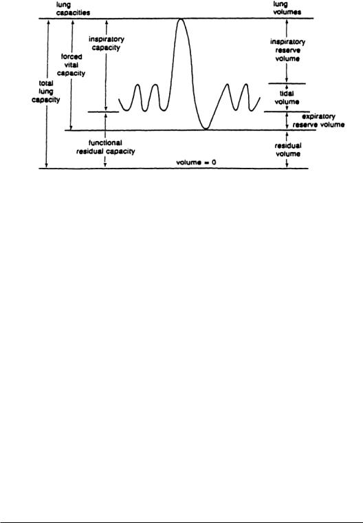
FIGURE 7.3 Lung capacities and lung volumes.
always less than tidal volume.The extent of this difference in volume depends primarily on the anatomic dead space, the 150to 160-ml internal volume of the conducting airway passages.The term dead is quite appropriate, since it represents wasted respiratory effort; i.e., no significant gas exchange occurs across the thick walls of the trachea, bronchi, and bronchiolus. Since normal tidal volume at rest is usually about 500 ml of air per breath, one can easily calculate that because of the presence of this dead space, about 340 to 350 ml of fresh air actually penetrates the alveoli and becomes involved in the gas exchange process. An additional 150 to 160 ml of stale air exhaled during the previous breath is also drawn into the alveoli.
The term volume is used for elemental differences of lung volume, whereas the term capacity is used for combination of lung volumes.Figure 7.3 illustrates the interrelationship between each of the following lung volumes and capacities:
1.Total lung capacity (TLC): The amount of gas contained in the lung at the end of maximal inspiration.
2.Forced vital capacity (FVC): The maximal volume of gas that can be forcefully expelled after maximal inspiration.
3.Inspiratory capacity (IC):The maximal volume of gas that can be inspired from the resting expiratory level.
4.Functional residual capacity (FRC):The volume of gas remaining after normal expiration. It will be noted that functional residual capacity (FRC) is the same as the resting volume.There is a small difference, however, between resting volume and FRC because FRC is measured while the patient breathes, whereas resting volume is measured with no breathing. FRC is properly defined only at end-expiration at rest and not during exercise.
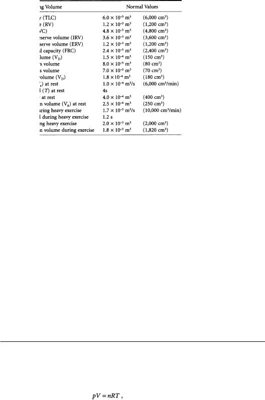
These volumes and specific capacities, represented in Fig. 7.3, have led to the development of specific tests (that will be discussed below) to quantify the status of the pulmonary system.Typical values for these volumes and capacities are provided in Table 7.2.
7.3 Perfusion of the Lung
For gas exchange to occur properly in the lung, air must be delivered to the alveoli via the conducting airways, gas must diffuse from the alveoli to the capillaries through extremely thin walls, and the same gas must be removed to the cardiac atrium by blood flow.This three-step process involves (1) alveolar
Respiratory System |
7-7 |
TABLE 7.2 Typical LungVolumes for Normal, Healthy Males
Source: Adapted and used with permission from Forster et al. [1986].
ventilation, (2) the process of diffusion, and (3) ventilatory perfusion, which involves pulmonary blood flow.Obviously,an alveolus that is ventilated but not perfused cannot exchange gas.Similarly,a perfused alveolus that is not properly ventilated cannot exchange gas.The most efficient gas exchange occurs when ventilation and perfusion are matched.
There is a wide range of ventilation-to-perfusion ratios that naturally occur in various regions of the lung [Johnson, 1991]. Blood flow is somewhat affected by posture because of the effects of gravity. In the upright position, there is a general reduction in the volume of blood in the thorax, allowing for larger lung volume. Gravity also influences the distribution of blood, such that the perfusion of equal lung volumes is about five times greater at the base compared with the top of the lung [Astrand and Rodahl,1970].There is no corresponding distribution of ventilation;hence the ventilation-to-perfusion ratio is nearly five times smaller at the top of the lung (Table 7.3). A more uniform ventilation-to- perfusion ratio is found in the supine position and during exercise [Jones, 1984b].
Blood flow through the capillaries is not steady. Rather, blood flows in a halting manner and may even be stopped if intraalveolar pressure exceeds intracapillary blood pressure during diastole. Mean blood flow is not affected by heart rate [West,1977],but the highly distensible pulmonary blood vessels admit more blood when blood pressure and cardiac output increase.During exercise,higher pulmonary blood pressures allow more blood to flow through the capillaries. Even mild exercise favors more uniform perfusion of the lungs [Astrand and Rodahl,1970].Pulmonary artery systolic pressures increases from 2670 N/m2 (20 mmHg) at rest to 4670 N/m2 (35 mmHg) during moderate exercise to 6670 N/ m2 (50 mmHg) at maximal work [Astrand and Rodahl, 1970].
7.4 Gas Partial Pressures
The primary purpose of the respiratory system is gas exchange. In the gas-exchange process, gas must diffuse through the alveolar space, across tissue, and through plasma into the red blood cell, where it finally chemically joins to hemoglobin.A similar process occurs for carbon dioxide elimination.
As long as intermolecular interactions are small, most gases of physiologic significance can be considered to obey the ideal gas law: