

Biotechnology for Biomedical Engineers - Martin L. Yarmush et al
.pdf9-14 Biotechnology for Biomedical Engineers
F(ab)’2fragment consists of two Fab arms held together by disulfide bonds in the hinge region and was first generated by pepsin digestion. Finally, the Fv fragment, which consists of just the two N-terminal variable domains,also was first generated by proteolytic digestion and is now more commonly generated via antigen-engineering techniques.
The other fragments listed in Fig. 9.2 are of genetic origin. These include the sFv (single-chain antigen), the VH domain (single-domain antigen), multivalent sFvs (miniantibodies), and multivalent Fabs. The multivalent constructs can either be monospecific or bispecific. The single-chain antigen (SCA, sFv) consists of the two variable domains linked together by a long flexible polypeptide linker [Bird et al,1988;Huston et al.,1988].The sFv is an attempt to stabilize the Fv fragment,which is known to dissociate at low concentrations into its individual domains due to the low-affinity constant for the VH-VL interaction. Two different constructs have been made: the VL-VH construct where the linker extends from the C terminus of the VL domain to the N terminus of the VH domain, and the VH-VL construct, where the linker runs from the C terminus of theVH domain to the N terminus of theVL. The linker is usually about 15 amino acids long (the length required to span the distance between the two domains) and has no particular sequence requirements other than to minimize potential interferences in the folding of the individual domains.The so-called universal linker used by many workers in the area is (GGGGS)3. Other strategies to stabilize the Fv fragment include chemical cross-linking of the two domains and disulfidelinked domains [Glockshuber et al., 1990]. Chemical cross-linking via glutaraldehyde has been demonstrated to be effective in stabilizing the Fv fragment in one instance; here, the crosslinking was carried out in the presence of the hapten (phosphorylcholine) to avoid modification of the binding site, an approach that may not be feasible with protein antigens. In the disulfide-linked sFv, Cys residues are introduced at suitable locations in the framework region of the Fv so as to form a natural interdomain disulfide bond. This strategy was shown to be much more effective in stabilizing the Fv fragment against thermal denaturation than either the single-chain antigen approach or chemical crosslinking for the one case where all three approaches were tested [Glockshuber et al., 1990].
The single-domain antigen consists of just theVH domain and has been shown by some to possess antigen-binding function on its own in the absence of theVL domain [Ward et al., 1989].There is some skepticism regarding this approach due to the rather high potential for nonspecific binding (the removal of the VL domain exposes a very hydrophobic surface), poor solubility, and somewhat compromised selectivity. For example, while the Fv fragment retains its ability to distinguish between related antigenic species, the single-domain antigen does not.
Miniantibodies consist of sFv fragments held together by so-called dimerization handles.sFv fragments are fused via a flexible hinge region to several kinds of amphipathic helices,which then acts as dimerization devices [Pack and Pluckthun, 1992].Alternately, the two sFvs can be fused via a long polypeptide linker, similar to the one linking the individual domains of each Fv but longer to maintain the relative orientation of the two binding sites.Multivalent Fabs use a somewhat similar approach with the dimerization handles comprising of zippers from the transcription factors jun and fos [Kostelny et al., 1992].
In addition to the constructs described above, a whole new set of genetically engineered fusion proteins with antibodies has been described [Wright et al., 1992].Antibody fusion proteins are made by replacing a part of the antibody molecule with a fusion partner such as an enzyme or a toxin that confers a novel function to the antibody. In some cases, such as immunotoxins, the variable regions of the antibody are retained in order to retain antigen binding and specificity, while the constant domains are deleted and replaced by a toxin such as ricin or Pseudomonas exotoxin. Alternately, the constant regions are retained (thereby retaining the effector functions) and the variable regions replaced with other targeting proteins (such as CD4 for AIDS therapy and IL-2 for cancer therapy).
9.5 Applications of Monoclonal Antibodies and Fragments
The majority of applications for which monoclonal antibodies have been used can be divided into three general categories:(1) purification,(2) diagnostic functions (whether for detecting cancer,analyzing
Monoclonal Antibodies and Their Engineered Fragments |
9-15 |
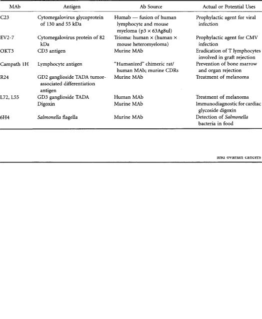
for toxins in food, or monitoring substance abuse by athletes), and (3) therapeutic functions. From the time that monoclonal antibody technology was introduced almost 20 years ago,application methodologies using whole antibody have gradually been transformed into methodologies using antibody fragments such as the Fab2, Fab’, and Fv fragments and even synthetic peptides of a CDR region. Antibody conjugates have come to include bound drugs,toxins,radioisotopes,lymphokines,and enzymes [Pietersz and McKenzie,1992] and are largely used in the treatment of cancer.Tables 9.1 and 9.2 list some typical examples of monoclonal antibodies and fragments used for diagnostic and therapeutic applications.
TABLE 9.1 Uses ofWhole MAb Derived from Hybridoma,Other Cell Fusions,and Genetically Engineered Cell Lines
TABLE 9.2 Immunoconjugates Having Potential for Cancer Therapy
9-16 Biotechnology for Biomedical Engineers
Thousands of murine monoclonal antibodies have been made to human carcinomas since the introduction of antibody technology, but very few, if any, of these monoclonal antibodies are entirely specific for malignant cells. In the vast majority of cases, these monoclonal antibodies define tumorassociated differentiation antigens (TADAs),which are either proteins,mucins,proteoglycans,or glycolipids (gangliosides).Examples ofTADA proteins are carcinoembryonic antigen (CEA) and a-fetoprotein (AFP), both well-known diagnostic markers. An anti-CEA antibody has been conjugated with the enzyme carboxypeptidase, which, in turn, activates a prodrug at the site of the tumor [Bagshawe et al., 1992].This strategy overcomes the inability of monoclonal antibodies conjugated with drugs to deliver a therapeutic dose. Examples of TADA gangliosides are those referred to as GD2 and GD3, for which the respective unmodified monoclonal antibodies have shown to be effective therapeutics,particularly when intralesionally administered [Irie et a., 1989]. An example of a TADA mucin is the antigens found in colorectal and ovarian carcinoma that react with the antibody 72.3.Chimeric monoclonal antibodies as well as fragments of antibody 72.3 have been constructed and tested [Khazaeli et al., 1991; King et al, 1992].
As mentioned above, monoclonal antibodies defining different TADAs have been used passively (unmodified) and as carriers of,for example,radioisotopes and enzymes.From the results brought forth to date, the passive mode of antibody therapy has produced relatively few remissions in patients, and in those cases where it has shown effects, it is likely that the ability of the antibody to mediate ADCC (antibody-dependent cellular cytotoxicity) and CDC (complement-dependent cytotoxicity) has contributed to the remission.For the case of modified monoclonal antibodies (e.g.,toxin and radioisotope conjugates), success of treatment is varied depending on the type of neoplasm. Antibody conjugate treatment of leukemia and lymphoma results in a relatively greater remission rate than that found in treatments of malignancies having solid tumors (i.e., carcinoma of the ovary, colon, and lung).
The rare case of complete remission for solid tumors is probably due to the inaccessibility of antibody to that tumor. Several barriers impeding access of antibody to cancer cells have been pointed out [Jain,1988].A few of these barriers include (1) the high interstitial fluid pressure in tumor nodules,
(2) heterogeneous or poor vascularization of tumors,and (3) the long distances extravasated monoclonal antibodies must travel in the interstial mesh of proteoglycans in the tumor. There also exists the possibility that tumor antigen shed from the surface is limiting antibody buildup. In the case of bound toxins or drugs, there is the added concern that organs such as the kidney and liver are quickly processing and eliminating the antibody conjugates. In this respect, immunoconjugates based on antibody fragments (such as the sFv) can be very advantageous. For example, it has been shown that the sFv exhibits rapid diffusion into the extravascular space, increased volume of distribution, enhance tumor penetration, and improved renal elimination. An assessment of solid tumor therapy with modified antibody has led Riethmüller et al. [1993] to recommend that current cancer therapy be directed toward minimal residual disease, the condition in which micrometastatic cells exist after curatively resecting solid tumors.
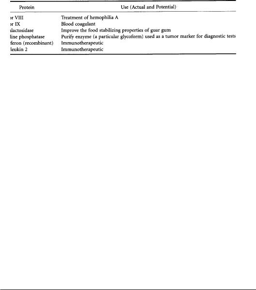
With regard to purification, the research literature is replete with examples of immunoaffinity purification of enzymes, receptors, peptides, and small organic molecules. In contrast, commercial applications of immunoaffinity chromatography, even on industrially or clinically relevant molecules, are far less widespread (Table 9.3). Despite its potential utility, immunoadsorption is an expensive process. A significant portion of the high cost is the adsorbent itself, which is related to the cost of materials, preparation, and most important, the antibody. In addition, the binding capacity of the immunoadsorbent declines with repeated use,and a systemic study has shown that significant deactivation can typically take place over 40 to 100 cycles [Antonsen et al,1991].A number of factors can contribute to this degradation, including loss of antibody, structural change of the support matrix, nonspecific absorption of contaminating proteins, incomplete antigen elution, and loss of antibody function. In most cases,this degradation is associated with repeated exposure to harsh elution conditions.Noteworthy commercial applications of immunoaffinity chromatography on useful molecules include the separation of factor VIII used to treat hemophilia A and factor IX, another coagulation factor in the bloodclotting cascade [Tharakan et al.,1992].The immunoaffinity purification step for factorVIII was one of several additional steps added to the conventional preparation methodology in which plasma
Monoclonal Antibodies and Their Engineered Fragments |
9-17 |
TABLE 9.3 Clinically or Industrially Relevant Proteins Purified by Immunoaffinity Chromatography
cryoprecipitates were heattreated.The new method contains a virus-inactivation procedure that precedes the immunoaffinity column,followed by an additional chromatographic step (ion exchange).The latter step serves to eliminate the eluting solvent and further reduce virus-inactivating compounds. The often-mentioned concern of antibody leakage from the column matrix did not appear to be a problem. Furthermore,with the relatively mild elution conditions used (40% ethylene glycol),one would expect little change in the antibody-binding capacity over many elution cycles.
Typical immunoaffinity matrices contain whole antibody as opposed to antibody fragments. Fragmentation of antibody by enzymatic means contributes additional steps to immunoadsorbent preparation and adds to the overall cost of the separation and is thus avoided. However, fragmentation can lead to a more efficient separation by enabling the orientation of antibody-binding sites on the surface of the immunomatrix [Prisyazhnoy et al,1988;Yarmush et al.,1992].Intact antibodies are bound in a random fashion, resulting in a loss of binding capacity upon immobilization. Recombinant antibody fragments could prove to be more useful for immunoaffinity applications due to the potential for production of large quantities of the protein at low cost and improved immobilization characteristics and stability [Spitznagel and Clark, 1993]. In what one could consider the ultimate fragment of a antibody, some investigators have utilized a peptide based on the CDR region of one of the chains (termed minimal recognition units) to isolate the antigen. Welling et al. [1990] have synthesized and tested a 13-residue synthetic peptide having a sequence similar to one hypervariable region of an antilysozyme antibody.
Important diagnostic uses of antibody include the monitoring in clinical laboratories of the cardiac glycoside digoxin and the detection of the Salmonella bacteria in foods (Table 9.1).These two examples highlight the fact that despite the exquisite specificity offered by monoclonal antibodies, detection is not failure-proof.Within digoxin immunoassays there are two possible interfering groups: endogenous digoxin-like substances and digoxin metabolites; moreover, several monoclonal antibodies many be necessary to avoid underor overestimating digoxin concentrations.In the case of bacteria detection in food, at least two antibodies (MOPC 467 myeloma protein and 6H4 antibody) are needed to detect all strains of Salmonella.
9.6 Summary
The domain structure of antibodies, both at the protein and genetic levels, facilitates the manipulation of antibody properties via genetic engineering (antibody engineering). Antibody engineering has shown tremendous potential for basic studies and industrial and medical applications. It has been used to explore fundamental questions about the effect of structure on antigen binding and on the biologic effector functions of the antibody molecules. A knowledge of the rules by which the particular sequences of amino acids involved in the binding surface are chosen in response to a particular antigenic determinant would enable the production of antibodies with altered affinities and specificities. Understanding the structures and mechanisms involved in the effector function of antibodies is already starting to result in the production of antibodies with novel biologic effector functions for use as diagnostic and therapeutic reagents. In addition, the production of antibodies via immunoglobulin gene expression has enabled the engineering of novel hybrid, chimeric, and mosaic genes using recombinant DNA techniques and the transfection and expression of these genetically engineered
9-18 |
Biotechnology for Biomedical Engineers |
genes in a number of different systems such as bacteria and yeast, plant cells, myeloma or hybridoma cells, and nonlymphoid mammalian cells.
References
Antonsen KP, Colton CK,Yarmush ML. 1991. Elution conditions and degradation mechanisms in longterm immunoadsorbent use.Biotechnol Prog 7:159.
Bagshawe KD,Sharma SK,Springer CJ,et al.1992.Antibody directed enzyme prodrug therapy (ADEPT).
Antibody Immunoconj Radiopharm 54:133.
Barbas CF, Kang AS, Lerner RA, Benkovic SJ. 1991.Assembly of combinatorial antibody libraries on phase surfaces:The gene III site. Proc Natl Acad Sci USA 88:7978.
Barbas CF, Bain JD,Hoekstra DM,Lerner RA.1992.Semisynthetic combinatorial antibody libraries:A chemical solution to the diversity problem. Proc Natl Acad Sci USA 89:4457.
Bender E,Woof JM,Atkin JD, et al.1993. Recombinant human antibodies: Linkage of an Fab fragment from a combinatorial library to an Fc fragment for expression in mammalian cell culture. Hum Antibod Hybridomas 4:74.
Better M, Chang CP, Robinson RR, Horwitz AH. 1988. Escherichia coli secretion of an active chimeric antibody fragment. Science 240:1041.
Better M, Horowitz AH. 1989. Expression of engineered antibodies and antibody fragments in microorganisms. Methods Enzymol 178:476.
Bird RE,Hardman KD,Jacobson JW,et al.1988.Single-chain antibody-binding proteins.Science 242:423. Boss MA,Kenten JH,Wood CR,Emtage JS.1984.Assembly of functional antibodies from immunoglobulin
heavy and light chains synthesized in E. coli. Nucleic Acids Res 12:3791.
Buchner J, Rudolph R. 1991. Renaturation, purification, and characterization of recombinant Fab fragments produced in Escherichia coli. Biotechnology 9:157.
Burton DR. 1991. Human and mouse monoclonal antibodies by repertoire cloning. Trends Biotechnol 9:169.
Burton DR,Barbas CF,Persson MAA,et al.1991.A large array of human monoclonal antibodies to type 1 human immunodeficiency virus from combinatorial libraries of asymptomatic seropositive individuals.Proc Natl Acad Sci USA 88:10134.
Cabilly S, Riggs AD, Pande H, et al. 1984. Generation of antibody activity from immunoglobulin polypeptide chains produced in Escherichia coli. Proc Natl Acad Sci USA 81:3273.
Caton AJ, Koprowski H. 1990. Influenza virus hemagglutinin-specific antibodies isolated from a combinatorial expression library are closely related to the immune response of the donor.Proc Natl Acad Sci USA 87:6450.
ClacksonT, Hoogenboom HR, Griffiths AD,Winter G. 1991. Making antibody fragments using phage display libraries.Nature 352:624.
Davies DR,Padlan EA,Sheriff S.1990.Antigen-antibody complexes.Annu Rev Biochem 59:439. Duchosal MA,Eming SA,Fischer P,et al.1992.Immunization of hu-PBL-SCID mice and the rescue of
human monoclonal Fab fragments through combinatorial libraries.Nature 355:258. Fanger MW, Morganelli PM,Guyre PM.1992.Bispecific antibodies. Crit Rev Immunol 12:101.
Glockshuber R, Malia M, Pfitzinger I, Pluckthun A. 1990, A comparison of strategies to stabilize immunoglobulin fragments. Biochemistry 29:1362.
Gram H, Lore-Anne M, Barbas CF, et al. 1992. In vitro selection and affinity maturation of antibodies from a naive combinatorial immunoglobulin library. Proc Natl Acad Sci USA 89:3576.
Hassemann CA,Capra JD.1990.High-level production of a functional immunoglobulin heterodimer in a baculovirus expression system. Proc Natl Acad Sci USA 87:3942.
Hiatt A, Cafferkey R, Bowdish K. 1989. Production of antibodies in transgenic plants. Nature 342:76. Hiatt A,Ma JK-C.1992. Monoclonal antibody engineering in plants. FEBS Lett 307:71. Hoogenboom HR, Griffiths AD, Johnson KS, et al. 1991. Multi-subunit proteins on the surface of
Monoclonal Antibodies and Their Engineered Fragments |
9-19 |
filamentous phage: Methodologies for displaying antibody (Fab) heavy and light chains. Nucleic Acids Res 19:4133.
Hoogenboom HR, Marks JD, Griffiths AD, Winter G. 1992. Building antibodies from their genes.
Immunol Rev 130:41.
Horowitz AH, Chang PC, Better M, et al. 1988. Secretion of functional antibody and Fab fragment from yeast cells. Proc Natl Acad Sci USA 85:8678.
Huse WD, Sastry L, Iverson SA, et al. 1989. Generation of a large combinatorial library of the immunoglobulin repertoire in phage lambda. Science 246:1275.
Huston JS, Levinson D, Mudgett-Hunter M, et al. 1988. Protein engineering of antibody binding sites: Recovery of specific activity in an anti-digoxin single-chain Fv analogue produced in Escherichia coli. Proc Natl Acad Sci USA 85:5879.
IshizakaT,Helm B,Hakimi J,et al.1986.Biological properties of a recombinant human immunoglobulin e-chain fragment. Proc Natl Acad Sci USA 83:8323.
Jain RK. 1988. Determinants of tumor blood flow:A review. Cancer Res 48:2641.
Kang AS,Barbas CF,Janda KD,et al.1991.Linkage of recognition and replication functions by assembling combinatorial antibody Fab libraries along phage surfaces. Proc Natl Acad Sci USA 88:4363.
Keneten J, Helm B, Ishizaka T, et al. 1984. Properties of a human immunoglobulin e-chain fragment synthesized in Escherichia coli. Proc Natl Acad Sci USA 81:2955.
Khazaeli MB,Saleh MN,LiuTP,Meredith RF.1991. Pharmacokinetics and immune response of 131I- chi-meric mouse/human B72.3 (human g4) monoclonal antibody in humans.Cancer Res 51:5461.
King DJ,Adair JR,Angal S,et al.1992.Expression,purification,and characterization of a mouse-human chimeric antibody and chimeric Fab’ fragment. Biochem J 281:317.
Kostelny SA, Cole MS,Tso JY. 1992. Formation of bispecific antibody by the use of leucine zippers. J Immunol 148:1547.
Köhler G, Milstein C. 1975. Continuous cultures of fused cells secreting antibody of predefined specificity.Nature 256:495.
Landsteiner K.1945.The Specificity of Serological Reactions. Cambridge,Mass,Harvard University Press. Marks JD,Hoogenboom HR,BonnertTP,et al.1991.Bypassing immunization:Human antibodies from
V-gene libraries displayed on phase. J Mol Biol 222:581.
Marks JD,Griffiths AD,Malmqvist M,et al.1992.Bypassing immunization:Building high affinity human antibodies by chain shuffling. Biotechnology 10:779.
McCafferty J,Griffiths AD,Winter G,Chriswell DJ.1990.Phage antibodies:Filamentous phage displaying antibody variable domains.Nature 348:552.
Morrison SL, OiVT. 1989. Genetically engineered antibody molecules. Adv Immunol 41:65.
Mullinax RL, Gross EA, Amberg JR, et al. 1990. Identification of human antibody fragment clones specific for tetanus toxoid in a bacteriophage l immunoexpression library. Proc Natl Acad Sci USA 87:8095.
Neil GA,Urnovitz HB.1988.Recent improvements in the production of antibody-secreting hybridoma cells.Trends Biotechnol 6:209.
Orlandi R, Gussow DH, Jones PT,Winter G. 1989. Cloning immunoglobulin variable domains for expression by the polymerase chain reaction. Proc Natl Acad Sci USA 86:3833.
Pack P,Pluckthun P.1992. Miniantibodies:Use of amphipathic helices to produce functional flexibility linked dimeric Fv fragments with high avidity in Escherichia coli. Biochemistry 31:1579.
Perrson MAA,Caothien RH,Burton DR.1991.Generation of diverse high-affinity human monoclonal antibodies by repertoire cloning. Proc Natl Acad Sci USA 88:2432.
Pietersz GA, McKenzie IFC. 1992. Antibody conjugates for the treatment of cancer. Immunol Rev 129:57.
Plückthun A.1992.Monoand bivalent antibody fragments produced in Escherichia coli:Engineering, folding and antigen-binding. Immunol Rev 130:150.
PrisyazhnoyVS, Fusek M,AlakhovYB. 1988. Synthesis of high-capacity immunoaffinity sorbents with
9-20 |
Biotechnology for Biomedical Engineers |
|
oriented immobilized immunoglobulins or their Fab’ fragments for isolation of proteins.J Chromatogr |
|
424:243. |
Putlitz JZ, Kubasek WL, Duchene M, et al. 1990.Antibody production in baculovirus-infected insect cells. Biotechnology 8:651.
Riethmüller G,Schneider-Gädicke E, Johnson JP. 1993.Monoclonal antibodies in cancer therapy. Curr Opin Immunol 5:732.
Sandhu JS.1992.Protein engineering of antibodies.Crit Rev Biotechnol 12:437.
Sastry L,Alting-Mees M,HuseWD,et al.1989.Cloning of the immunoglobulin repertoire in Escherichia coli for generation of monoclonal catalytic antibodies: Construction of a heavy chain variable region-specific cDNA library.Proc Natl Acad Sci USA 86:5728.
Skerra A, Plückthun A. 1988.Assembly of a functional immunoglobulin Fv fragment in Escherichia coli. Science 240:1038.
Skerra A.1993. Bacterial expression of immunoglobulin fragments.Curr Opin Biotechnol 5:255. SpitznagelTM, Clark DS. 1992. Surface density and orientation effects on immobilized antibodies and
antibody fragments. Biotechnology 11:825.
Tizard IR.1992.The genetic basis of antigen recognition.In Immunology:An Introduction, 3d ed.Orlando, Fla. Saunders Coolege Publishing.
Ward ES,Gussow D,Griffiths AD,et al.1989.Binding activities of a repertoire of single immunoglobulin variable domains secreted from Escherichia coli. Nature 341:544.
Wood CR,Boss MA,Kenten JH,et al.1985.The synthesis and in vivo assembly of functional antibodies in yeast.Nature 314:446.
Welling GW,GuertsT,Van Gorkum J,et al.1990.Synthetic antibody fragment as ligand in immunoaffinity chromatography.J.Chromatogr 512:337.
Wright A,Shin S-U,Morrison SL.1992.Genetically engineered antibodies:progress and prospects.Crit Rev Immunol 12:125.
Yarmush ML,Lu X,Yarmush D.1992.Coupling of antibody-binding fragments to solid-phase supports: Site-directed binding of F(ab’)2 fragments. J Biochem Biophys Methods 25:285.
Zebedee SL, Barbas CF,Yao-Ling H, et al. 1992. Human combinatorial libraries to hepatitis B surface antigen. Proc Natl Acad Sci USA 89:3175.

S.PatrickWalton
Center for Engineering in Medicine, Massachusetts General Hospital, Harvard Medical School, and Shriners Burns Hospital, Boston
Charles M.Roth
Center for Engineering in Medicine, Massachusetts General Hospital, Harvard Medical School, and Shriners Burns Hospital, Boston
Martin L.Yarmush
Center for Engineering in Medicine, Massachusetts General Hospital, Harvard Medical School, and Shriners Burns Hospital, Boston
10
Antisense Technology
10.1 |
Background |
10–1 |
10.2 |
Mechanisms of Inhibition |
10–3 |
10.3 |
Chemically Modified Oligonucleotides |
10–4 |
10.4 |
Oligonucleotide Synthesis and Purification |
10–7 |
10.5 |
Specificity of Oligonucleotides |
10–7 |
10.6 |
Oligonucleotide Delivery |
10–8 |
10.7 |
Potential Applications of Antisense |
|
|
Oligonucleotides |
10–10 |
|
Viral Diseases • Cancer |
|
10.8 |
InVivo Pharmacology |
10–11 |
10.9 |
Animal Models |
10–12 |
10.10 |
ClinicalTrials |
10–13 |
10.11 |
Summary |
10–13 |
Antisense molecules can selectively inhibit the expression of one gene among the 100,000 present in a typical human cell.This inhibition is likely based on simple Watson-Crick base-pairing interactions between nucleic acids and makes possible, in principle, the rational design of therapeutic drugs that can specifically inhibit any gene with a known sequence.The intervention into disease states at the level of gene expression may potentially make drugs based on antisense techniques significantly more efficient and specific than other standard therapies. Indeed, antisense technology is already an indispensable research tool and may one day be an integral part of future antiviral and anticancer therapies.
Zamecnik and Stephenson were the first to use antisense DNA to modulate gene expression.They constructed antisense oligonucleotides complementary to the 3¢ and 5¢ ends of Rous sarcoma virus (RSV) 35S RNA and added them directly to a culture of chick embryo fibroblasts infected with RSV [1]. Remarkably, viral replication was inhibited, indicating that the cells had somehow internalized the antisense DNA and that the DNA was somehow interrupting the viral life cycle.
Natural antisense inhibition was first observed in bacteria as a means of regulating the replication of plasmid DNA [2, 3]. RNA primers required for the initiation of replication were bound by (i.e., formed duplexes with) antisense RNA.The concentration of these RNA primers, and therefore the initiation of replication, was controlled by the formation of these duplexes. Shortly after this discovery, investigators developed antisense RNA constructs to control gene expression in mammalian cells. Antisense RNA, encoded on expression plasmids that were transfected into mouse cells, successfully blocked expression of target genes [4].These early successes launched what is now a significant effort to expand the use of antisense molecules for research and therapeutic purposes.
10.1 Background
Antisense oligonucleotides are DNA or RNA molecules whose sequences are complementary to RNA transcribed from the target gene.These molecules block the expression of a gene by interacting
0-8493-1811-4/03/$0.00+$1.50 |
10-1 |
© 2003 by CRC Press LLC |
10-2 |
Biotechnology for Biomedical Engineers |
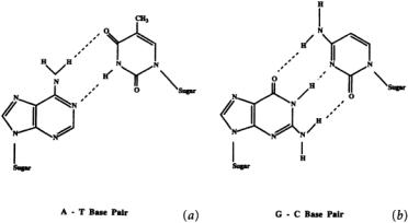
FIGURE 10.1 Watson-Crick base pairing interactions between adenosine and thymidine (A-T) and between guanosine and cytidine (G-C).The sugar is 2¢-deoxyribose.
with its RNA transcript.Antisense molecules are typically short, single-stranded oligonucleotides with a sequence complementary to a sequence within the target RNA transcript. The oligonucleotides bind to this sequence via Watson-Crick base pairing (adenosine binds to thymidine (DNA) or uracil (RNA), and guanosine binds to cytidine), form a DNA:RNA duplex, and block translation of the RNA (see Fig. 10.1).
Nucleic acids have been used in a variety of strategies to modulate gene expression. For example, antisense RNA, encoded by a plasmid transfected into the target cells, has been used to inhibit gene expression.Cells are transfected with a plasmid encoding the antisense RNA,the plasmid is transcribed, and the resulting antisense RNA transcript inhibits gene expression. Unfortunately, difficulties in controlling the expression of the transfected plasmid hinder the effective use of antisense RNA for therapeutic purposes. More recently, antisense RNA work has focused on the use of hammerhead ribozymes.These synthetic RNA oligonucleotides have the ability to cleave other RNA strands and they appear more therapeutically viable. Other methods for using nucleic acids to block gene expression are being explored, such as:
1.nucleic acids that competitively bind to proteins via their 3-dimensional structure (aptamers),
2.nucleic acids designed to prevent transcription by forming a DNA triplex with the target gene (antigene), and
3.nucleic acids that mimic binding sites for transcription and translation complexes (decoys).
Although promising, these approaches are preliminary and are beyond the scope of this chapter.The interested reader is directed to reviews concerning these topics [5–7]. Only antisense DNA molecules, which mimic the antisense strand of the target gene and thus hybridize to RNA transcribed from the gene, will be discussed in detail here. For a recent review on antisense RNA, see Rossi [8].
Many technical issues limit the therapeutic usefulness of current antisense oligonucleotides; for instance, they are highly susceptible to degradation by nucleases. Also, our understanding of the mechanism of antisense inhibition must improve before the widespread development of therapeutically useful antisense molecules is possible.Advances in oligonucleotide chemistry have begun to resolve the issue of their intracellular stability; however, the reaction of natural systems to these unnatural species has not been fully explored. Many oligonucleotides still lack adequate specificity for in vivo use, and their delivery to cells is often non-specific and inefficient.The impact of these problems and approaches to solving them will be discussed. In addition, potential applications of antisense techniques to antiviral and anticancer therapies and their evaluation in animal models and clinical trials will also be highlighted.
Antisense Technology |
10-3 |
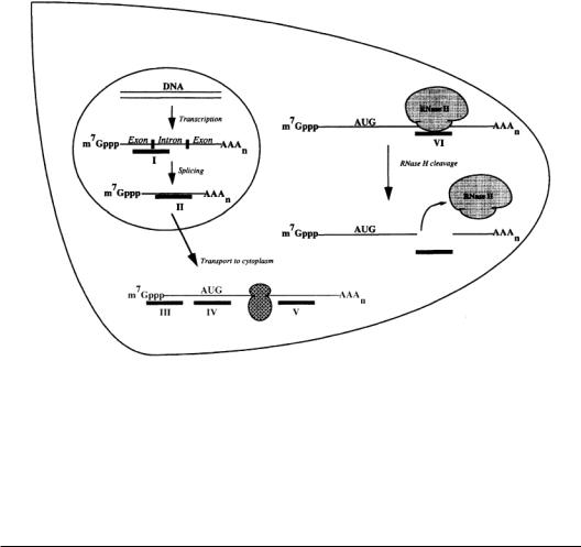
FIGURE 10.2 Several possible sequence specific sites of antisense inhibition.Antisense oligodeoxynucleotides are represented by black bars. Antisense oligodeoxynucleotides can interfere with (I) splicing, (II) transport of the nascent mRNA from the nucleus to the cytoplasm, (III) binding of initiation factors, (IV) assembly of ribosomal subunits at the start codon or (V) elongation of translation. Inhibition of capping and polyadenylation is also possible. (VI) Antisense oligodeoxynucleotides that activate RNase H (e.g., oligonucleotides with phosphodiester and phosphorothioate backbones) can also inhibit gene expression by binding to their target mRNA, catalyzing the RNase H cleavage of the mRNA into segments that are rapidly degraded by exonucleases.
10.2 Mechanisms of Inhibition
The inhibition of gene expression by antisense molecules is believed to occur by a combination of two mechanisms:ribonuclease H (RNase H) degradation of the RNA and steric hindrance of the processing of the RNA [9].Some oligonucleotides,after hybridizing to the target RNA,activate RNase H,which specifically recognizes an RNA:DNA duplex and cleaves the RNA strand.The antisense oligonucleotide is not degraded and is free to bind to and catalyze the destruction of other target RNA transcripts [10]. Oligonucleotides that activate RNase H, therefore, are potentially capable of destroying many RNA transcripts in their lifetime, which suggests that lower concentrations of these oligonucleotides may be sufficient to significantly inhibit gene expression [9].
However, not all oligonucleotides activate RNase H enzymes. Some oligonucleotides inhibit gene expression by interfering with the RNA’s normal life cycle. Newly transcribed RNA must be spliced, polyadenylated, and transported to the cytoplasm before being translated into protein. In theory, virtually any step in an RNA’s life cycle can be blocked by an oligonucleotide (see Fig. 10.2). Oligonucleotides that do not activate RNase H can block RNA maturation by binding to sites on the RNA important for posttranscriptional processing or for ribosomal assembly and translation initiation [11]. Oligonucleotides designed to block the elongation of translation by binding to coding sequences downstream from the initiation codon region have rarely succeeded, presumably because ribosomes destabilize and read through DNA:RNA duplexes [11]. Inhibition by both RNase H degradation and steric hindrance was demonstrated in a study in which the expression of an intracellular adhesion molecule (ICAM-1) was blocked using two different oligonucleotides [12].