

Biotechnology for Biomedical Engineers - Martin L. Yarmush et al
.pdfTABLE 11.3 Common CloningVectors
(shorter) fragments of DNA will travel through the pores more rapidly than the larger (longer) fragments, thus effecting separation. Agarose, a highly purified derivative of agar, is commonly used to separate relatively large fragments of DNA (100 to 50,000 base pairs) with modest resolution (50 to 100 base pairs), while cross-linked polyacrylamide is used to separate smaller fragments (10 to 1,000 base pairs) with single base-pair resolution. Fragment sizes are generally estimated by comparison with standards run in another lane of the same gel. Electrophoresis is used extensively as both an analytical and a preparative tool in molecular biology.
Enzymatic DNA Sequencing. In the late 1970s,Sanger and coworkers [4] reported a procedure employing DNA polymerase to obtain DNA sequence information from unknown cloned fragments. While significant improvements and modifications have been made since that time, the basic technique remains the same: DNA polymerase is used to synthesize a complementary copy of an unknown single-stranded DNA (the template) in the presence of the four DNA monomers (deoxynucleotide triphosphates, or dNTPs). DNA polymerase requires a double-stranded starting point, so a singlestranded DNA (the primer) is hybridized at a unique site on the template (usually in the vector), and it is at this point that DNA synthesis is initiated. Key to the sequencing process is the use of a modified monomer, a dideoxynucleotide triphosphate (ddNTP), in each reaction. The ddNTP lacks the 3'- hydroxyl functionality (it has been replaced by a hydrogen) necessary for phosphodiester bond formation, and its incorporation thus blocks further elongation of the growing chain by polymerase.Four reactions are carried out, each containing all four dNTPs and one of the four ddNTPs. By using the proper ratios of dNTPs to ddNTP, each reaction generates a nested set of fragments, each fragment beginning at exactly the same point (the primer) and terminating with a particular ddNTP at each base complementary to that ddNTP in the template sequence. The products of the reactions are then separated by electrophoresis in four lanes of a polyacrylamide slab gel. Since conventional sequencing procedures utilize radiolabeling (incorporation of a small amount of 32P- or 35S-labeled dNTP by the polymerase), visualization of the gel is achieved by exposing it to film.The sequence can be obtained from the resulting autoradiogram, which appears as a series of bands (often termed a ladder) in each of the four lanes. Each band is composed of fragments of a single size, the shortest fragments being at the bottom of the gel and the longest at the top.Adjacent bands represent a single base pair difference, so the sequence is determined by reading up the ladders in the four lanes and noting which lane contains the band with the next largest sized fragments.The enzymatic sequencing process is diagrammed in Fig. 11.2. It should be noted that although other methods exist, the enzymatic sequencing technique is currently the most commonly used DNA sequencing procedure due to its simplicity and reliability. Polymerase Chain Reaction (PCR). PCR [5] is an in vitro procedure for amplifying particular DNA sequences up to 108-fold that is utilized in an ever-increasing variety of ways in genome analysis.The sequence to be amplified is defined by a pair of single-stranded primers designed to hybridize to unique sites flanking the target sequence on opposite strands. DNA polymerase in the presence of the four dNTPs is used to synthesize a complementary DNA copy across the target sequence starting at the two primer sites.The amplification procedure is performed by repeating the following cycle 25 to 50 times (see Fig. 11.3). First, the double-stranded target DNA is denatured at high temperature (94 to 96°C). Second, the mixture is cooled, allowing the primers to anneal to their complementary sites on the target single strands. Third, the temperature is adjusted for optimal DNA polymerase activity, initiating synthesis.Since the primers are complementary to the newly synthesized strands as well as the
11-4
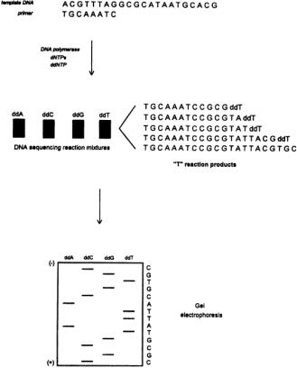
FIGURE 11.2 Enzymatic DNA sequencing.A synthetic oligonucleotide primer is hybridized to its complementary site on the template DNA. DNA polymerase and dNTPs are then used to synthesize a complementary copy of the unknown portion of the template in the presence of a chain-terminating ddNTP (see text).A nested set of fragments beginning with the primer sequence and ending at every ddNTP position is produced in each reaction (the ddTTP reaction products are shown). Four reactions are carried out, one for each ddNTP.The products of each reaction are then separated by gel electrophoresis in individual lanes, and the resulting ladders are visualized.The DNA sequence is obtained by reading up the set of four ladders, one base at a time, from smallest to largest fragment.
original target,each cycle of denaturation/annealing/synthesis effectively doubles the amount of target sequence present in the reaction, resulting in a 2² amplification (n—number of cycles). The initial implementation of PCR utilized a polymerase that was unstable at the high temperatures required for denaturation, thus requiring manual addition of polymerase prior to the synthesis step of every cycle. An important technological development was the isolation of DNA polymerase from a thermophilic bacterium, Thermus aquaticus (Taq), which can withstand the high denaturation temperatures [6]. Additionally, the high optimal synthesis temperature (70 to 72°C) of Taq polymerase improves the specificity of the amplification process by reducing spurious priming from annealing of the primers to nonspecific secondary sites in the target.
While PCR can be performed successfully manually, it is a tedious process, and numerous thermal cycling instruments have become commercially available.Modern thermal cyclers are programmable and capable of processing many samples at once, using either small plastic tubes or microtiter plates, and are characterized by accurate and consistent temperature control at all sample positions, rapid temperature ramping, and minimal temperature over/undershoot. Temperature control is provided by a variety of means (Peltier elements,forced air,water) using metal blocks or water or air baths.Speed,precise temperature control, and high sample throughput are the watchwords of current thermal cycler design.
PCR technology is commonly used to provide sufficient material for cloning from genomic DNA sources, to identify and characterize particular DNA sequences in an unknown mixture, to rapidly
11-5
11-6 |
Biotechnology for Biomedical Engineers |
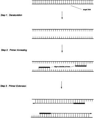
FIGURE 11.3 The first cycle in the polymerase chain reaction. In step 1, the double-stranded target DNA is thermally denatured to produce single-stranded species.A pair of synthetic primers, flanking the specific region of interest,are annealed to the single strands to form initiation sites for DNA synthesis by polymerase (step 2).Finally, complementary copies of each target single strand are synthesized by polymerase in the presence of dNTPs, thus doubling the amount of target DNA initially present (step 3). Repetition of this cycle effectively doubles the target population, affording one million-fold or greater amplification of the initial target sequence.
produce templates for DNA sequencing from very small amounts of target DNA,and in cycle sequencing, a modification of the enzymatic sequencing procedure that utilizes Taq polymerase and thermal cycling to amplify the products of the sequencing reactions.
Chemical Synthesis of Oligodeoxynucleotides. The widespread use of techniques based on DNA polymerase,such as enzymatic DNA sequencing and the PCR,as well as of numerous other techniques utilizing short, defined-sequence, single-stranded DNAs in genome analysis, is largely due to the ease with which small oligodeoxynucleotides can be obtained.The chemical synthesis of oligonucleotides has become a routine feature of both individual biology laboratories and core facilities. The most widely used chemistry for assembling short (<100 base pair) oligonucleotides is the phosphoramidite approach [7], which has developed over the past 15 years or so.This approach is characterized by rapid, high-yield reactions and stable reagents. Like modern peptide synthesis chemistry, the approach relies on the tethering of the growing DNA chain to a solid support (classically glass or silica beads, more recently cross-linked polystyrene) and is cyclic in nature. At the end of the assembly, the desired oligonucleotide is chemically cleaved from the support, generally in a form that is sufficiently pure for its immediate use in a number of applications.The solid phase provides two significant advantages: It allows for the use of large reagent excesses, driving the reactions to near completion in accord with the laws of mass action while reducing the removal of these excesses following the reactions to a simple matter of thorough washing, and it enables the reactions to be performed in simple, flow-through cartridges, making the entire synthesis procedure easily automatable. Indeed, a number of chemical DNA synthesis instruments (“gene machines”) are commercially available, capable of synthesizing one

Tools for Genome Analysis |
11-7 |
to several oligonucleotides at once.Desired sequences are programmed through a keyboard or touchpad, reagents are installed, and DNA is obtained a few hours later. Improvements in both chemistry and instrument design have been aimed at increasing synthesis throughput (reduced cycle times, increased number of simultaneous sequence assemblies), decreasing scale (most applications in genome analysis require subnanomole quantities of any particular oligonucleotide), and concomitant with these two, reducing cost per oligonucleotide.
11.3 Tools for Genome Analysis
Physical Mapping. In the analysis of genomes,it is often useful to begin with a less complex mixture than an entire genome DNA sample.Individual chromosomes can be obtained in high purity using a technology known as chromosome sorting [8], a form of flow cytometry. A suspension of chromosomes stained with fluorescent dye is flowed past a laser beam.As a chromosome enters the beam, appropriate optics detect the scattered and emitted light. Past the beam, the stream is acoustically broken into small droplets.The optical signals are used to electronically trigger the collection of droplets containing chromosomes by electrostatically charging these droplets and deflecting them into a collection medium using a strong electric field. Chromosomes can be differentiated by staining the suspension with two different dyes that bind in differing amounts to the various chromosomes and looking at the ratio of the emission intensity of each dye as it passes the laser/detector. Current commercial chromosome sorting instrumentation is relatively slow, requiring several days to collect sufficient material for subsequent analysis.
As mentioned previously, whole genomes or even chromosomes cannot yet be analyzed as intact entities. As such, fractionation of large nucleic acids into smaller fragments is necessary to obtain the physical material on which to perform genetic analysis.Fractionation can be achieved using a variety of techniques: limited or complete digestion by restriction enzymes, sonication, or physical shearing through a small orifice. These fragments are then cloned into an appropriate vector, the choice of which depends on the size range of fragments involved (see Table 11.3).The composite set of clones derived from a large nucleic acid is termed a library. In general,it is necessary to produce several libraries in different cloning vectors containing different-sized inserts.This is necessary because the mapping of clones is facilitated by larger inserts, while the sequencing of clones requires shorter inserts.
The library-generating process yields a very large number of clones having an almost random distribution of insert endpoints in the original fragment.It would be very costly to analyze all clones in a library, and unnecessary as well. Instead, a subset of overlapping clones is selected whose inserts span the entire starting fragment.These clones must be arrayed in the linear order in which they are found in the starting fragment; the process for doing this is called physical mapping. The conventional method for physically mapping clones uses restriction enzymes to cleave each clone at enzyme-specific sites, separating the products of the digestion by electrophoresis, and comparing the resulting patterns of restriction fragment sizes for different clones to find similarities. Clones exhibiting a number of the samesized fragments likely possess the same subsequence and thus overlap.Clearly,the longer the inserts contained in the library, the faster a large genetic region can be covered by this process, since fewer clones are required to span the distance.Physical mapping also provides landmarks,the enzyme cleavage sites in the sequence, that can be used to provide reference points for the mapping of genes and other functional sequences. Mapping by restriction enzyme digestion is simple and reliable to perform; however,manual map assembly from the digest data is laborious,and significant effort is currently being expended in the development of robust and accurate map assembly software.
Normal agarose gel electrophoresis can effectively separate DNA fragments less than 10,000 base pairs and fragments between 10,000 and 50,000 base pairs less effectively under special conditions. However, the development of very large insert cloning vectors, such as the yeast artificial chromosome [9],necessitated the separation of fragments significantly larger than 10,000 base pairs to allow for use in physical mapping. In order to address this issue, a technology called pulsed-field gel electrophoresis (PFGE) was developed. Unlike conventional electrophoresis, in which the electric field remains essentially
11-8 Biotechnology for Biomedical Engineers
constant, homogeneous, and unidirectional during a separation, PFGE utilizes an electric field that periodically changes its orientation.The principle of PFGE is thought to be as follows:When DNA molecules are placed in an electric field, the molecules elongate in the direction of the field and then begin to migrate through the gel pores. When the field is removed, the molecules relax to a more random coiled state and stop moving. Reapplication of the field in another orientation causes the DNA to change its conformation in order to align in that direction prior to migration. The time required for this conformational change to occur has been found to be very dependent on the size of the molecules,with larger molecules reorienting more slowly than small ones.Thus longer DNAs move more slowly under the influence of the constantly switching electric field than shorter ones, and sizebased separation occurs. PFGE separations of molecules as large as 10 million base pairs have been demonstrated.Numerous instruments for PFGE have been constructed,differing largely in the strategy employed to provide electric field switching [10].
Physical maps based on restriction sites are of limited long-term utility, since they require the provision of physical material from the specific library from which the map was derived in order to be utilized experimentally.A more robust landmarking approach based on the PCR has been developed recently [11], termed sequence-tagged site (STS) content mapping. An STS is a short, unique sequence in a genome that can be amplified by the PCR. Clones in a library are screened for the presence of a particular STS using PCR; if the STS is indeed unique in the genome, then clones possessing that STS are reliably expected to overlap.Physical mapping is thus reduced to choosing and synthesizing pairs of PCR primers that define unique sequences in the genome. Additionally, since STSs are defined by pairs of primer sequences, they can be stored in a database and are thus universally accessible. DNA Sequencing. Early in the development of tools for large-scale DNA analysis, it was recognized that one of the most costly and time-consuming processes was the accumulation of DNA sequence information. Two factors, the use of radioisotopic labels and the manual reading and recording of DNA sequence films, made it impossible to consider genome-scale (106 to 109 base pairs) sequence analysis using the conventional techniques.To address this, several groups embarked on the development of automated DNA sequencing instruments [12–14].Today, automated DNA sequencing is one of the most highly advanced of the technologies for genome analysis, largely due to the extensive effort expended in instrument design, biochemical organization, and software development.
Key to the development of these instruments was the demonstration that fluorescence could be employed in the place of autoradiography for detection of fragments in DNA sequencing gels and that the use of fluorescent labels enabled the acquisition of DNA sequence data in an automated fashion in real time.Two approaches have been demonstrated: the “single-color, four-lane” approach and the “fourcolor,single-lane” approach.The former simply replaces the radioisotopic label used in conventional enzymatic sequencing with a fluorescent label, and the sequence is determined by the order of fluorescent bands in the four lanes of the gel.The latter utilizes a different-colored label for each of the four sequencing reactions (thusA might be “blue,” C,“green,” G,“yellow,” andT,“red”).The four basespecific reactions are performed separately and upon completion are combined and electrophoresed in a single lane of the gel, and the DNA sequence is determined from the temporal pattern of fluorescent colors passing the detector. For a fixed number of gel lanes (current commercial automated DNA sequencers have 24 to 36), the four-color approach provides greater sample throughput than the single-color approach. Instruments employing the four-color technology are more widely used for genome analysis at present, and as such, this strategy will be discussed more fully.
In order to utilize fluorescence as a detection strategy for DNA sequencing, a chemistry had to be developed for the specific incorporation of fluorophores into the nested set of fragments produced in the enzymatic sequencing reactions.The flexibility of chemical DNA synthesis provided a solution to this problem.A chemistry was developed for the incorporation of an aliphatic primary amine in the last cycle of primer synthesis (i.e., at the 5¢ terminus) using standard DNA synthesis protocols [15, 16].This amine was then conjugated with any of several of readily available amine-reactive fluorochromes that had been developed previously for the labeling of proteins to produce the desired labeled sequencing primers.The purified dye-primer was demonstrated to perform well in DNA sequencing, exhibiting
Tools for Genome Analysis |
11-9 |
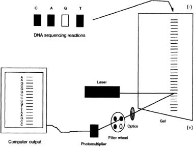
FIGURE 11.4 Schematic illustration of automated fluorescence-based DNA sequencing using the “four-color, single-lane” approach.The products of each of the four enzymatic sequencing reactions are “color-coded” with a different fluorescent dye, either through the use of dye-labeled primers or dye-terminators. The four reaction mixtures are then combined and the mixture separated by gel electrophoresis.The beam of an argon ion laser is mechanically scanned across the width of the gel near its bottom to excite the labeled fragments to fluorescence. The emitted light is collected through a four-color filter wheel onto a photomultiplier tube.The color of each fluorescing band is determined automatically by a computer from the characteristic four-point spectrum of each dye, and the order of colors passing the detector is subsequently translated into DNA sequence information.
both efficient extension by the polymerase and the necessary single-base resolution in the electrophoretic separation [12].A set of four spectrally discriminable, amine-reactive fluorophores has been developed [16] for DNA sequencing.
While dye-primers are relatively easy to obtain by this method, they are costly to prepare in small quantities and require sophisticated chromatographic instrumentation to obtain products pure enough for sequencing use.Thus they are generally prepared in large amounts and employed as vector-specific “universal” primers, as in situations in which a very large number of inserts cloned in a given vector need to be sequenced [17]. For occasions where small amounts of sample-specific primers are needed, as in the sequencing of products from the PCR, a simpler and more economical alternative is the use of dideoxynucleotides covalently coupled to fluorescent dyes (so-called dye-terminators), since these reagents allow the use of conventional unlabeled primers [18].
Special instrumentation (Fig. 11.4) has been developed for the fluorescence-based detection of nucleic acids in DNA sequencing gels. An argon ion laser is used to excite the fluorescent labels in order to provide sufficient excitation energy at the appropriate wavelength for high-sensitivity detection. The laser beam is mechanically scanned across the width of the gel near its bottom in order to interrogate all lanes.As the labeled DNA fragments undergoing electrophoresis move through the laser beam, their emission is collected by focusing optics onto a photomultiplier tube located on the scanning stage. Between the photomultiplier tube and the gel is a rotating four-color filter wheel.The emitted light from the gel is collected through each of the four filters in the wheel in turn, generating a continuous four-point spectrum of the detected radiation.The color of the emission of the passing bands is determined from the characteristic four-point spectrum of each fluorophore,and the identified color is then translated into DNA sequence using the associated dye/base pairings.Sequence acquisition and data analysis are handled completely by computer; the system is sufficiently sophisticated that the operator can load the samples on the gel, activate the electrophoresis and the data acquisition, and
11-10 |
Biotechnology for Biomedical Engineers |
return the next day for the analyzed data.The current commercial implementation of this technology produces about 450 to 500 bases per lane of analyzed DNA sequence information at an error rate of a few percent in a 12to 14-hour period.
The rate of data production of current DNA sequencers is still too low to provide true genomescale analytical capabilities,although projects in the few hundred kilobase pair range have been accomplished. Improvements such as the use of gel-filled capillaries [19] or ultrathin slab gels (thicknesses on the order of 50 to 100 µm as opposed to the conventional 200 to 400 µm) [20,21] are currently being explored.The improved heat dissipation in these thin-gel systems allows for the use of increased electric field strengths during electrophoresis, with a concomitant reduction in run time of fivefold or so.
The greatly increased throughput of the automated instruments over manual techniques has resulted in the generation of a new bottleneck in the DNA sequencing process that will only be exacerbated by higher-throughput systems: the preparation of sufficient sequencing reaction products for analysis. This encompasses two preparative processes, the preparation of sequencing templates and the performance of sequencing reactions. Automation of the latter process has been approached initially through the development of programmable pipetting robots that operate in the 96-well microtiter plate format commonly used for immunoassays, since a 96-well plate will accommodate sequencing reactions for 24 templates. Sequencing robots of this sort have become important tools for large-scale sequencing projects.Template preparation has proven more difficult to automate. No reliable system for selecting clones, infecting and culturing bacteria, and isolating DNA has been produced, although several attempts are in progress. It is clear that unlike in the case of the sequencing robots, where instrumentation has mimicked manual manipulations with programmable mechanics, successful automation of template preparation will require a rethinking of current techniques with an eye toward process automation.Furthermore,in order to obtain true genome-scale automation,the entire sequencing procedure from template preparation through data acquisition will need to be reengineered to minimize, if not eliminate,operator intervention using the principles of systems integration,process management, and feedback control.
Genetic Mapping. Simply stated, genetic mapping is concerned with identifying the location of genes on chromosomes. Classically, this is accomplished using a combination of mendelian and molecular genetics called linkage analysis, a complete description of which is too complex to be fully described here (but see Watson et al. [1]). However, an interesting approach to physically locating clones (and the genes contained within them) on chromosomes is afforded by a technique termed fluorescence in situ hybridization (FISH) [22]. Fluorescently labeled DNA probes derived from cosmid clones can be hybridized to chromosome spreads,and the location of the probe-chromosome hybrid can be observed using fluorescence microscopy. Not only can clones be mapped to particular chromosomes in this way, but positions and distances relative to chromosomal landmarks (such as cytogenetic bands, telomere, or centromere) can be estimated to as little as 50,000 base pairs in some cases, although 1 million base pairs or larger is more usual. The technique is particularly useful when two or more probes of different colors are used to order sequences relative to one another.
Computation. Computation plays a central role in genome analysis at a variety of levels, and significant efforts has been expanded on the development of software and hardware tools for biologic applications. A large effort has been expended in the development of software that will rapidly assemble a large contiguous DNA sequence from the many smaller sequences obtained from automated instruments. This assembly process is computationally demanding, and only recently have good software tools for this purpose become readily available.Automated sequencers produce 400 to 500 base pairs of sequence per template per run.However,in order to completely determine the linear sequence of a 50,000-base- pair cosmid insert (which can be conceptually represented as a linear array of 100 adjacent 500-base- pair templates), it is necessary to assemble sequence from some 300 to 1000 clones to obtain the redundancy of data needed for a high-accuracy finished sequence, depending on the degree to which the clones can be preselected for sequencing based on a previously determined physical map.Currently, the tools for acquiring and assembling sequence information are significantly better than those for physical mapping; as such, most large-scale projects employ strategies that emphasize sequencing at the

Tools for Genome Analysis |
11-11 |
expense of mapping [23]. Improved tools for acquiring and assembling mapping data are under development, however, and it remains to be seen what effect they will have on the speed and cost of obtaining finished sequence on a genome scale relative to the current situation.
Many software tools have been developed in the context of the local needs of large-scale projects. These include software for instrument control (data acquisition and signal processing), laboratory informationmanagement systems, and local data-handling schemes. The development of process approaches to the automation of genome analysis will necessitate the continued development of tools of these types.
The final outcome of the analysis of any genome will be a tremendous amount of sequence information that must be accessible to researchers interested in the biology of the organism from which it was derived. Frequently, the finished genomic sequence will be the aggregate result of the efforts of many laboratories. National and international information resources (databases) are currently being established worldwide to address the issues of collecting, storing, correlating, annotating, standardizing, and distributing this information. Significant effort is also being expended to develop tools for the rapid analysis of genome sequence data that will enable biologists to find new genes and other functional genetic regions, compare very large DNA sequences for similarity, and study the role of genetic variation in biology. Eventually, as the robust tools for predicting protein tertiary structure and function from primary acid sequence data are developed, genome analysis will extend to the protein domain through the translation of new DNA sequences into their functional protein products.
11.4 Conclusions
Genome analysis is a large-scale endeavor whose goal is the complete understanding of the basic blueprint of lifeThe scale of even the smallest genomes of biologic interest is too large to be effectively analyzed using the traditional tools of molecular biology and genetics. Over the past 10 years,a suite of biochemical techniques and bioanalytical instrumentation has been developed that has allowed biologists to begin to probe large genetic regions, although true genome-scale technology is still in its infancy. It is anticipated that the next 10 years will see developments in the technology for physical mapping, DNA sequencing, and genetic mapping that will allow for a 10to 100-fold increase in our ability to analyze genomes, with a concomitant decrease in cost, through the application of process-based principles and assembly-line approaches.The successful realization of a true genome analysis capability will require the close collaborative efforts of individuals from numerous disciplines in both science and engineering.
Acknowledgments
I would like to thank Dr. Leroy Hood,Dr. Maynard Olson, Dr. BarbaraTrask, Dr.Tim Hunkapiller, Dr. Deborah Nickerson, and Dr. Lee Rowen for the useful information, both written and verbal, that they provided during the preparation of this chapter.
References
1.Watson JD, Gilman M,Witkowski J, Zoller M (eds). 1992. Recombinant DNA, 2d ed. New York, Scientific American Books,WH Freeman.
2.Lewin B. 1987. Genes III, 3d ed. NewYork,Wiley.
3.Olson MV. 1993.The human genome project. Proc Natl Acad Sci USA 90:4338.
4.Sanger F, Nicklen S, Coulson AR. 1977. DNA sequencing with chain-terminating inhibitors. Proc NatlAcad Sci USA 74:5463.
5.Saiki RK, Scharf SJ, Faloona F, et al. 1985. Enzymatic amplification of betaglobin sequences and restriction site analysis for diagnosis of sickle cell anemia. Science 230:1350.
11-12 |
Biotechnology for Biomedical Engineers |
6.Saiki RK, Gelfand DH, Stoffel S, et al. 1988. Primer-directed enzymatic amplification of DNA with a thermostable DNA polymerase. Science 239:487.
7.Gait MJ (ed). 1984. Oligonucleotide Synthesis:A Practical Approach. Oxford, England, IRL Press.
8.Engh Gvd. 1993. New applications of flow cytometry. Curr Opin Biotechnol 4:63.
9.Burke DT,Carle GF,Olson MV.1987.Cloning of large segments of exogenous DNA into yeast by means of artificial chromosome vectors.Science 236:806.
10.Lai E, Birren BW, Clark SM, Hood L. 1989. Pulsed field gel electrophoresis.Biotechniques 7:34.
11.Olson M, Hood L, Cantor C, Botstein D. 1989.A common language for physical mapping of the human genome. Science 245:1434.
12.Smith LM,Sanders JZ,Kaiser RJ,et al.1986.Fluorescence detection in automated DNA sequence analysis. Nature 321:674.
13.Prober JM,Trainor GL, Dam RJ, et al. 1987.A system for rapid DNA sequencing with fluorescent chain terminating dideoxynucleotides.Science 238:336.
14.AnsorgeW,Sproat B,Stegemann J,et al.1987.Automated DNA sequencing:Ultrasensitive detection of fluorescent bands during electrophoresis. Nucleic Acid Res 15:4593.
15.Smith LM, Fung S, Hunkapiller MW, et al. 1985.The synthesis of oligonucleotides containing an aliphtic amino group at the 5' terminus: Synthesis of fluorescent DNA primers for use in DNA sequencing.NucleicAcids Res 15:2399.
16.Connell C,Fung S,Heiner C,et al.1987.Biotechniques 5:342.
17.Kaiser R,HunkapillerT,Heiner C,Hood L.1993.Specific primer-directed DNA sequence analysis using automated fluorescence detection and labeled primers. Methods Enzymol 218:122.
18.Lee LG, Connell CR,Woo SL, et al. 1992. DNA sequencing with dye-labeled terminators andT7 DNA polymerase: Effect of dyes and dNTPs on incorporation of dye-terminators and probability analysis of termination fragments.Nucleic Acids Res 20:2471.
19.Mathies RA, Huang XC. 1992. Capillary array electrophoresis: an approach to high-speed, highthroughput DNA sequencing. Nature 359:167.
20.Brumley RL Jr,Smith LM.1991.Rapid DNA sequencing by horizontal ultrathin gel electrophoresis.
Nudeic Acid Res 19:4121.
21.Stegemann J, Schwager C, Erfle H, et al. 1991. High speed on-line DNA sequencing on ultrathin slab gels. Nucleic Acid Res 19:675.
22.Trask BJ. 1991. Gene mapping by in situ hybridization.Curr Opin Genet Dev 1:82.
23.Hunkapiller T, Kaiser RJ, Koop BF, Hood L. 1991. Large-scale and automated DNA sequencing.
Science 254:59.

12
Vaccine Production
|
12.1 |
Antigen Cultivation |
|
|
12–2 |
|
|
|
Microbial Cultivation |
• |
Virus Cultivation |
|
|
John G.Aunins |
12.2 |
Downstream Processing |
|
12–6 |
||
|
Purification Principles |
• |
Purification Examples |
|
||
Merck Research Laboratories |
|
|
||||
12.3 |
Formulation and Delivery |
12–9 |
||||
Ann L.Lee |
||||||
|
Live Organisms • Subunit Antigens |
|
||||
Merck Research Laboratories |
12.4 |
Future Trends |
|
|
12–11 |
|
David B.Volkin |
|
Vaccine Cultivation • |
Downstream Processing • |
Vaccine |
||
Merck Research Laboratories |
|
Adjuvants and Formulation |
|
|||
|
12.5 |
Conclusions |
|
|
12–13 |
|
Vaccines are biologic preparations that elicit immune system responses that protect an animal against pathogenic organisms.The primary component of the vaccine is an antigen, which can be a weakened (attenuated) version of an infectious pathogen or a purified molecule derived from the pathogen. Upon oral administration or injection of a vaccine, the immune system generates humoral (antibody) and cellular (cytotoxic, or killer T cell) responses that destroy the antigen or antigen-infected cells. When properly administered, the immune response to a vaccine has a long-term memory component, which protects the host against future infections.Vaccines often contain adjuvants to enhance immune response, as well as formulation agents to preserve the antigen during storage or upon administration, to provide proper delivery of antigen, and to minimize side reactions.
Table 12.1 presents a simple classification scheme for vaccines according to the type of organism and antigen, Live, attenuated whole-organism vaccines have been favored for simplicity of manufacture and for the strong immune response generated when the organism creates a subclinical infection before being overwhelmed.These are useful when the organism can be reliably attenuated in pathogenicity (while maintaining immunogenicity) or when the organism is difficult to cultivate ex vivo in large quantities and hence large amounts of antigen cannot be prepared.Conversely,subunit antigen vaccines are used when it is easy to generate large amounts of the antigen or when the whole organisms is not reliably attenuated.Since there is no replication in vivo, subunit vaccines rely on administrating relatively large amounts of antigen mass and are almost always adjuvanted to try to minimize the antigen needed. Subunit preparations have steadily gained favor,since biologic,engineering,and analytical improvements make them easily manufactured and characterized to a consistent standard. Passive vaccines are antibody preparations from human blood serum.These substitute for the patient’s humoral response for immunesuppressed persons, for postexposure prophylaxis of disease, and for high-infection risk situations where immediate protection is required, such as for travelers or medical personnel. Even more so than subunit vaccines, large amounts of antibodies are required.These vaccines do not provide long-term immune memory.
The organism and the nature of the antigen combine to determine the technologies of manufacture for the vaccine.Vaccine production generally involves growing the organism or its antigenic parts (cultivation), treating it to purify and/or detoxify the organism and antigen (downstream processing),
0-8493-1811-4/03/$0.00+$1.50 |
12-1 |
© 2003 by CRC Press LLC |