

Biotechnology for Biomedical Engineers - Martin L. Yarmush et al
.pdf4-6 Biotechnology for Biomedical Engineers
produces perhaps 50,000 output fibers, and is fairly constant in cytoarchitectonics whether at the center of vision, where it processes approximately 30 minutes of arc, or at the far periphery, where it processes 7 to 8 degrees of arc.
The topographic mapping of the visual field onto the cortex suffers an abrupt discontinuity between the left and right hemispheres, and yet our perception of the visual scene suffers no obvious rift in the midline. This is due to the corpus callosum, an enormous tract containing at least 200 million axons, that connects the two hemispheres. The posterior portion of the corpus callosum connects the two halves ofV1, linking cells that have similar orientations and whose receptive fields overlap in the vertical midline.Thus a perceptually seamless merging of left and right visual fields is achieved. Higher levels of the visual system are likewise connected across the corpus callosum.This is demonstrated, for example, by the clinical observation that cutting the corpus callosum prevents a subject from verbally describing objects in the left field of view (the right hemisphere). Speech, which normally involves the left hemisphere, cannot process visual objects from the right hemisphere without the corpus callosum.
By merging the information from both eyes,V1 is capable of analyzing the distance to an object. Many cues for depth are available to the visual system, including occlusion, parallax (detected by the convergence of the eyes), optical focusing of the lens, rotation of objects, expected size of objects, shape based on perspective, and shadow casting. Stereopsis, which uses the slight difference between images due to the parallax between the two eyes, was first enunciated in 1838 by Sir Charles Wheatstone and is probably the most important cue [Wheatstone, 1838]. Fixating on an object causes it to fall on the two foveas. Other objects that are nearer become outwardly displaced on the two retinas, while objects that are farther away become inwardly displaced.About 2 degrees of horizontal disparity is tolerated, with fusion by the visual system into a single object. Greater horizontal disparity results in double vision. Almost no vertical displacement (a few minutes of arc) is tolerated. Physiologic experiments have revealed a particular class of complex cells inV1 that are disparity tuned.They fall into three general classes. One class fires only when the object is at the fixation distance, another only when the object is nearer,and a third only when it is farther away [Poggio andTalbot,1981].Severing the corpus callosum leads to a loss of stereopsis in the vertical midline of the visual field.
When the inputs to the two retinas cannot be combined, one or the other image is rejected.This phenomenon is known as retinal rivalry and can occur in a piecewise manner or can even lead to blindness in one eye.The general term amblyopia refers to the partial or complete loss of eyesight not caused by abnormalities in the eye.The most common form of amblyopia is caused by strabismus, in which the eyes are not aimed in a parallel direction but rather are turned inward (cross-eyed) or outward (walleyed).This condition leads to habitual suppression of vision from one of the eyes and sometimes to blindness in that eye or to alternation, in which the subject maintains vision in both eyes by using only one eye at a time. Cutting selected ocular muscles in kittens causes strabismus, and the kittens respond by alternation, preserving functional vision in both eyes. However, the number of cells in the cortex displaying binocular responses is greatly reduced. In humans with long-standing alternating strabismus, surgical repair making the eyes parallel again does not bring back a sense of depth. Permanent damage has been caused by the subtle condition of the images on the two retinas not coinciding.This may be explained by the Hebb model for associative learning, in which temporal association between inputs strengthens synaptic connections [Hebb, 1961].
Further evidence that successful development of the visual system depends on proper input comes from clinical experience with children who have cataracts at birth. Cataracts constitute a clouding of the lens, permitting light, but not images, to reach the retina. If surgery to remove the cataracts is delayed until the child is several years old, the child remains blind even though images are restored to the retina. Kittens and monkeys whose eyelids are sewn shut during a critical period of early development stay blind even when the eyes are opened. Physiologic studies in these animals show very few cells responding in the visual cortex. Other experiments depriving more specific elements of an image, such as certain orientations or motion in a certain direction, yield a cortex without the corresponding cell type.
Vision System |
4-7 |
Color
Cones,which dominate the fovea,can detect wavelengths between 400 and 700 nm.The population of cones in the retina can be divided into three categories, each containing a different pigment.This was established by direct microscopic illumination of the retina [Wald,1974;Marks et al,1964].The pigments have a bandwidth on the order of 100 nm, with significant overlap, and with peak sensitivities at 560 nm (yellow-green), 530 nm (blue-green), and 430 nm (violet).These three cases are commonly known as red, green, and blue. Compared with the auditory system, whose array of cochlear sensors can discriminate thousands of different sonic frequencies, the visual system is relatively impoverished with only three frequency parameters.Instead,the retina expends most of its resolution on spatial information. Color vision is absent in many species, including cats, dogs, and some primates, as well as in most nocturnal animals, since cones are useless in low light.
By having three types of cones at a given locality on the retina,a simplified spectrum can be sensed and represented by three independent variables,a concept known as trichromacy.This model was developed by ThomasYoung and Hermann von Helmholtz in the 19th century before neurobiology existed and does quite well at explaining the retina [Young,1802;Helmholtz,1889].The model is also the underlying basis for red-green-blue (RGB) video monitors and color television [Ennes, 1981]. Rods do not help in discriminating color, even though the pigment in rods does add a fourth independent sensitivity peak.
Psychophysical experimentation yields a complex,redundant map between spectrum and perceived color,or hue, including not only the standard red, orange, yellow,green, and blue but hues such as pink, purple, brown, and olive green that are not themselves in the rainbow. Some of these may be achieved by introducing two more variables: saturation, which allows for mixing with white light, and intensity, which controls the level of color.Thus three variables are still involved: hue, saturation, and intensity.
Another model for color vision was put forth in the 19th century by Ewald Hering [Hering, 1864]. This theory also adheres to the concept of trichromacy,espousing three independent variables.However, unlike theYoung-Helmholtz model, these variables are signed; they can be positive, negative, or zero. The resulting three axes are red-green, yellow-blue, and black-white.The Hering model is supported by the physiologic evidence for the center/surround response, which allows for positive as well as negative information. In fact, two populations of cells, activated and suppressed along the red-green and yellowblue axes, have been found in monkey LGN.Yellow is apparently detected by a combination of red and green cones.
The Hering model explains, for example, the perception of the color brown, which results only when orange or yellow is surrounded by a brighter color.It also accounts for the phenomenon of color constancy, in which the perceived color of an object remains unchanged under differing ambient light conditions provided background colors are available for comparison. Research into color constancy was pioneered in the laboratory of Edwin Land [Land and McCann, 1971].As David Hubel says, “We require color borders for color, just as we require luminance borders for black and white” [Hubel, 1988, p. 178].As one might expect, when the corpus callosum is surgically severed, color constancy is absent across the midline.
Color processing inV1 is confined to small circular areas, known as blobs, in which double-opponent cells are found.They display a center/surround behavior based on the red-green and yellow-blue axes but lack orientation selectivity.TheV1 blobs were first identified by their uptake of certain enzymes, and only later was their role in color vision discovered [Livingstone and Hubel, 1984].The blobs are especially prominent in layers 2 and 3, which receive input from the P cells of the LGN.
Higher Cortical Centers
How are the primitive elements of image processing so far discussed united into an understanding of the image? BeyondV1 are many higher cortical centers for visual processing, at least 12 in the occipital lobe and others in the temporal and parietal lobes. Area V2 receives axons from both the blob and interblob areas ofV1 and performs analytic functions such as filling in the missing segments of an edge.
4-8 Biotechnology for Biomedical Engineers
V2 contains three areas categorized by different kinds of stripes: thick stripes that process relative horizontal position and stereopsis, thin stripes that process color without orientations, and pale stripes that extend the process of end-stopped orientation cells.
BeyondV2,higher centers have been labeledV3,V4,V5,etc.Four parallel systems have been delineated [Zeki, 1992], each system responsible for a different attribute of vision, as shown in Fig. 4.1. This is obviously an oversimplification of a tremendously complex system.
Corroborative clinical evidence supports this model.For example,lesions inV4 lead to achromatopsia, in which a patient can only see gray and cannot even recall colors. Conversely, a form of poisoning, carbon monoxide chromatopsia, results when theV1 blobs andV2 thin stripes selectively survive exposure to carbon monoxide thanks to their rich vasculature, leaving the patient with a sense of color but not of shape.A lesion inV5 leads to akinetopsia, in which objects disappear.
As depicted in Fig. 4.1, all visual information is processed through V1 and V2, although discrete channels within these areas keep different types of information separate.A total lesion ofV1 results in the perception of total blindness. However, not all channels are shown in Fig. 4.1, and such a “totally blind” patient may perform better than randomly when forced to guess between colors or between motion in different directions.The patient with this condition, called blindsight, will deny being able to see anything [Weiskrantz, 1990].
Area V1 preserves retinal topographic mapping and shows receptive fields, suggesting a piecewise analysis of the image,although a given area ofV1 receives sequential information from disparate areas of the visual environment as the eyes move.V2 and higher visual centers show progressively larger receptive fields and less defined topographic mapping but more specialized responses. In the extreme of specialization, neurobiologists joke about the “grandmother cell,” which would respond only to a particular face. No such cell has yet been found. However, cortical regions that respond to faces in general have been found in the temporal lobe. Rather than a “grandmother cell,” it seems that faceselective neurons are members of ensembles for coding facts [Gross and Sergen, 1992].
Defining Terms
Binocular convergence:The response of a single neuron to the same location in the visual field of each eye. Color constancy:The perception that the color of an object remains constant under different lighting conditions.
Even though the spectrum reaching the eye from that object can be vastly different,other objects in the field of view are used to compare.
Cytoarchitectonics:The organization of neuron types into layers as seen by various staining techniques under the microscope.Electrophysiologic responses of individual cells can be correlated with their individual layer.
Magnification:The variation in amount of retinal area represented per unit area ofV1 from the fovea to the peripheral vision.Even though the fovea takes up an inordinate percentage ofV1 compared with the rest of the visual field, the scale of the cellular organization remains constant.Thus the image from the fovea is,in effect,magnified before processing.
Receptive field:The area in the visual field that evokes a response in a neuron.Receptive fields may respond to specific stimuli such as illuminated bars or edges with particular directions of motion,etc.
Stereopsis:The determination of distance to objects based on relative displacement on the two retinas because of parallax.
Topographic mapping:The one-to-one correspondence between location on the retina and location within a structure in the brain.Topographic mapping further implies that contiguous areas on the retina map to contiguous areas in the particular brain structure.
References
Belliveau JH, Kwong KK et al. 1992. Magnetic resonance imaging mapping of brain function: Human visual cortex. Invest Radiol 27(suppl 2):S59.
Vision System |
4-9 |
Cohen MS, Bookheimer SY. 1994. Localization of brain function using magnetic resonance imaging.
Trends Neurosci 17(7):268.
Daniel PM, Whitteridge D. 1961. The representation of the visual field on the cerebral cortex in monkeys. J Physiol 159:203.
Edelman GM.1978.Group selection and phasic reentrant signalling:A theory of higher brain function. In GM Edelman andVB Mountcastle (eds),The Mindful Brain, pp 51–100,Cambridge,MIT Press.
Ennes HE.1981.NTSC color fundamentals.In Television Broadcasting: Equipment, Systems, and Operating Fundamentals. Indianapolis, Howard W. Sams & Co.
Felleman DJ,V Essen DC. 1991. Distributed hierarchical processing in the primate cerebral cortex.
Cerebral Cortex 1(1):1.
Gross CG, Sergen J. 1992. Face recognition. Curr Opin Neurobiol 2(2):156. Hebb DO. 1961. The Organization of Behavior. NewYork,Wiley. Helmholtz H. 1889, Popular Scientific Lectures. London, Longmans.
Hering E. 1864. Outlines of a Theory of Light Sense. Cambridge, Harvard University Press. Hubel DH. 1995. Eye, Brain, and Vision. NewYork, Scientific American Library.
Land EH, McCann JJ. 1971. Lightness and retinex theory. J. Opt Soc Am 61:1.
Livingstone MS, Hubel DH. 1984. Anatomy and physiology of a color system in the primate visual cortex. J Neurosci 4:309.
MarksWB, DobelleWH, MacNichol EF. 1964.Visual pigments of single primate cones.Science 143:1181. Marr D. 1982. Vision. San Francisco,WH Freeman.
MountcastleVB. 1957. Modality and topographic properties of single neurons of cat’s somatic sensory cortex. J Neurophysiol 20(3):408.
Poggio GF, Talbot WH. 1981. Mechanisms of static and dynamic stereopsis in foveal cortex of the rhesus monkey. J Physiol 315:469.
Wald G. 1974. Proceedings:Visual pigments and photoreceptors—Review and outlook. Exp Eye Res 18(3):333.
Weiskrantz L. 1990.The Ferrier Lecture: Outlooks for blindsight: explicit methodologies for implicit processors. Proc R Soc Lond B239:247.
Wheatstone SC. 1838. Contribution to the physiology of vision. Philosoph Trans R Soc Lond
Young T. 1802.The Bakerian Lecture: On the theory of lights and colours. Philosoph Trans R Soc Lond 92:12.
Zeki S. 1992.The visual image in mind and brain. Sci Am, Sept. 1992, p. 69.
Zeki S,Watson JD, Lueck CJ, et al. 1991.A direct demonstration of functional specialization in human visual cortex. J Neurosci 11(3):641.
Further Reading
An excellent introductory text about the visual system is Eye, Brain, and Vision, by Nobel laureate, David H. Hubel (1995, Scientific American Library, New York). A more recent general text with a thorough treatment of color vision, as well as the higher cortical centers, is A Vision of the Brain, by Semir Zeki (1993, Blackwell Scientific Publications, Oxford).
Other useful texts with greater detail about the nervous system are From Neuron to Brain, by Nicholls, Martin,Wallace,and Kuffler (3rd ed.,1992,Sinauer Assoc,Sunderland,Mass.), The Synaptic Organization of the Brain, by Shepherd (4th ed., 1998, Oxford Press, NewYork), and Fundamental Neuroanatomy, by Nauta and Feirtag (1986, Freeman, NewYork).
A classic text that laid the foundation of computer vision by Vision, by David Marr (1982,Freeman, New York). Other texts dealing with the mathematics of image processing and image analysis are
Digital Image Processing, by Pratt (1991,Wiley, New York), and Digital Imaging Processing and Computer Vision, by Schalkoff (1989,Wiley, NewYork).

5
Auditory System
5.1 |
Physical and PsychologicalVariables |
5-1 |
|
Acoustics • Psychoacoustics |
|
5.2 |
The Peripheral Auditory System |
5-2 |
Ben M.Clopton
The External Ear • The Middle Ear • The Inner Ear • The |
|
Basilar Membrane • Spiral Ganglion Cells and the Auditory |
|
Nerve |
|
5.3 The Central Auditory System |
5-7 |
University of Washington |
|
Overview • Neural Bases of Processing |
|
Francis A.Spelman |
5.4 |
Pathologies |
5-11 |
University of Washington |
5.5 |
Models of Auditory Function |
5-12 |
The auditory system can be divided into two large subsystems, peripheral and central.The peripheral auditory system converts the condensations and rarefactions that produce sound into neural codes that are interpreted by the central auditory system as specific sound tokens that may affect behavior.
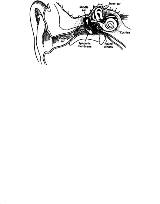
The peripheral auditory system is subdivided into the external ear, the middle ear, and the inner ear (Fig. 5.1).The external ear collects sound energy as pressure waves that are converted to mechanical motion at the eardrum. This motion is transformed across the middle ear and transferred to the inner ear, where it is frequency-analyzed and converted into neural codes that are carried by the eighth cranial nerve, or auditory nerve, to the central auditory system.
Sound information, encoded as discharges in an array of thousands of auditory nerve fibers, is processed in nuclei that make up the central auditory system.The major centers include the cochlear nuclei (CN), the superior olivary complex (SOC), the nuclei of the lateral lemniscus (NLL), the inferior colliculi
(IC), the medial geniculate body (MGB) of the thalamus, and the auditory cortex (AC).The CN, SOC, and NLL are brainstem nuclei;the IC is at the midbrain level;and the MGB and AC constitute the auditory thalamocortical system.
While interesting data have been collected from groups other than mammals, this chapter will emphasize the mammalian auditory system. This chapter ignores the structure and function of the vestibular system.While a few specific references are included, most are general in order to provide a more introductory entry into topics.
5.1 Physical and Psychological Variables
Acoustics
Sound is produced by time-varying motion of the particles in air.The motions can be defined by their pressure variations or by their volume velocities. Volume velocity is defined as the average particle velocity produced across a cross-sectional area and is the acoustic analog of electric current. Pressure is the acoustic analog of voltage. Acoustic intensity is the average rate of the flow of energy through a unit area normal to the direction of the propagation of the sound wave. It is the product of the acoustic pressure and the
0-8493-1811-4/03/$0.00+$ 1.50 |
|
© 2003 by CRC Press LLC |
5-1 |
5-2 |
Biotechnology for Biomedical Engineers |
FIGURE 5.1The peripheral auditory system showing the ear canal,tympanic membrane,middle ear and ossicles, and the inner ear consisting of the cochlea and semicircular canals of the vestibular system. Nerves communicating with the brain are also shown.
volume velocity and is analogous to electric power.Acoustic impedance, the analog of electrical impedance, is the complex ratio of acoustic pressure and volume velocity. Sound is often described in terms of either acoustic pressure or acoustic intensity [Kinsler and Frey, 1962].
The auditory system has a wide dynamic range, i.e., it responds to several decades of change in the magnitude of sound pressure.Because of this wide dynamic range,it is useful to describe the independent variables in terms of decibels, where acoustic intensity is described by dB=10 log(I/I0), where I is the
reference intensity, or equivalently for acoustic pressure, dB=20 log(P/P ), where P |
0 |
|
is the reference |
||
pressure. |
0 |
0 |
|
|
|
Psychoacoustics
Physical variables, such as frequency and intensity, may have correlated psychological variables, such as pitch and loudness. Relationships between acoustic and psychological variables,the subject of the field of psychoacoustics, are generally not linear and may be very complex,but measurements of human detection and discrimination can be made reliably. Humans without hearing loss detect tonal frequencies from 20 Hz to 20 kHz.At 2 to 4 kHz their dynamic range, the span between threshold and pain,is approximately 120 dB.The minimum threshold for sound occurs between 2 and 5 kHz and is about 20 µPa.At the low end of the auditory spectrum, threshold is 80 dB higher, while at the high end, it is 70 dB higher. Intensity differences of 1 dB can be detected, while frequency differences of 2 to 3 Hz can be detected at frequencies below about 3 kHz [Fay, 1988].
5.2 The Peripheral Auditory System
The External Ear
Ambient sounds are collected by the pinna, the visible portion of the external ear, and guided to the middle ear by the external auditory meatus, or ear canal.The pinna acquires sounds selectively due to its geometry and the sound shadowing effect produced by the head. In those species whose ears can be moved voluntarily through large angles, selective scanning of the auditory environment is possible.
The ear canal serves as an acoustic waveguide that is open at one end and closed at the other.The open end at the pinna approximates a short circuit (large volume velocity and small pressure variation),
Auditory System |
5-3 |
while that at the closed end is terminated by the tympanic membrane (eardrum).The tympanic membrane has a relatively high acoustic impedance compared with the characteristic impedance of the meatus and looks like an open circuit.Thus the ear canal can resonate at those frequencies for which its length is an odd number of quarter wavelengths.The first such frequency is at about 3 kHz in the human.The meatus is antiresonant for those frequencies for which its length is an integer number of half wavelengths. For a discussion of resonance and antiresonance in an acoustic waveguide, see a text on basic acoustics, e.g., Kinsler and Frey [1962].
The acoustic properties of the external ear produce differences between the sound pressure produced at the tympanic membrane and that at the opening of the ear canal.These differences are functions of frequency, with larger differences found at frequencies between 2 and 6 kHz than those below 2 kHz. These variations have an effect on the frequency selectivity of the overall auditory system.
The Middle Ear
Anatomy
Tracing the acoustic signal, the boundaries of the middle ear include the tympanic membrane at the input and the oval window at the output.The middle ear bones,the ossicles, lie between. Pressure relief for the tympanic membrane is provided by the eustachian tube.The middle ear is an air-filled cavity.
The Ossicles
The three bones that transfer sound from the tympanic membrane to the oval window are called the malleus (hammer), incus (anvil), and stapes (stirrup).The acoustic impedance of the atmospheric source is much less than that of the aqueous medium of the load.The ratio is 3700 in an open medium, or 36 dB [Kinsler and Frey, 1962].The ossicles comprise an impedance transformer for sound, producing a mechanical advantage that allows the acoustic signal at the tympanic membrane to be transferred with low loss to the round window of the cochlea (inner ear). The air-based sound source produces an acoustic signal of low-pressure and high-volume velocity, while the mechanical properties of the inner ear demand a signal of high-pressure and low-volume velocity.
The impedance transformation is produced in two ways:The area of the tympanic membrane is greater than that of the footplate of the stapes, and the lengths of the malleus and incus produce a lever whose length is greater on the side of the tympanic membrane than it is on the side of the oval window. In the human, the mechanical advantage is about 22:1 [Dobie and Rubel, 1989] and the impedance ratio of the transformer is 480, 27 dB, changing the mismatch from 3700:1 to about 8:1.
This simplified discussion of the function of the ossicles holds at low frequencies, those below 2 kHz. First, the tympanic membrane does not behave as a piston at higher frequencies but can support modes of vibration.Second,the mass of the ossicles becomes significant.Third,the connections between the ossicles is not lossless,nor can the stiffness of these connections be ignored.Fourth,pressure variations in the middle ear cavity can change the stiffness of the tympanic membrane. Fifth, the cavity of the middle ear produces resonances at acoustic frequencies.
Pressure Relief
The eustachian tube is a bony channel that is lined with soft tissue.It extends from the middle ear to the nasopharynx and provides a means by which pressure can be equalized across the tympanic membrane. The function is clearly observed with changes in altitude or barometric pressure.A second function of the eustachian tube is to aerate the tissues of the middle ear.
The Inner Ear
The mammalian inner ear is a spiral structure,the cochlea (snail),consisting of three fluid-filled chambers, or scalae,the scala vestibuli, the scala media, and the scala tympani (Fig. 5.2).The stapes footplate introduces mechanical displacements into the scala vestibuli through the oval window at the base of the cochlea.At the other end of the spiral,the apex of the cochlea,the scala vestibuli and the scala tympani communicate
5-4 |
Biotechnology for Biomedical Engineers |
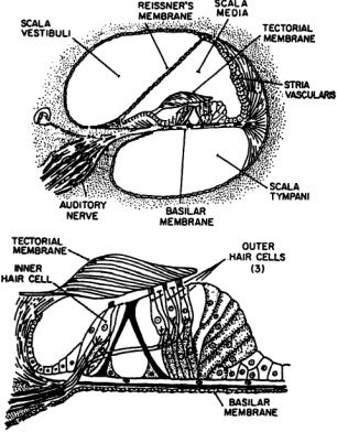
FIGURE 5.2 Cross-section of one turn of the cochlea showing the scala vestibuli, scala media, and scala tympani. Reissner’s membrane separates the SM and SV,while the basilar membrane and organ of Corti separate the SM and ST.
by an opening, the helicotrema. Both are filled with an aqueous medium, the perilymph.The scala media spirals between them and is filled with endolymph, a medium that is high in K+ and low in Na+.The scala media is separated from the scala vestibuli by Reissner’s membrane, which is impermeable to ions,and the scala media is separated from the scala tympani by the basilar membrane (BM) and organ of Corti. The organ of Corti contains the hair cells that transduce acoustic signals into neural signals, the cells that support the hair cells, and the tectorial membrane to which the outer hair cells are attached.The BM provides the primary filter function of the inner ear and is permeable so that the cell bodies of the hair cells are bathed in perilymph.
Fluid and tissue displacements travel from the footplate of the stapes along the cochlear spiral from base to apex. Pressure relief is provided for the incompressible fluids of the inner ear by the round window membrane; e.g., if a transient pressure increase at the stapes displaces its footplate inward, there will be a compensatory outward displacement of the round window membrane.
The Basilar Membrane
Physiology
The BM supports the hair cells and their supporting cells (see Fig. 5.2). Sound decomposition into its frequency components is a major code of the BM.A transient sound, such as a click, initiates a traveling wave of displacement in the BM, and this motion has frequency-dependent characteristics that arise from properties of the membrane and its surrounding structures [Bekesy, 1960].The membrane’s width varies as it traverses the cochlear duct: It is narrower at its basal end than at its apical end. It is stiffer at the base than at the apex, with stiffness varying by about two orders of magnitude [Dobie and Rubel, 1989].The
Auditory System |
5-5 |
membrane is a distributed structure, which acts as a delay line, as suggested by the nature of the traveling wave [Lyon and Mead, 1989].The combination of mechanical properties of the BM produces a structure that demonstrates a distance-dependent displacement when the ear is excited sinusoidally.The distance from the apex to the maximum displacement is logarithmically related to the frequency of a sinusoidal tone [LePage, 1991].
Tuning is quite sharp for sinusoidal signals.The slope of the tuning curve is much greater at the high-frequency edge than at the low-frequency edge, with slopes of more than 100 dB per octave at the high edge and about half that at the low edge [Lyon and Mead, 1989].The filter is sharp, with a 10dB bandwidth of 10 to 25% of the center frequency.
The auditory system includes both passive and active properties.The outer hair cells (see below) receive efferent output from the brain and actively modify the characteristics of the auditory system. The result is to produce a “cochlear amplifier,” which sharpens the tuning of the BM [Lyon and Mead, 1989], as well as adding nonlinear properties to the system [Geisler, 1992; Cooper and Rhode, 1992], along with otoacoustic emissions [LePage, 1991].
The Organ of Corti
The organ of Corti is attached to the BM on the side of the aqueous fluid of the scala media. It is comprised of the supporting cells for the hair cells, the hair cells themselves, and the tectorial membrane. The cilia of the inner hair cells (IHCs) do not contact the tectorial membrane, while those of the outer hair cells (OHCs) do. Both IHCs and OHCs have precise patterns of stereocilia at one end, which are held within the tectorial plate next to the overlying tectorial membrane.The IHCs synapse with spiral ganglion cells, the afferent neurons, while the OHCs synapse with efferent neurons. Both IHCs and OHCs are found along the length of the organ of Corti.The IHCs are found in a single line,numbering between about 3000 and 4000 in human.There are three lines of OHCs, numbering about 12,000 in human [Nadol, 1988].
Inner Hair Cells
The stereocilia of the IHCs are of graded,decreasing length from one side of the cell where a kinocilium is positioned early in ontogeny. If the cilia are deflected in a direction toward this position, membrane channels are further opened to allow potassium to enter and depolarize the cell [Hudspeth, 1987]. Displacement in the other direction reduces channel opening and produces a relative hyperpolarization [Hudspeth and Corey, 1977].These changes in intracellular potential modulate transmitter release at the base of the IHCs.
The IHCs are not attached to the tectorial membrane,so their response to motion of the membrane is proportional to the velocity of displacement rather than to displacement itself, since the cilia of the hair cells are bathed in endolymph.When the membrane vibrates selectively in response to a pure tone, the stereocilia are bent atop a small number of hair cells,which depolarize in response to the mechanical event.Thus, the tonotopic organization of the BM is transferred to the hair cells and to the rest of the auditory system.The auditory system is organized tonotopically, i.e., in order of frequency, because the frequency ordering of the cochlea is mapped through successive levels of the system. While this organization is preserved throughout the system, it is much more complex than a huge set of finely tuned filters.
Hair cells in some species exhibit frequency tuning when isolated [Crawford and Fettiplace, 1985], but mammalian hair cells exhibit no tuning characteristics. The tuning of the mammalian auditory system depends on the mechanical characteristics of the BM as modified by the activity of the OHCs.
Outer Hair Cells
The OHCs have cilia that are attached to the tectorial membrane. Since their innervation is overwhelmingly efferent, they do not transfer information to the brain but are modulated in their mechanical action by the brain.There are several lines of evidence that lead to the conclusion that the OHCs play an active role in the processes of the inner ear. First, OHCs change their length in response