

Biotechnology for Biomedical Engineers - Martin L. Yarmush et al
.pdf1-8
TABLE 1.4Venous System
Biotechnology for Biomedical Engineers
Total systemic blood volume: 4394 ml—84.5% of total blood volume; 19.5% in arteries (~3:2 large:small), 5.9% in capillaries, 74.6% in veins (~3:1 large:small); 63% of volume is in vessels greater than 1 mm internal diameter Total pulmonary blood volume: 468 ml—9.0% of total blood volume; 31.8% in arteries, 22.2% in capillaries, 46% in veins; 58.3% of volume is in vessels greater than 1 mm internal diameter; remainder of blood in heart, about 338 ml (6.5% of total blood volume)

An Outline of Cardiovascular Structure and Function |
1-9 |
(9% elastin, 6% collagen, by weight) to 1:2 in small tributaries (5% elastin, 10% collagen)—and the amount of smooth muscle tissue increases from 7.5% by weight of large arteries (the remaining 7.5% consisting of various extractives) to 15% in small tributaries. By the time one reaches the capillaries, one encounters single-cell-thick endothelial tubes—devoid of any smooth muscle tissue, elastin, or collagen—downstream of which the vascular wall gradually “reassembles itself,” layer by layer, as it directs blood back to the heart through the venous system (Table 1.4).
Blood vessel structure is directly related to function.The thick-walled large arteries and main distributing branches are designed to withstand the pulsating 80-to-130-mmHg blood pressures that they must endure.The smaller elastic conducting vessels need only operate under steadier blood pressures in the range 70 to 90 mmHg,but they must be thin enough to penetrate and course through organs without unduly disturbing the anatomic integrity of the mass involved.Controlling arterioles operate at blood pressures between 45 and 70 mmHg but are heavily endowed with smooth muscle tissue (hence their referred to as muscular vessels) so that they may be actively shut down when flow to the capillary bed they service is to be restricted (for whatever reason),and the smallest capillary resistance vessels (which operate at blood pressures on the order of 10 to 45 mmHg) are designed to optimize conditions for transport to occur between blood and the surrounding interstitial fluid.Traveling back up the venous side,one encounters relatively steady blood pressures continuously decreasing from around 30 mmHg all the way down to near zero, so these vessels can be thin-walled without disease consequence. However, the low blood pressure,slower,steady (time-dependent) flow,thin walls,and larger caliber that characterize the venous system cause blood to tend to “pool” in veins, allowing them to act somewhat like reservoirs. It is not surprising,then,that at any given instant,one normally finds about two-thirds of the total human blood volume residing in the venous system, the remaining one-third being divided among the heart (6.5%), the microcirculation (7% in systemic and pulmonary capillaries), and the arterial system (19.5 to 20%).
In a global sense, then, one can think of the human cardiovascular system—using an electrical analogy—as a voltage source (the heart), two capacitors (a large venous system and a smaller arterial system), and a resistor (the microcirculation taken as a whole). Blood flow and the dynamics of the system represent electrical inductance (inertia), and useful engineering approximations can be derived from such a simple model.The cardiovascular system is designed to bring blood to within a capillary size of each and every one of the more than 1014 cells of the body—but which cells receive blood at any given time,how much blood they get,the composition of the fluid coursing by them,and related physiologic considerations are all matters that are not left up to chance.
1.4 Cardiovascular Control
Blood flows through organs and tissues either to nourish and sanitize them or to be itself processed in some sense—e.g.,to be oxygenated (pulmonary circulation),stocked with nutrients (splanchnic circulation), dialyzed (renal circulation),cooled (cutaneous circulation),filtered of dilapidated red blood cells (splenic circulation), and so on. Thus any given vascular network normally receives blood according to the metabolic needs of the region it perfuses and/or the function of that region as a blood treatment plant and/or thermoregulatory pathway. However, it is not feasible to expect that our physiologic transport system can be “all things to all cells all of the time”—especially when resources are scarce and/or time is a factor.Thus the distribution of blood is further prioritized according to three basic criteria:(1) how essential the perfused region is to the maintenance of life itself (e.g.,we can survive without an arm,a leg, a stomach,or even a large portion of our small intestine but not without a brain,a heart,and at least one functioning kidney and lung, (2) how essential the perfused region is in allowing the organism to respond to a life-threatening situation (e.g., digesting a meal is among the least of the body’s concerns in a “fight-or-flight” circumstance), and (3) how well the perfused region can function and survive on a decreased supply of blood (e.g.,some tissues—like striated skeletal and smooth muscle—have significant anaerobic capability;others—like several forms of connective tissue—can function quite effectively at a significantly decreased metabolic rate when necessary;some organs—like the liver—are larger than they

1-10 |
Biotechnology for Biomedical Engineers |
really need to be; and some anatomic structures—like the eyes, ears,and limbs—have duplicates, giving them a built-in redundancy).
Within this generalized prioritization scheme, control of cardiovascular function is accomplished by mechanisms that are based either on the inherent physicochemical attributes of the tissues and organs themselves—so-called intrinsic control—or on responses that can be attributed to the effects on cardiovascular tissues of other organ systems in the body (most notably the autonomic nervous system and the endocrine system)—so-called extrinsic control. For example, the accumulation of wastes and depletion of oxygen and nutrients that accompany the increased rate of metabolism in an active tissue both lead to an intrinsic relaxation of local precapillary sphincters (rings of muscle)— with a consequent widening of corresponding capillary entrances—which reduces the local resistance to flow and thereby allows more blood to perfuse the active region. On the other hand, the extrinsic innervation by the autonomic nervous system of smooth muscle tissues in the walls of arterioles allows the central nervous system to completely shut down the flow to entire vascular beds (such as the cutaneous circulation) when this becomes necessary (such as during exposure to extremely cold environments).
In addition to prioritizing and controlling the distribution of blood, physiologic regulation of cardiovascular function is directed mainly at four other variables: cardiac output, blood pressure, blood volume, and blood composition. From Equation (1.1) we see that cardiac output can be increased by increasing the heart rate (a chronotropic effect), increasing the end-diastolic volume (allowing the heart to fill longer by delaying the onset of systole), decreasing the end-systolic volume (an inotropic effect), or doing all three things at once. Indeed, under the extrinsic influence of the sympathetic nervous system and the adrenal glands, HR can triple—to some 240 beats/min if necessary—EDV can increase by as much as 50%—to around 200 ml or more of blood—and ESV and decrease a comparable amount (the cardiac reserve)—to about 30 to 35 ml or less.The combined result of all three effects can lead to over a sevenfold increase in cardiac output—from the normal 5 to 5.5 liters/min to as much as 40 to 41 liters/min or more for very brief periods of strenuous exertion.
The control of blood pressure is accomplished mainly by adjusting at the arteriolar level the downstream resistance to flow—an increased resistance leading to a rise in arterial backpressure, and vice versa.This effect is conveniently quantified by a fluid-dynamic analogue to Ohm’s famous E—IR law in electromagnetic theory, voltage drop E being equated to fluid pressure drop DP, electric current/ corresponding to flow—cardiac output (CO)—and electric resistance R being associated with an analogous vascular “peripheral resistance” (PR).Thus one may write
(1.4)
Normally,the total systemic peripheral resistance is 15 to 20 mmHg/liter/min of flow but can increase significantly under the influence of the vasomotor center located in the medulla of the brain, which controls arteriolar muscle tone.
The control of blood volume is accomplished mainly through the excretory function of the kidney. For example, antidiuretic hormone (ADH) secreted by the pituitary gland acts to prevent renal fluid loss (excretion via urination) and thus increases plasma volume, whereas perceived extracellular fluid overloads such as those which result from the peripheral vasoconstriction response to cold stress lead to a sympathetic/adrenergic receptor-induced renal diuresis (urination) that tends to decrease plasma volume—if not checked, to sometimes dangerously low dehydration levels. Blood composition, too, is maintained primarily through the activity of endocrine hormones and enzymes that enhance or repress specific biochemical pathways. Since these pathways are too numerous to itemize here, suffice it to say that in the body’s quest for homeostasis and stability, virtually nothing is left to chance, and every biochemical end can be arrived at through a number of alternative means. In a broader sense, as the organism strives to maintain life, it coordinates a wide variety of different functions, and central to its ability to do just that is the role played by the cardiovascular system in transporting mass, energy, and momentum.
An Outline of Cardiovascular Structure and Function |
1-11 |
Defining Terms
Atrioventricular (AV) node: A highly specialized cluster of neuromuscular cells at the lower portion of the right atrium leading to the interventricular septum; the AV node delays sinoatrial, (SA) node-generated electrical impulses momentarily (allowing the atria to contract first) and then conducts the depolarization wave to the bundle of His and its bundle branches.
Autonomic nervous system: The functional division of the nervous system that innervates most glands, the heart, and smooth muscle tissue in order to maintain the internal environment of the body.
Cardiac muscle: Involuntary muscle possessing much of the anatomic attributes of skeletal voluntary muscle and some of the physiologic attributes of involuntary smooth muscle tissue; SA nodeinduced contraction of its interconnected network of fibers allows the heart to expel blood during systole.
Chronotropic:Affecting the periodicity of a recurring action,such as the slowing (bradycardia) or speeding up (tachycardia) of the heartbeat that results from extrinsic control of the SA node.
Endocrine system: The system of ductless glands and organs secreting substances directly into the blood to produce a specific response from another “target” organ or body part.
Endothelium: Flat cells that line the innermost surfaces of blood and lymphatic vessels and the heart. Homeostasis: A tendency to uniformity or stability in an organism by maintaining within narrow
limits certain variables that are critical to life.
Inotropic: Affecting the contractility of muscular tissue, such as the increase in cardiac power that results from extrinsic control of the myocardial musculature.
Precapillary sphincters: Rings of smooth muscle surrounding the entrance to capillaries where they branch off from upstream metarterioles. Contraction and relaxation of these sphincters close and open the access to downstream blood vessels, thus controlling the irrigation of different capillary networks.
Sinoatrial (SA) node: Neuromuscular tissue in the right atrium near where the superior vena cava joins the posterior right atrium (the sinus venarum); the SA node generates electrical impulses that initiate the heartbeat, hence its nickname the cardiac “pacemaker.”
Stem cells: A generalized parent cell spawning descendants that become individually specialized.
Acknowledgments
The author gratefully acknowledges the assistance of Professor Robert Hochmuth in the preparation of Table 1.1 and the Radford Community Hospital for their support of the Biomedical Engineering Program atVirginia Tech.
References
Bhagavan NV. 1992. Medical Biochemistry. Boston, Jones and Bartlett.
Beall HPT, Needham D, Hochmuth RM. 1993.Volume and osmotic properties of human neutrophils. Blood 81(10):2774–2780.
Caro CG, Pedley TJ, Schroter RC, Seed WA. 1978. The Mechanics of the Circulation. NewYork, Oxford University Press.
Chandran KB. 1992. Cardiovascular Biomechanics. NewYork, NewYork University Press.
Frausto da Silva JJR,Williams RJP. 1993. The Biological Chemistry of the Elements. New York, Oxford University Press/Clarendon.
Dawson TH. 1991. Engineering Design of the Cardiovascular System of Mammals. Englewood Cliffs, NJ, Prentice-Hall.
Duck FA. 1990. Physical Properties of Tissue. San Diego,Academic Press.
Kaley G,Altura BM (Eds).Microcirculation,Vol I (1977),Vol II (1978),Vol III (1980). Baltimore,University Park Press.
1-12 Biotechnology for Biomedical Engineers
Kessel RG,Kardon RH.1979.Tissue and Organs—AText-Atlas of Scanning Electron Microscopy. San Francisco, WH Freeman.
Lentner C (Ed). Geigy Scientific Tables, Vol 3: Physical Chemistry, Composition of Blood, Hematology and Somatometric Data, 8th ed. 1984. New Jersey, Ciba-Geigy.
———Vol 5: Heart and Circulation, 8th ed. 1990. New Jersey, Ciba-Geigy.
Schneck DJ. 1990. Engineering Principles of Physiologic Function. NewYork, NewYork University Press. Tortora GJ, Grabowski SR. 1993. Principles of Anatomy and Physiology, 7th ed. New York,
HarperCollins.

2
Endocrine System
|
2.1 |
Endocrine System: Hormones, Signals, and Communication |
|
|
|
Between Cells and Tissues |
2-1 |
|
2.2 |
Hormone Action at the Cell Level: Signal Recognition, Signal |
|
|
|
Transduction, and Effecting a Physiological Response |
2-4 |
|
|
Hormones Acting at the Cell Surface • Hormones Acting |
|
Derek G. Cramp |
|
within the Cell |
|
City University, London |
2.3 |
Endocrine System: Some Other Aspects of Regulation and |
|
Ewart R. Carson |
|
Control |
2–6 |
|
Negative Feedback • Positive Feedback • Rhythmic |
|
|
City University, London |
|
Endocrine Control |
|
The body, if it is to achieve optimal performance, must possess mechanisms for sensing and responding appropriately to numerous biologic cues and signals in order to control and maintain its internal environment. This complex role is effected by the integrative action of the endocrine and neural systems.The endocrine contribution is achieved through a highly sophisticated set of communication and control systems involving signal generation, propagation, recognition, transduction, and response. The signal entities are chemical messengers or hormones that are distributed through the body by the blood circulatory system to their respective target organs to modify their activity in some fashion.
Endocrinology has a comparatively long history, but real advances in the understanding of endocrine physiology and mechanisms of regulation and control only began in the late 1960s with the introduction of sensitive and relatively specific analytical methods;these enabled low concentrations of circulating hormones to be measured reliably,simply,and at relatively low cost.The breakthrough came with the development and widespreadadoptionofcompetitiveproteinbindingandradioimmunoassaysthatsupersededexistingcumbersome bioassay methods.Since then,knowledge of the physiology of individual endocrine glands and of the neural control of the pituitary gland and the overall feedback control of the endocrine system has progressed and is growing rapidly.Much of this has been accomplished by applying to endocrinological research the methods developed in cellular and molecular biology and recombinant DNA technology.At the same time,theoretical and quantitative approaches using mathematical modeling complemented experimental studies have been of value in gaining a greater understanding of endocrine dynamics.
2.1 Endocrine System: Hormones, Signals, and Communication Between Cells and Tissues
Hormones are synthesized and secreted by specialized endocrine glands to act locally or at a distance, having been carried in the bloodstream (classic endocrine activity) or secreted into the gut lumen (lumocrine activity) to act on target cells that are distributed elsewhere in the body.Hormones are chemically diverse, physiologically potent molecules that are the primary vehicle for intercellular communication with the
0-8493-1811-4/03/$0.00+$ 1.50 |
|
© 2003 by CRC Press LLC |
2-1 |
2-2 |
Biotechnology for Biomedical Engineers |
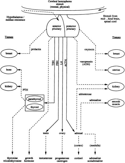
FIGURE 2.1 Representation of the forward pathways of pituitary and target gland hormone release and action: tropic hormones; tissue-affecting hormones.
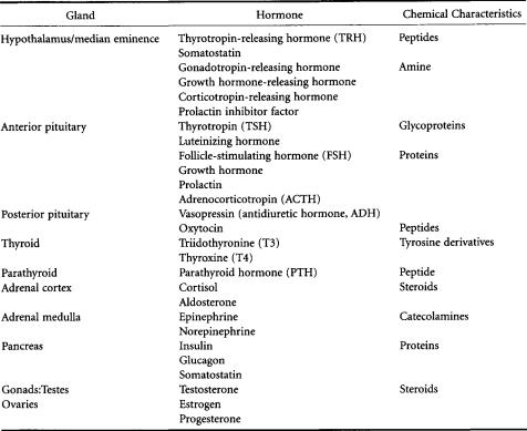
capacity to override the intrinsic mechanisms of normal cellular control.They can be classified broadly into three groups according to their physicochemical characteristics: (1) steroid hormones produced by chemical modification of cholesterol, (2) peptide and protein hormones,and (3) those derived from the aromatic amino acid tyrosine.The peptide and protein hormones are essentially hydrophilic and are therefore able to circulate in the blood in the free state; however, the more hydrophobic lipidderived molecules have to be carried in the circulation bound to specific transport proteins. Figure 2.1 and Table 2.1 show, in schematic and descriptive form respectively, details of the major endocrine glands of the body and the endocrine pathways.
Endocrine System |
2-3 |
TABLE 2.1 Main Endocrine Glands and the Hormones They Produce and Release
The endocrine and nervous system are physically and functionally linked by a specific region of the brain called the hypothalamus, which lies immediately above the pituitary gland,to which it is connected by an extension called the pituitary stalk. The integrating function of the hypothalamus is mediated by cells that possess the properties of both nerve and processes that carry electrical impulses and on stimulation can release their signal molecules into the blood. Each of the hypothalamic neurosecretory cells can be stimulated by other nerve cells in higher regions of the brain to secrete specific peptide hormones or release factors into the adenohypophyseal portal vasculature.These hormones can then specifically stimulate or suppress the secretion of a second hormone from the anterior pituitary.
The pituitary hormones in the circulation interact with their target tissues, which, if endocrine glands, are stimulated to secrete further (third) hormones that feed back to inhibit the release of the pituitary hormones. It will be seen from Fig.2.1 and Table 2.1 that the main targets of the pituitary are the adrenal cortex, the thyroid, and the gonads.These axes provide good examples of the control of pituitary hormone release by negative-feedback inhibition;e.g.,adrenocorticotropin (ACTH),luteinizing hormone (LH), and follicle-stimulating hormone (FSH) are selectively inhibited by different steroid hormones, as is thyrotropin (TSH) release by the thyroid hormones.
In the case of growth hormone (GH) and prolactin, the target tissue is not an endocrine gland and thus does not produce a hormone; then the feedback control is mediated by inhibitors. Prolactin is under dopamine inhibitory control,whereas hypothalamic releasing and inhibitory factors control GH release. The two posterior pituitary (neurohypophyseal) hormones, oxytocin and vasopressin, are synthesized in the supraoptic and paraventricular nuclei and are stored in granules at the end of the nerve fibers in the posterior pituitary. Oxytocin is subsequently secreted in response to peripheral stimuli from the cervical stretch receptors or the suckling receptors of the breast. In a like manner, antidiuretic hormone (ADH, vasopressin) release is stimulated by the altered activity of hypothalamic osmoreceptors responding to changes in plasma solute concentrations.

2-4 |
Biotechnology for Biomedical Engineers |
It will be noted that the whole system is composed of several endocrine axes with the hypothalamus, pituitary, and other endocrine glands together forming a complex hierarchical regulatory system.There is no doubt that the anterior pituitary occupies a central position in the control of hormone secretion and,because of its important role,was often called the “conductor of the endocrine orchestra.” However, the release of pituitary hormones is mediated by complex feedback control, so the pituitary should be regarded as having a permissive role rather than having the overall control of the endocrine system.
2.2Hormone Action at the Cell Level: Signal Recognition, Signal Transduction, and Effecting a Physiological Response
The ability of target glands or tissues to respond to hormonal signals depends on the ability of the cells to recognize the signal.This function is mediated by specialized proteins or glycoproteins in or on the cell plasma membrane that are specific for a particular hormone, able to recognize it, bind it with high affinity, and react when very low concentrations are present. Recognition of the hormonal signal and activation of the cell surface receptors initiates a flow of information to the cell interior that triggers a chain of intracellular events in a preprogrammed fashion that produces a characteristic response. It is useful to classify the site of such action of hormones into two groups: those that act at the cell surface without, generally, traversing the cell membrane and those that actually enter the cell before effecting a response.In the study of this multistep sequence,two important events can be readily studied,namely, the binding of the hormone to its receptor and activation of cytoplasmic effects. However, it is some of the steps between these events, such as receptor activation and signal generation, that are still relatively poorly defined. One method employed in an attempt to elucidate the intermediate steps has been to use ineffective mutant receptors, which when assayed are either defective in their hormone-binding capabilities or in effector-activation and thus unable to transduce a meaningful signal to the cell. But, the difficulty with these studies has been to distinguish receptor-activation and signal-generation defects from hormone-binding and effector-activation defects.
Hormones Acting at the Cell Surface
Most peptide and protein hormones are hydrophilic and thus unable to traverse the lipid-containing cell membrane and must therefore act through activation of receptor proteins on the cell surface.When these receptors are activated by the binding of an extracellular signal ligand,the ligand-receptor complex initiates a series of protein interactions within or adjacent to the inner surface of the plasma membrane, which in turn brings about changes in intracellular activity.This can happen in one of two ways.The first involves the so-called second messenger, by altering the activity of a plasma membrane-bound enzyme,which in turn increases (or sometimes decreases) the concentration of an intracellular mediator. The second involves activation of other types of cell surface receptors, which leads to changes in the plasma membrane electrical potential and the membrane permeability,resulting in altered transmembrane transport of ions or metabolites. If the hormone is thought of as the “first messenger,” cyclic adenosine monophosphate (cAMP) can be regarded as the “second messenger”;capable of triggering a cascade of intracellular biochemical events that can lead either to a rapid secondary response, such as altered ion transport, enhanced metabolic pathway flux, steroidogenesis or to a slower response, such as DNA, RNA, and protein synthesis resulting in cell growth or cell division.
The peptide and protein hormones circulate at very low concentrations relative to other proteins in the blood plasma.These low concentrations are reflected in the very high affinity and specificity of the receptor sites, which permits recognition of the relevant hormones amid the profusion of protein molecules in the circulation.Adaptation to a high concentration of a signal ligand in a time-dependent reversible manner enables cells to respond to changes in the concentration of a ligand instead of to its absolute concentration.The number of receptors in a cell is not constant; synthesis of receptors may be
Endocrine System |
2-5 |
induced or repressed by other hormones or even by their own hormones. Adaptation can occur in several ways. Ligand binding can inactivate a cell surface receptor either by inducing its internalization and degradation or by causing the receptor to adopt an inactive conformation. Alternatively, it may result from the changes in one of the nonreceptor proteins involved in signal transduction following receptor activation. Downregulation is the name given to the process whereby a cell decreases the number of receptors in response to intense or frequent stimulation and can occur by degradation or more temporarily by phosphorylation and sequestration. Upregulation is the process of increasing receptor expression either by other hormones or in response to altered stimulation.
The cell surface receptors for peptide hormones are linked functionally to a cell membrane-bound enzyme that acts as the catalytic unit. This receptor complex consists of three components: (1) the receptor itself that recognizes the hormone, (2) a regulatory protein called a G-protein that binds guanine nucleotides and is located on the cytosolic face of the membrane, and (3) adenylate cyclase, which catalyzes the conversion of ATP to cyclic AMP.As the hormone binds at the receptor site, it is coupled through a regulatory protein, which acts as a transducer, to the enzyme adenyl cyclase, which catalyzes the formation of cAMP from adenosine triphosphate (ATP). The G-protein consists of 3 subunits, which in the unstimulated state form a heterotrimer to which a molecule of GDP is bound. Binding of the hormone to the receptor causes the subunit to exchange its GDP for a molecule of GTP (guanine triphosphate), which then dissociates from the subunits. This in turn decreases the affinity of the receptor for the hormone and leads to its dissociation. The GTP subunit not only activates adenylate cyclase, but also has intrinsic GTPase activity and slowly converts GTP back to GDP,thus allowing the subunits to reassociate and so regain their initial resting state.There are hormones, such as somatostatin, that possess the ability to inhibit AMP formation but still have similarly structured receptor complexes.The G-protein of inhibitory complexes consists of an inhibitory subunit complexed with a subunit thought to be identical to the subunits of the stimulatory G-protein. But, it appears that a single adenylate cyclase molecule can be simultaneously regulated by more than one G-protein enabling the system to integrate opposing inputs.
The adenylate cyclase reaction is rapid, and the increased concentration of intracellular cAMP is shortlived, since it is rapidly hydrolyzed and destroyed by the enzyme cAMP phosphodiesterase that terminates the hormonal response.The continual and rapid removal of cAMP and free calcium ions from the cytosol makes for both the rapid increase and decrease of these intracellular mediators when the cells respond to signals. Rising cAMP concentrations affect cells by stimulating cAMP-dependent protein kinases to phosphorylate specific target proteins. Phosphorylation of proteins leads to conformational changes that enhance their catalytic activity,thus providing a signal amplification pathway from hormone to effector. These effects are reversible because phosphorylated proteins are rapidly dephosphorylated by protein phosphatases when the concentration of cAMP falls. A similar system involving cyclic GMP, although less common and less well studied, plays an analogous role to that of cAMP. The action of thyrotropinreleasing hormone (TRH), parathyroid hormone (PTH), and epinephrine is catalyzed by adenyl cyclase, and this can be regarded as the classic reaction.
However, therearevariantmechanisms. Inthephosphatidylinositol-diacylglycerol(DAG)/inositoltriphosphate (IP3) system,some surface receptors are coupled through another G-protein to the enzyme phospholipase C, whichcleavesthemembranephospholipidtoformDAGandIP3orphospholipaseD, whichcleavesphosphatidyl choline to DAG via phosphatidic acid. DAG causes the calcium, phospholiddependent protein kinase C to translocate to the the cell membrane from the cytosolic cell compartment,becoming 20 times more active in the process.IP3 mobilizes calcium from storage sites associated with the plasma and intracellular membranes thereby contributing to the activation of protein kinase C as well as other calcium dependent processes.DAG is cleared from the cell either by conversion to phosphatidic acid which may be recycled to phospholipid or it may be broken down to fatty acids and glycerol.The DAG derived from phosphatidylinositol usually contains arachidonic acid esterified to the middle carbon of glycerol.Arachidonic acid is the precursor of the prostaglandins and leukotrienes that are biologically active eicosanoids.
Thyrotropin and vasopressin modulate an activity of phospholipase C that catalyzes the conversion of phosphatidylinositol to diacylglycerol and inositol, 1, 4, 5-triphosphate, which act as the second