

Biotechnology for Biomedical Engineers - Martin L. Yarmush et al
.pdf3-8 |
Biotechnology for Biomedical Engineers |
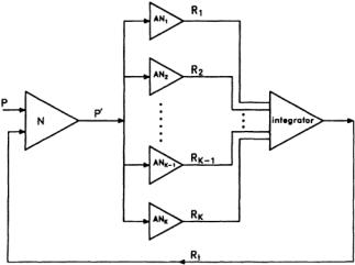
FIGURE 3.2 An ALOPEX system.The stimulus is presented on the CRT.The observer or any pattern-recognition device (PRD) faces the CRT; the subject’s response is sent to the ALOPEX interface unit, where it is recorded and integrated, and the final response is sent to the computer. The computer calculates the values of the new pattern to be presented on the CRT according to the ALOPEX algorithm, and the process continues until the desired pattern appears on the CRT.At this point, the response is considered to be optimal and the process stops.
stimulus the next time it is presented.In a way,then, the brain creates mental “images” (independent of the stimulus) that tend to modify the representation of the stimulus in the brain.
This section describes some efforts in which different methods have been used in trying to address the difficult task of feedback loops in the brain. However, no attempt will be made to explain or even postulate where these feedback loops might be located.If one considers the brain as a huge set of neural nets, then one question has been debated for many years:What is the role of the individual neuron in the net, and what is the role of each network in the holistic process of the brain? More specifically,does the neuron act as an analyzer or a detector of specific features,or does it merely reflect the characteristic response of a population of cells of which it happens to be a member?What invariant relationships exist between sensory input and the response of a single neuron, and how much can be “read” about the stimulus parameters from the record of a single EP? In turn, then, how much feedback can one use from a single EP in order to influence the stimulus, and how successful can that influence be? Many physiologists express doubts that simultaneous observations of large numbers of individual neuronal activities can be readily interpreted.The main question we are asking is:Can a feedback process influence and modulate the stimuli patterns so that they appear optimal? If this is proven to be true, it would mean that we can reverse the pattern-recognition process, and instead of recognizing a pattern, we would be able to create a pattern from a vast variety of possible patterns.It would be like creating a link between our brain and a computer; equivalent to a brain-computer system network. Figure 3.2 is a schematic representation of such a process involved in what we call the feedback loop of the system.The patternrecognition device (PRD) is connected to an ALOPEX system (a computer algorithm and an image processor in this case) and faces a display monitor where different intensity patterns can be shown.Thin arrows representing response information and heavy arrows representing detailed pattern information are generated by the computer and relayed by the ALOPEX system to the monitor. ALOPEX is a set of algorithms described in detail elsewhere in this handbook.If this kind of arrangement is used for the determination of visual receptive fields of neurons, then the PRD is nothing more than the brain of an experimental animal.This way the neuron under investigation does its own selection of the best stimulus or trigger feature and reverses the role of the neuron from being a feature extractor to becoming a feature generator, as mentioned earlier.The idea is to find the response of the neuron to a stimulus and use this response as a positive feedback in the directed evaluation of the initially random pattern.Thus the cell involved filters out the key trigger features from the stimulus and reinforces them with the feedback.
As a generalization of this process, one might consider that a neuron N receives a visual input from a pattern P, which is transmitted in a modified form P’, to an analyzer neuron AN (or even a complex
Nervous System |
3-9 |
FIGURE 3.3 Diagammatic representation of the ALOPEX “inverse” pattern-recognition scheme. Each neuron represents a feature analyzer that responds to the stimulus with a scalar quantity R called the response. R is then fed back to the system,and the pattern is modified accordingly.This process continues until there is a close correlation between the desired output and the original pattern.
of neurons), as shown in Fig. 3.3.The analyzer responds with a scalar variable R that is then fed back to the system,and the pattern is modified accordingly.The process continues in small steps until there is an almost perfect correlation between the original pattern (template) and the one that neuron N indirectly created.This integrator sends the response back to the original modifier.The integrator need not be a linear summator. It could take any nonlinear form, a fact that is a more realistic representation of the visual cortex. One can envision the input patterns as templates preexisting in the memory of the system, a situation that might come about with visual experience. For a “naive” system, any initial pattern will do.As experience is gained, the patterns become less random. If one starts with a pattern that has some resemblance to one of the preexisting patterns, evolution will take its course. In nature, there might exist a mechanism similar to that ofALOPEX.By filtering the characteristics most important for the survival of the species, changes would be triggered. Perception, therefore, could be considered to be an interaction between sensory inputs and past experience in the form of templates stored in the memory of the perceiver and specific to the perceiver’s needs. These templates are modifiable with time and adjusted accordingly to the input stimuli.With this approach,the neural nets and ensembles of nets under observation generate patterns that describe their thinking and memory properties. The normal flow of information is reversed and controls the afferent systems.
The perception processes as well as feature extraction or suppression of images or objects can be ascribed to specific neural mechanisms due to some sensory input or even due to some “wishful thinking” of the PRD. If it is true that the association cortex is affecting the sensitivity of the sensory cortex, then an ALOPEX mechanism is what one needs to close the loop for memory and learning.
Memory and Learning
If we try to define what memory is,we will face the fact that memory is not a single mental faculty but is rather composed of multiple abilities mediated by separate and distinct brain systems. Memory for a recent event can be expressed explicitly as a conscious recollection or implicitly as a facilitation of test performance without conscious recollection.The major distinction between these two memories is that explicit or declarative memory depends on limbic and diencephalic structures and provides the basis for recollection of events, while implicit or nondeclarative memory supports skills and habit learning, single conditioning, and the well-researched phenomenon of priming.
3-10 |
Biotechnology for Biomedical Engineers |
Declarative memory refers to memory of recent events and is usually assessed by tests of recall or recognition for specific single items.When the list of items becomes longer, a subject not only learns about each item on the list but also makes associations about what all these items have in common; i.e., the subject learns about the category that the items belong to. Learning leads to changes that increase or decrease the effectiveness of impulses arriving at the junctions between neurons, and the cumulative effect of these changes constitutes memory.Very often a particular pattern of neural activity leads to a result that occurs some time after the activity has ended. Learning then requires some means of relating the activity that is to be changed to the evaluation that can be made only by the delayed consequence. This phenomenon in physics is called hysteresis and refers to any modifications of future actions due to past actions. Learning then could be defined as change in any neuronal response resulting from previous experiences due to an external stimulus. Memory, in turn, would be the maintenance of these changes over time.The collection of neural changes representing memory is commonly known as the engram, and a major part of recent work has been to identify and locate engrams in the brain,since specific parts of the nervous system are capable of specific types of learning.The view of memory that has recently emerged is that information storage is tied to specific processing areas that are engaged during learning. The brain is organized so that separate regions of neocortex simultaneously carry out computations on specific features or characteristics of the external stimulus, no matter how complex that stimulus might be.If the brain learns specific properties or features of the stimulus, then we talk about the nonassociative memory. Associated with this type of learning is the phenomenon of habituation, in which if the same stimulus is presented repeatedly, the neurons respond less and less, while the introduction of a new stimulus increases the sensitization of the neuron. If the learning includes two related stimuli, then we talk about associative learning.This type of learning includes two types: classic conditioning and operant conditioning.The first deals with relationships among stimuli, while the latter deals with the relationship of the stimulus to the animal’s own behavior. In humans, there exist two types of memory: short-term and long-term memories.The best way to study any physiologic process in humans,and especially memory, is to study its pathology.The study of amnesia has provided strong evidence distinguishing between these types of memory.Amnesic patients can keep a short list of numbers in mind for several minutes if they pay attention to the task. The difficulty comes when the list becomes longer, especially if the amount to be learned exceeds the brain capacity of what can be held in immediate memory. It could be that this happens because more systems have to be involved and that temporary information storage may occur within each brain area where stable changes in synaptic efficacy can eventually develop. Plasticity within existing pathways can account for most of the observations, and short-term memory occurs too quickly for it to require any major modifications of neuronal pathways. The capacity of long-term memory requires the integrity of the medial temporal and diencephalic regions in conjunction with neurons for storage of information.Within the domain of long-term memory, amnesic patients demonstrate intact learning and retention of certain motor, perceptual, and cognitive skills and intact priming effects.These patients do not exhibit any learning deficits but have no conscious awareness of prior study sessions or recognition of previously presented stimuli.
Priming effects can be tested by presenting words and then providing either the first few letters of the word or the last part of the word for recognition by the patient. Normal subjects, as expected, perform better than amnesic subjects.However,if these patients are instructed to “read” the incomplete word instead of memorizing it, then they perform as well as the normal individuals.Also, these amnesic patients perform well if words are cued by category names.Thus priming effects seem to be independent of the processes of recall and recognition memory, which is also observed in normal subjects.All this evidence supports the notion that the brain has organized its memory functions around fundamentally different information storage systems. In perceiving a word, a preexisting array of neurons is activated that have concurrent activities that produce perception, and priming is one of these functions.
Memory is not fixed immediately after learning but continues to grow toward stabilization over a period of time.This stabilization is called consolidation of memory. Memory consolidation is a dynamic feature of long-term memory, especially the declarative memory, but it is neither an automatic process with fixed lifetime nor is it determined at the time of learning. It is rather a process of reorganization
Nervous System |
3-11 |
of stored information.As time passes, some not yet consolidated memories fade out by remodeling the neural circuitry that is responsible for the original representation or by establishing new representations, since the original one can be forgotten.
The problems of learning and memory are studied continuously and with increased interest these days, especially because artificial systems such as neural networks can be used to mimic functions of the nervous system.
References
Cowan WM, Cuenod M (Eds). 1975. Use of Axonal Transport for Studies of Neuronal Connectivity. New York, Elsevier.
Deutsch S, Micheli-Tzanakou E. 1987. Neuroelectric Systems. NewYork, NYU Press. Ganong WF. 1989. Review of Medical Physiology, 14th ed. Norwalk, CT,Appleton and Lange. Hartzell HC. 1981. Mechanisms of slow postsynaptic potentials. Nature 291:593.
McMahon TA. 1984. Muscles, Reflexes and Locomotion. Princeton, NJ, Princeton University Press. Partridge LD,Partridge DL.1993.The Nervous System:Its Function and Interaction with theWorld. Cambridge,
MA, MIT Press.
Shepherd GM. 1978. Microcircuits in the nervous system. Sci Am 238(2):92–103.

4
Vision System
George Stetten |
4.1 |
Fundamentals ofVision Research |
4-1 |
4.2 |
A ModularView of theVision System |
4-1 |
|
Duke University |
|
The Eyes • The Retina • Optic Chiasm • Superior Colliculus • |
|
|
|
LGN • AreaV1 • Color • Higher Cortical Centers |
|
David Marr, an early pioneer in computer vision, defined vision as extracting “…from images of the external world,a description that is useful for the viewer and not cluttered with irrelevant information” [Marr, 1982]. Advances in computers and video technology in the past decades have created the expectation that artificial vision should be realizable.The nontriviality of the task is evidenced by the continuing proliferation of new and different approaches to computer vision without any observable application in our everyday lives. Actually, computer vision is already offering practical solutions in industrial assembly and inspection, as well as for military and medical applications, so it seems we are beginning to master some of the fundamentals.However, we have a long way to go to match the vision capabilities of a 4-year-old child. In this chapter we will explore what is known about how nature has succeeded at this formidable task—that of interpreting the visual world.
4.1 Fundamentals of Vision Research
Research into biologic vision systems has followed several distinct approaches.The oldest is psychophysics, in which human and animal subjects are presented with visual stimuli and their responses recorded. Important early insights also were garnered by correlating clinical observations of visual defects with known neuroanatomic injury. In the past 50 years, a more detailed approach to understanding the mechanisms of vision has been undertaken by inserting small electrodes deep within the living brain to monitor the electrical activity of individual neurons and by using dyes and biochemical markers to track the anatomic course of nerve tracts. This research has led to a detailed and coherent, if not complete, theory of a visual system capable of explaining the discrimination of form, color, motion, and depth.This theory has been confirmed by noninvasive radiologic techniques that have been used recently to study the physiologic responses of the visual system,including positron emission tomography [Zeki et al., 1991] and functional magnetic resonance imaging [Belliveau et al., 1992; Cohen and Bookheimer, 1994], although these noninvasive techniques provide far less spatial resolution and thus can only show general regions of activity in the brain.
4.2 A Modular View of the Vision System
The Eyes
Movement of the eyes is essential to vision,not only allowing rapid location and tracking of objects but also preventing stationary images on the retina, which are essentially invisible. Continual movement of the image on the retina is essential to the visual system.
0-8493-1811-4/03/$0.00+$1.50 © |
|
2003 by CRC Press LLC |
4-1 |
4-2 |
Biotechnology for Biomedical Engineers |
The eyeball is spherical and therefore free to turn in both horizontal and vertical directions. Each eye is rotated by three pairs of mutually opposing muscles, innervated by the oculomotor nuclei in the brainstem.The eyes are coordinated as a pair in two useful ways: turning together to find and follow objects and turning inward to allow adjustment for parallax as objects become closer.The latter is called convergence.
The optical portion of the eye,which puts an image on the retina,is closely analogous to a photographic or television camera.Light enters the eye,passing through a series of transparent layers—the cornea,the aqueous humor, the lens, and the vitreous body—to eventually project on the retina.
The cornea, the protective outer layer of the eye,is heavily innervated with sensory neurons,triggering the blink reflex and tear duct secretion in response to irritation.The cornea is also an essential optical element, supplying two thirds of the total refraction in the eye. Behind the cornea is a clear fluid, the aqueous humor, in which the central aperture of the iris, the pupil, is free to constrict or dilate.The two actions are accomplished by opposing sets of muscles.
The lens, a flexible transparent object behind the iris, provides the remainder of refraction necessary to focus an image on the retina.The ciliary muscles surrounding the lens can increase the lens’ curvature, thereby decreasing its focal length and bringing nearer objects into focus.This is called accommodation. When the ciliary muscles are at rest, distant objects are in focus.There are no contradictory muscles to flatten the lens.This depends simply on the elasticity of the lens, which decreases with age. Behind the lens is the vitreous humor, consisting of a semigelatinous material filling the volume between the lens and the retina.
The Retina
The retina coats the back of the eye and is therefore spherical, not flat, making optical magnification constant at 3.5 degrees of scan angle per millimeter.The retina is the neuronal front end of the visual system, the image sensor. In addition, it accomplishes the first steps in edge detection and color analysis before sending the processed information along the optic nerve to the brain.The retina contains five major classes of cells, roughly organized into layers.The dendrites of these cells each occupy no more than 1 to 2 mm2 in the retina, limiting the extent of spatial integration from one layer of the retina to the next.
First come the receptors, which number approximately 125 million in each eye and contain the lightsensitive pigments responsible for converting photons into chemical energy. Receptor cells are of two general varieties: rods and cones.The cones are responsible for the perception of color,and they function only in bright light.When the light is dim, only rods are sensitive enough to respond. Exposure to a single photon may result in a measurable increase in the membrane potential of a rod.This sensitivity is the result of a chemical cascade, similar in operation to the photo multiplier tube, in which a single photon generates a cascade of electrons.All rods use the same pigment,whereas three different pigments are found in three separate kinds of cones.
Examination of the retina with an otoscope reveals its gross topography.The yellow circular area occupying the central 5 degrees of the retina is called the macula lutea, within which a small circular pit called the fovea may be seen. Detailed vision occurs only in the fovea, where a dense concentration of cones provides visual activity to the central 1 degree of the visual field.
On the inner layer of the retina is a layer of ganglion cells, whose axons make up the optic nerve, the output of the retina.They number approximately 1 million, or less than 1% of the number of receptor cells. Clearly, some data compression has occurred in the space between the receptors and the ganglion cells.Traversing this space are the bipolar cells, which run from the receptors through the retina to the ganglion cells.Bipolar cells exhibit the first level of information processing in the visual system;namely, their response to light on the retina demonstrates “center/surround” receptive fields;that is,a small dot on the retina elicits a response, while the area surrounding the spot elicits the opposite response.If both the center and the surround are illuminated, the net result is no response.Thus bipolar cells respond only at the border between dark and light areas. Bipolar cells come in two varieties, on-center and offcenter, with the center respectively brighter or darker than the surround.
Vision System |
4-3 |
The center response of bipolar cells results from direct contact with the receptors.The surround response is supplied by the horizontal cells, which run parallel to the surface of the retina between the receptor layer and the bipolar layer,allowing the surrounding area to oppose the influence of the center. The amacrine cells, a final cell type, also run parallel to the surface but in a different layer, between the bipolar cells and the ganglion cells, and are possibly involved in the detection of motion.
Ganglion cells, since they are triggered by bipolar cells, also have center/surround receptive fields and come in two types, on-center and off-center. On-center ganglion cells have a receptive field in which illumination of the center increases the firing rate and a surround where it decreases the rate. Off-center ganglion cells display the opposite behavior. Both types of ganglion cells produce little or no change in firing rate when the entire receptive field is illuminated, because the center and surround cancel each other. As in many other areas of the nervous system, the fibers of the optic nerve use frequency encoding to represent a scalar quantity.
Multiple ganglion cells may receive output from the same receptor, since many receptive fields overlap. However, this does not limit overall spatial resolution, which is maximum in the fovea, where two points separated by 0.5 minutes of arc may be discriminated. This separation corresponds to a distance on the retina of 2.5 µm, which is approximately the center-to-center spacing between cones. Spatial resolution falls off as one moves away from the fovea into the peripheral vision,where resolution is as low as 1 degree of arc.
Several aspects of this natural design deserve consideration.Why do we have center/surround receptive fields? The ganglion cells, whose axons make up the optic nerve, do not fire unless there is meaningful information, i.e., a border, falling within the receptive field. It is the edge of a shape we see rather than its interior.This represents a form of data compression. Center/surround receptive fields also allow for relative rather than absolute measurements of color and brightness.This is essential for analyzing the image independent of lighting conditions.Why do we have both on-center and off-center cells? Evidently, both light and dark are considered information.The same shape is detected whether it is lighter or darker than the background.
Optic Chiasm
The two optic nerves, from the left and right eyes, join at the optic chiasm, forming a hemidecussation, meaning that half the axons cross while the rest proceed uncrossed.The resulting two bundles of axons leaving the chiasm are called the optic tracts.The left optic tract contains only axons from the left half of each retina. Since the images are reversed by the lens, this represents light from the right side of the visual field. The division between the right and left optic tracts splits the retina down the middle, bisecting the fovea.The segregation of sensory information into the contralateral hemispheres corresponds to the general organization of sensory and motor centers in the brain.
Each optic tract has two major destinations on its side of the brain:(1) the superior colliculus and (2) the lateral geniculate nucleus (LGN). Although topographic mapping from the retina is scrambled within the optic tract, it is reestablished in both major destinations so that right, left, up, and down in the image correspond to specific directions within those anatomic structures.
Superior Colliculus
The superior colliculus is a small pair of bumps on the dorsal surface of the midbrain.Another pair, the inferior colliculus, is found just below it. Stimulation of the superior colliculus results in contralateral eye movement. Anatomically, output tracts from the superior colliculus run to areas that control eye and neck movement. Both the inferior and superior colliculi are apparently involved in locating sound. In the bat, the inferior colliculus is enormous, crucial to that animal’s remarkable echolocation abilities. The superior colliculus processes information from the inferior colliculus, as well as from the retina, allowing the eyes to quickly find and follow targets based on visual and auditory cues.
Different types of eye movements have been classified.The saccade (French,for “jolt”) is a quick motion of the eyes over a significant distance. The saccade is how the eyes explore an image, jumping from
4-4 |
Biotechnology for Biomedical Engineers |
landmark to landmark, rarely stopping in featureless areas. Nystagmus is the smooth pursuit of a moving image, usually with periodic backward saccades to lock onto subsequent points as the image moves by. Microsaccades are small movements, several times per second, over 1 to 2 minutes of arc in a seemingly random direction. Microsaccades are necessary for sight; their stabilization leads to effective blindness.
LGN
The thalamus is often called “the gateway to the cortex” because it processes much of the sensory information reaching the brain.Within the thalamus, we find the lateral geniculate nucleus (LGN),a peanutsized structure that contains a single synaptic stage in the major pathway of visual information to higher centers.The LGN also receives information back from the cortex,so-called reentrant connections,as well as from the nuclei in the brainstem that control attention and arousal.
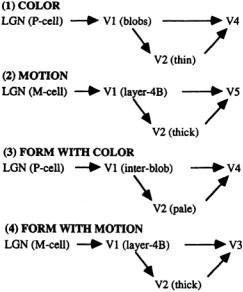
The cells in the LGN are organized into three pairs of layers. Each pair contains two layers, one from each eye. The upper two pairs consist of parvocellular cells (P cells) that respond with preference to different colors.The remaining lower pair consists of magnocellular cells (M cells) with no color preference (Fig. 4.1). The topographic mapping is identical for all six layers; i.e., passing through the layers at a given point yields synapses responding to a single area of the retina. Axons from the LGN proceed to the primary visual cortex in broad bands,the optic radiations, preserving this topographic mapping and displaying the same center/surround response as the ganglion cells.
FIGURE 4.1Visual pathways to cortical areas showing the separation of information by type. The lateral geniculate nucleus (LGN) and areas V1 and V2 act as gateways to more specialized higher areas.
Area V1
The LGN contains approximately 1.5 million cells. By comparison, the primary visual cortex, or striate cortex, which receives the visual information from the LGN, contains 200 million cells. It consists of a thin (2-mm) layer of gray matter (neuronal cell bodies) over a thicker collection of white matter (myelinated axons) and occupies a few square inches of the occipital lobes.The primary visual cortex has been called area 17 from the days when the cortical areas were first differentiated by their cytoarchitectonics (the microscopic architecture of their layered neurons). In modern terminology, the primary visual cortex is often called visual area 1, or simplyV1.
Destroying any small piece ofV1 eliminates a small area in the visual field,resulting in scotoma, a local blind spot. Clinical evidence has long been available that a scotoma may result from injury, stroke, or tumor in a local part ofV1. Between neighboring cells inVl’s gray matter,horizontal connections are at most 2 to 5 mm in length.Thus, at any given time, the image from the retina is analyzed piecemeal in V1.Topographic mapping from the retina is preserved in great detail. Such mapping is seen elsewhere in the brain, such as in the somatosensory cortex [Mountcastle, 1957]. Like all cortical surfaces,V1 is a highly convoluted sheet,with much of its area hidden within its folds.If unfolded,V1 would be roughly pear-shaped,with the top of the pear processing information from the fovea and the bottom of the pear processing the peripheral vision. Circling the pear at a given latitude would correspond roughly to circling the fovea at a fixed radius.
Vision System |
4-5 |
The primary visual cortex contains six layers,numbered 1 through 6.Distinct functional and anatomic types of cells are found in each layer. Layer 4 contains neurons that receive information from the LGN. Beyond the initial synapses,cells demonstrate progressively more complex responses.The outputs ofV1 project to an area known as visual area 2 (V2), which surrounds V1, and to higher visual areas in the occipital, temporal, and parietal lobes as well as to the superior colliculus. V1 also sends reentrant projections back to the LGN. Reentrant projections are present at almost every level of the visual system [Felleman and Essen, 1991; Edelman, 1978].
Cells in V1 have been studied extensively in animals by inserting small electrodes into the living brain (with surprisingly little damage) and monitoring the individual responses of neurons to visual stimuli.Various subpopulations of cortical cells have thus been identified. Some, termed simple cells, respond to illuminated edges or bars at specific locations and at specific angular orientations in the visual field.The angular orientation must be correct within 10 to 20 degrees for the particular cell to respond.All orientations are equally represented. Moving the electrode parallel to the surface yields a smooth rotation in the orientation of cell responses by about 10 degrees for each 50 µm that the electrode is advanced.This rotation is subject to reversals in direction, as well as “fractures,” or sudden jumps in orientation.
Other cells,more common than simple cells,are termed complex cells. Complex cells respond to a set of closely spaced parallel edges within a particular receptive field. They may respond specifically to movement perpendicular to the orientation of the edge.Some prefer one direction of movement to the other. Some complex and simple cells are end-stopped, meaning they fire only if the illuminated bar or edge does not extend too far.Presumably,these cells detect corners,curves,or discontinuities in borders and lines. End-stopping takes place in layers 2 and 3 of the primary visual cortex. From the LGN through the simple cells and complex cells, there appears to be a sequential processing of the image. It is probable that simple cells combine the responses of adjacent LGN cells and that complex cells combine the responses of adjacent simple cells.
A remarkable feature in the organization ofV1 is binocular convergence, in which a single neuron responds to identical receptive fields in both eyes,including location,orientation,and directional sensitivity to motion. It does not occur in the LGN, where axons from the left and right eyes are still segregated into different layers. Surprisingly, binocular connections to neurons are present in V1 at birth. Some binocular neurons are equally weighted in terms of responsiveness to both eyes, while others are more sensitive to one eye than to the other. One finds columns containing the latter type of cells in which one eye dominates, called ocular dominance columns, in uniform bands approximately 0.5 mm wide everywhere in V1. Ocular dominance columns occur in adjacent pairs, one for each eye, and are prominent in animals with forward-facing eyes, such as cats,chimpanzees, and humans.They are nearly absent in rodents and other animals whose eyes face outward.
The topography of orientation-specific cells and of ocular dominance columns is remarkably uniform throughoutV1,which is surprising because the receptive fields near the fovea are 10 to 30 times smaller than those at the periphery. This phenomenon is called magnification. The fovea maps to a greater relative distance on the surface ofV1 than does the peripheral retina, by as much as 36-fold [Daniel and Whitteridge, 1961]. In fact, the majority ofV1 processes only the central 10 degrees of the visual field. Both simple and complex cells in the foveal portion can resolve bars as narrow as 2 minutes of arc. Toward the periphery, the resolution falls off to 1 degree of arc.
As an electrode is passed down through the cortex perpendicular to the surface,each layer demonstrates receptive fields of characteristic size, the smallest being at layer 4, the input layer. Receptive fields are larger in other layers due to lateral integration of information. Passing the electrode parallel to the surface of the cortex reveals another important uniformity toV1. For example, in layer 3, which sends output fibers to higher cortical centers, one must move the electrode approximately 2 mm to pass from one collection of receptive fields to another that does not overlap.An area approximately 2 mm across thus represents the smallest unit piece ofV1,i.e.,that which can completely process the visual information. Indeed, it is just the right size to contain a complete set of orientations and more than enough to contain information from both eyes. It receives a few tens of thousands of fibers from the LGN,