

Biotechnology for Biomedical Engineers - Martin L. Yarmush et al
.pdf2-6 |
Biotechnology for Biomedical Engineers |
messengers.They mobilize bound intracellular calcium and activate a protein kinase, which in turn alters the activity of other calcium-dependent enzymes within the cell.
Increased concentrations of free calcium ions affect cellular events by binding to and altering the molecular conformation of calmodulin; the resulting calcium ion-calmodulin complex can activate many different target proteins, including calcium ion-dependent protein kinases. Each cell type has a characteristic set of target proteins that are so regulated by cAMP-dependent kinases and/or calmodulin that it will respond in a specific way to an alteration in cAMP or calcium ion concentrations.In this way, cAMP or calcium ions act as second messengers in such a way as to allow the extracellular signal not only to be greatly amplified but, just as importantly, also to be made specific for each cell type.
The action of the important hormone insulin, which regulates glucose metabolism, depends on the activation of the enzyme tyrosine kinase catalyzing the phosphorylation of tyrosyl residues of proteins. This effects changes in the activity of calcium-sensitive enzymes, leading to enhanced movement of glucose and fatty acids across the cell membrane and modulating their intracellular metabolism.The binding of insulin to its receptor site has been studied extensively; the receptor complex has been isolated and characterized. It was such work that highlighted the interesting aspect of feedback control at the cell level, downregulation: the ability of peptide hormones to regulate the concentration of cell surface receptors.After activation, the receptor population becomes desensitized, or “downregulated”; leading to a decreased availability of receptors and thus a modulation of transmembrane events.
Hormones Acting Within the Cell
Steroid hormones are small hydrophobic molecules derived from cholesterol that are solubilized by binding reversibly to specify carrier proteins in the blood plasma. Once released from their carrier proteins, they readily pass through the plasma membrane of the target cell and bind, again reversibly, to steroid hormone receptor proteins in the cytosol. This is a relatively slow process when compared to protein hormones. The latter second messenger-mediated phosphorylationdephosphorylation reactions modify enzymatic processes rapidly with the physiological consequences becoming apparent in seconds or minutes and are as rapidly reversed. Nuclearmediated responses, on the other hand, lead to transcription/translation dependent changes that are slow in onset and tend to persist since reversal is dependent on degradation of the induced proteins. The protein component of the steroid hormonereceptor complex has an affinity for DNA in the cell nucleus, where it binds to nuclear chromatin and initiates the transcription of a small number of genes. These gene products may, in turn, activate other genes and produce a secondary response, thereby amplifying the initial effect of the hormone. Each steroid hormone is recognized by a separate receptor protein, but this same receptor protein has the capacity to regulate several different genes in different target cells. This, of course, suggests that the nuclear chromatin of each cell type is organized so that only the appropriate genes are made available for regulation by the hormone-receptor complex. The thyroid hormone triiodothyronine (T3) also acts, though by a different mechanism than the steroids, at the cell nucleus level to initiate genomic transcription. The hormonal activities of GH and prolactin influence cellular gene transcription and translation of messenger RNA by complex mechanisms.
2.3Endocrine System: Some Other Aspects of Regulation and Control
From the foregoing sections it is clear that the endocrine system exhibits complex molecular and metabolic dynamics that involve many levels of control and regulation. Hormones are chemical signals released from a hierarchy of endocrine glands and propagated through the circulation to a hierarchy of cell types.The integration of this system depends on a series of what systems engineers call “feedback loops”; feedback is a reflection of mutual dependence of the system variables: variable x affects variable
Endocrine System |
2-7 |
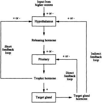
FIGURE 2.2 Illustration of the complexity of hormonal feedback control (+ indicates a positive or augmenting effect;—indicates a negative or inhibiting effect).
y, and y affects x. Further, it is essentially a closed-loop system in which the feedback of information from the system output to the input has the capacity to maintain homeostasis. A diagrammatic representation of the ways in which hormone action is controlled is shown in Fig. 2.2. One example of this control structure arises in the context of the thyroid hormones. In this case,TRH, secreted by the hypothalamus, triggers the anterior pituitary into the production of TSH. The target gland is the thyroid, which produces T3 and thyroxine (T4).The complexity of control includes both direct and indirect feedback of T3 and T4, as outlined in Fig. 2.2, together with TSH feedback on to the hypothalamus.
Negative Feedback
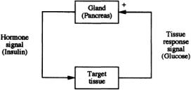
If an increase in y causes a change in x,which in turn tends to decrease y, feedback is said to be negative; in other words, the signal output induces a response that feeds back to the signal generator to decrease its output.This is the most common form of control in physiologic systems,and examples are many.For instance, as mentioned earlier, the anterior pituitary releases trophic or stimulating hormones that act on peripheral endocrine glands, such as the adrenals or thyroid or to gonads, to produce hormones that act back on the pituitary to decrease the secretion of the trophic hormones.These are examples of what is called long-loop feedback (see Fig. 2.2). (Note: the adjectives long and short reflect the spatial distance or proximity of effector and target sites.) The trophic hormones of the pituitary are also regulated by feedback action at the level of their releasing factors. Ultrashort-loop feedback is also described.There are numerous examples of short-loop feedback as well, the best being the reciprocal relation between insulin and blood glucose concentrations, as depicted in Fig. 2.3. In this case, elevated glucose concentration (and positive rate of change, implying not only proportional but also derivative control) has a positive effect on the pancreas, which secretes insulin in response. This has an inhibiting effect on glucose metabolism, resulting in a reduction of blood glucose toward a normal concentration; in other words, classic negative-feedback control.
2-8 |
Biotechnology for Biomedical Engineers |
FIGURE 2.3The interaction of insulin as an illustration of negative feedback within a hormonal control system.
Positive Feedback
If increase in y causes a change in x that tends to increase y, feedback is said to be positive; in other words, a further signal output is evoked by the response it induces or provokes.This is intrinsically an unstable system, but there are physiologic situations where such control is valuable. In the positive feedback situation, the signal output will continue until no further response is required. Suckling provides an example; stimulation of nipple receptors by the suckling child provokes an increased oxytocin release from the posterior pituitary with a corresponding increase in milk flow. Removal of the stimulus causes cessation of oxytocin release.
Rhythmic Endocrine Control
Many hormone functions exhibit rhythmically in the form of pulsatile release of hormones.The most common is the approximately 24-h cycle (circadian or diurnal rhythm).For instance,blood sampling at frequent intervals has shown that ACTH is secreted episodically, each secretory burst being followed 5 to 10 min later by cortisol secretion. These episodes are most frequent in the early morning, with plasma cortisol concentrations highest around 7 to 8 AM and lowest around midnight. ACTH and cortisol secretion vary inversely, and the parallel circadian rhythm is probably due to a cyclic change in the sensitivity of the hypothalamic feedback center to circulating cortisol.Longer cycles are also known, e.g., the infradian menstrual cycle.
It is clear that such inherent rhythms are important in endocrine communication and control, suggesting that its physiologic organization is based not only on the structural components of the system but also on the dynamics of their interactions.The rhythmic, pulsatile nature of release of many hormones is a means whereby time-varying signals can be encoded, thus allowing large quantities of information to be transmitted and exchanged rapidly in a way that small,continuous changes in threshold levels would not allow.
References
Inevitably, our brief exposition has been able to touch upon an enormous subject only by describing some of the salient features of this fascinating domain, but it is hoped that it may nevertheless stimulate a further interest. However, not surprisingly the endocrinology literature is massive, and it is suggested that anyone wishing to read further go initially to one of the many excellent textbooks and go on from there.Those we have found useful include:
Goodman HM. 1994. Basic Medical Endocrinology, 2nd ed., NewYork, Raven Press.
Greenspan FS, Strewler GJ (Eds.). 1997. Basic and Clinical Endocrinology, Appleton and Lange. 5th ed. O’Malley BW, Birnbaumer L, Hunter T. (Eds.). 1998. Hormones and Signaling. (Vol. 1).Academic Press. Wilson JD, Foster DW, Kronenberg HM (Eds.). 1998. Williams Textbook of Endocrinology, 9th ed.,WB
Saunders.

3
Nervous System
|
3.1 |
Definitions |
3-1 |
|
|
3.2 |
Functions of the Nervous System |
3-3 |
|
Evangelia |
3.3 |
Representation of Information in the Nervous System |
3-4 |
|
Micheli-Tzanakou |
3.4 |
Lateral Inhibition |
3-6 |
|
3.5 |
Higher Functions of the Nervous System |
3-7 |
||
Rutgers University |
||||
|
Pattern Recognition • Memory and Learning |
|
||
|
|
|
The nervous system, unlike other organ systems, is concerned primarily with signals, information encoding and processing, and control rather than manipulation of energy. It acts like a communication device whose components use substances and energy in processing signals and in reorganizing them, choosing, and commanding,as well as in developing and learning.A central question that is often asked is how nervous systems work and what are the principles of their operation. In an attempt to answer this question, we will, at the same time, ignore other fundamental questions, such as those relating to anatomic or neurochemical and molecular aspects.We will concentrate rather on relations and transactions between neurons and their assemblages in the nervous system.We will deal with neural signals (encoding and decoding), the evaluation and weighting of incoming signals, and the formulation of outputs. A major part of this chapter is devoted to higher aspects of the nervous system, such as memory and learning, rather than individual systems, such as vision and audition, which are treated extensively elsewhere in this handbook.
3.1 Definitions
Nervous systems can be defined as organized assemblies of nerve cells as well as nonneryous cells. Nerve cells,or neurons, are specialized in the generation,integration, and conduction of incoming signals from the outside world or from other neurons and deliver them to other excitable cells or to effectors such as muscle cells.Nervous systems are easily recognized in higher animals but not in the lower species, since the defining criteria are difficult to apply.
A central nervous system (CNS) can be distinguished easily from a peripheral nervous system (PNS), since it contains most of the motor and nucleated parts of neurons that innervate muscles and other effectors.The PNS contains all the sensory nerve cell bodies, with some exceptions, plus local plexuses, local ganglia, and peripheral axons that make up the nerves. Most sensory axons go all the way into the CNS,while the remaining sensory axons relay in peripheral plexuses.Motor axons originating in the CNS innervate effector cells.
The nervous system has two major roles: (1) to regulate, acting homeostatically in restoring some conditions of the organism after some external stimulus, and (2) to act to alter a preexisting condition by replacing it or modifying it. In both cases—regulation or initiation of a process—learning can be superimposed. In most species, learning is a more or less adaptive mechanism, combining and timing species-characteristic acts with a large degree of evolution toward perfection.
0-8493-1811-4/03/$0.00+$1.50 © |
|
2003 by CRC Press LLC |
3-1 |
3-2 Biotechnology for Biomedical Engineers
The nervous system is a complex structure for which realistic assumptions have led to irrelevant oversimplifications. The nervous system can be broken down into four components: sensory transducers, neurons, axons, and muscle fibers. Each of these components gathers, processes, and transmits information impinging on it from the outside world, usually in the form of complex stimuli. The processing is carried out by exitable tissues—neurons, axons, sensory receptors, and muscle fibers. Neurons are the basic elements of the nervous system. If put in small assemblies or clusters, they form neuronal assemblies or neuronal networks communicating with each other either chemically via synaptic junctions or electrically via tight junctions.The main characteristics of a cell are the cell body, or soma, which contains the nucleus, and a number of processes originating from the cell body, called the dendrites, which reach out to surroundings to make contacts with other cells.These contacts serve as the incoming information to the cell, while the outgoing information follows a conduction path, the axon.The incoming information is integrated in the cell body and generates its action potential at the axon hillock. There are two types of outputs that can be generated and therefore two types of neurons: those that generate graded potentials that attenuate with distance and those that generate action potentials. The latter travel through the axon, a thin, long process that passively passes the action potential or rather a train of action potentials without any attenuation (all- or-none effect). A number of action potentials is often called a spike train. A threshold built into the hillock, depending on its level, allows or stops the generation of the spike train. Axons usually terminate on other neurons by means of synaptic terminals or boutons and have properties similar to those of an electric cable with varying diameters and speeds of signal transmission. Axons can be of two types: myelinated or unmyelinated. In the former case, the axon is surrounded by a thick fatty material, the myelin sheath, which is interrupted at regular intervals by gaps called the nodes of Ranvier. These nodes provide for the saltatory conduction of the signal along the axon. The axon makes functional connections with other neurons at synapses on either the cell body, the dendrites, or the axons.There exist two kinds of synapses: excitatory and inhibitory, and as the names imply, they either increase the firing frequency of the postsynaptic neurons or decrease it, respectively.
Sensory receptors are specialized cells that, in response to an incoming stimulus, generate a corresponding electrical signal, a graded receptor potential. Although the mechanisms by which the sensory receptors generate receptor potentials are not known exactly,the most plausible scenario is that an external stimulus alters the membrane permeabilities.The receptor potential, then, is the change in intracellular potential relative to the resting potential.
It is important to notice here that the term receptor is used in physiology to refer not only to sensory receptors but also, in a different sense, to proteins that bind neurotransmitters, hormones, and other substances with great affinity and specificity as a first step in starting up physiologic responses. This receptor is often associated with nonneural cells that surround it and form a sense organ. The forms of energy converted by the receptors include mechanical, thermal, electromagnetic, and chemical energy. The particular form of energy to which a receptor is most sensitive is called its adequate stimulus. The problem of how receptors convert energy into action potentials in the sensory nerves has been the subject of intensive study. In the complex sense organs such as those concerned with hearing and vision, there exist separate receptor cells and synaptic junctions between receptors and afferent nerves. In other cases, such as the cutaneous sense organs, the receptors are specialized.Where a stimulus of constant strength is applied to a receptor repeatedly,the frequency of the action potentials in its sensory nerve declines over a period of time.This phenomenon is known as adaptation; if the adaptation is very rapid, then the receptors are called phasic, otherwise, they are called tonic.
Another important issue is the coding of sensory information. Action potentials are similar in all nerves, although there are variations in their speed of conduction and other characteristics.However, if the action potentials were the same in most cells, then what makes the visual cells sensitive to light and not to sound and the touch receptors sensitive to touch and not to smell? And how can we tell if these sensations are strong or not? These sensations depend on what is called the doctrine of specific nerve energies, which has been questioned over time by several researchers. No matter where a particular sensory pathway is stimulated along its course to the brain, the sensation produced is referred to the

Nervous System |
3-3 |
location of the receptor.This is the law of projections. An example of this law is the “phantom limb” in which an amputee complains about an itching sensation in the amputated limb.
3.2 Functions of the Nervous System
The basic unit of integrated activity is the reflex arc. This arc consists of a sense organ, afferent neuron, one or more synapses in a central integrating station (or sympathetic ganglion), an efferent neuron, and an effector.The simplest reflex arc is the monosynaptic one, which has only one synapse between the afferent and efferent neurons.With more than one synapse,the reflex arc is called polysynaptic. In each of these cases, activity is modified by both spatial and temporal facilitation, occlusion, and other effects.
In mammals, the concentration between afferent and efferent somatic neurons is found either in the brain or in the spinal cord.The Bell-Magendie law dictates that in the spinal cord the dorsal roots are sensory, while the ventral roots are motor.The action potential message that is carried by an axon is eventually fed to a muscle,to a secretory cell,or to the dendrite of another neuron.If an axon is carrying a graded potential, its output is too weak to stimulate a muscle, but it can terminate on a secretory cell or dendrite.The latter can have as many as 10,000 inputs. If the endpoint is a motor neuron, which has been found experimentally in the case of fibers from the primary endings,then there is a lag between the time when the stimulus was applied and when the response is obtained from the muscle.This time interval is called the reaction time and in humans is approximately 20 ms for a stretch reflex.The distance from the spinal cord can be measured, and since the conduction velocities of both the efferent and afferent fibers are known, another important quality can be calculated: the central delay.This delay is the portion of the reaction time that was spent for conduction to and from the spinal cord.It has been found that muscle spindles also make connections that cause muscle contraction via polysynaptic pathways, while the afferents from secondary endings make connections that excite extensor muscles.When a motor neuron sends a burst of action potentials to its skeletal muscle,the amount of contraction depends largely on the discharge frequency but also on many other factors,such as the history of the load on the muscle and the load itself.The stretch error can be calculated from the desired motion minus the actual stretch.If this error is then fed back to the motor neuron,its discharge frequency is modified appropriately. This corresponds to one of the three feedback loops that are available locally.Another loop corrects for overstretching beyond the point that the muscle or tendon may tear. Since a muscle can only contract, it must be paired with another muscle (antagonist) in order to effect the return motion. Generally speaking,a flexor muscle is paired with an extensor muscle that cannot be activated simultaneously.This means that the motor neurons that affect each one of these are not activated at the same time. Instead, when one set of motor neurons is activated, the other is inhibited, and vice versa.When movement involves two or more muscles that normally cooperate by contracting simultaneously, the excitation of one causes facilitation of the other synergistic members via cross-connections.All these networks form feedback loops. An engineer’s interpretation of how these loops work would be to assume dynamic conditions,as is the case in all parts of the nervous system.This has little value in dealing with stationary conditions, but it provides for an ability to adjust to changing conditions.
The nervous system, as mentioned earlier, is a control system of processes that adjust both internal and external operations.As humans, we have experiences that change our perceptions of events in our environment.The same is true for higher animals, which, besides having an internal environment the status of which is of major importance, also share an external environment of utmost richness and variety. Objects and conditions that have direct contact with the surface of an animal directly affect the future of the animal. Information about changes at some point provides a prediction of possible future status.The amount of information required to represent changing conditions increases as the required temporal resolution of detail increases.This creates a vast amount of data to be processed by any finite system. Considering the fact that the information reaching sensory receptors is too extensive and redundant, as well as modified by external interference (noise), the nervous system has a tremendously difficult task to accomplish. Enhanced responsiveness to a particular stimulus can be produced by structures that either increase the energy converging on a receptor or increase the effectiveness of

3-4 |
Biotechnology for Biomedical Engineers |
coupling of a specific type of stimulus with its receptor. Different species have sensory systems that respond to stimuli that are important to them for survival. Often one nervous system responds to conditions that are not sensed by another nervous system.The transduction,processing,and transmission of signals in any nervous system produce a survival mechanism for an organism but only after these signals have been further modified by effector organs.Although the nerve impulses that drive a muscle, as explained earlier, are discrete events, a muscle twitch takes much longer to happen, a fact that allows for their responses to overlap and produce a much smoother output. Neural control of motor activity of skeletal muscle is accomplished entirely by the modification of muscle excitation, which involves changes in velocity, length, stiffness, and heat production.The important of accurate timing of inputs and the maintenance of this timing across several synapses is obvious in sensory pathways of the nervous system. Cells are located next to other cells that have overlapping or adjacent receptor or motor fields. The dendrites provide important and complicated sites of interactions as well as channels of variable effectiveness for excitatory inputs,depending on their position relative to the cell body.Among the best examples are the cells of the medial superior olive in the auditory pathway.These cells have two major dendritic trees extending from opposite poles of the cell body. One receives synaptic inhibitory input from the ipsilateral cochlear nucleus, the other from the contralateral nucleus that normally is an excitatory input.These cells deal with the determination of the azimuth of a sound.When a sound is present on the contralateral side, most cells are excited, while ipsilateral sounds cause inhibition. It has been shown that the cells can go from complete excitation to full inhibition with a difference of only a few hundred milliseconds in arrival time of the two inputs.
The question then arises:How does the nervous system put together the signals available to it so that a determination of output can take place?To arrive at an understanding of how the nervous system intergrades incoming information at a given moment of time, we must understand that the processes that take place depend both on cellular forms and a topologic architecture and on the physiologic properties that relate input to output.That is,we have to know the transfer functions or coupling functions. Integration depends on the weighting of inputs. One of the important factors determining weighting is the area of synaptic contact.The extensive dendrites are the primary integrating structures. Electronic spread is the means of mixing,smoothing,attenuating,delaying,and summing postsynaptic potentials.The spatial distribution of input is often not random but systematically restricted.Also,the wide variety of characteristic geometries of synapses is no doubt important not only for the weighting of different combinations of inputs.When repeated stimuli are presented at various intervals at different junctions, increasing synaptic potentials are generated if the intervals between them are not too short or too long.This increase is due to a phenomenon called facilitation. If the response lasts longer than the interval between impulses, such that the second response rises from the residue of the first, then it is temporal summation. If, in addition, the response increment due to the second stimulus is larger than the preceding one,then it is facilitation.Facilitation is an important function of the nervous system and is found in quite different forms and durations ranging from a few milliseconds to tenths of seconds.Facilitation may grade from forms of sensitization to learning, especially at long intervals.A special case is the socalled posttetanic potentiation that is the result of highfrequency stimulation for long periods of time (about 10 seconds).This is an interesting case, since no effects can be seen during stimulation,but afterwards,any test stimulus at various intervals creates a marked increase in response up to many times more than the “tetanic” stimulus.Antifacilitation is the phenomenon where a decrease of response from the neuron is observed at certain junctions due to successive impulses. Its mechanism is less understood than facilitation.Both facilitation and antifacilitation may be observed on the same neuron but in different functions of it.
3.3 Representation of Information in the Nervous System
Whenever information is transferred between different parts of the nervous system,some communication paths have to be established, and some parameters of impulse firing relevant to communication must be set up.Since what is communicated is nothing more than impulses—spike trains—the only basic variables
Nervous System |
3-5 |
in a train of events are the number and intervals between spikes.With respect to this, the nervous system acts like a pulse-coded analog device,since the intervals are continuously graded.There exists a distribution of interval lengths between individual spikes, which in any sample can be expressed by the shape of the interval histogram.If one examines different examples,their distributions differ markedly.Some histograms look like Poisson distributions;some others exhibit gaussian or bimodal shapes.The coefficient of variation— expressed as the standard deviation over the mean—in some cases is constant, while in others it varies. Some other properties depend on the sequence of longer and shorter intervals than the mean. Some neurons show no linear dependence; some others show positive or negative correlations of successive intervals.If a stimulus is delivered and a discharge from the neuron is observed,a poststimulus time histogram can be used, employing the onset of the stimulus as a reference point and averaging many responses in order to reveal certain consistent features of temporal patterns. Coding of information can then be based on the average frequency, which can represent relevant gradations of the input. Mean frequency is the code in most cases, although no definition of it has been given with respect to measured quantities, such as averaging time, weighting functions, and forgetting functions. Characteristic transfer functions have been found, which suggests that there are several distinct coding principles in addition to the mean frequency.Each theoretically possible code becomes a candidate code as long as there exists some evidence that is readable by the system under investigation.Therefore, one has to first test for the availability of the code by imposing a stimulus that is considered “normal.” After a response has been observed, the code is considered to be available.If the input is then changed to different levels of one parameter and changes are observed at the postsynaptic level,the code is called readable. However,only if both are formed in the same preparation and no other parameter is available and readable can the code be said to be the actual code employed. Some such parameters are
1.Time of firing
2.Temporal pattern
3.Number of spikes in the train
4.Variance of interspike intervals
5.Spike delays or latencies
6.Constellation code
The latter is a very important parameter, especially when used in conjunction with the concept of receptive fields of units in the different sensory pathways.The unit receptors do not need to have highly specialized abilities to permit encoding of a large number of distinct stimuli. Receptive fields are topographic and overlap extensively.Any given stimulus will excite a certain constellation of receptors and is therefore encoded in the particular set that is activated.A large degree of uncertainty prevails and requires the brain to operate probabilistically. In the nervous system there exists a large amount of redundancy, although neurons might have different thresholds. It is questionable, however, if these units are entirely equivalent, although they share parts of their receptive fields. The nonoverlapping parts might be of importance and critical to sensory function. On the other hand, redundancy does not necessarily mean unspecified or random connectivity. Rather, it allows for greater sensitivity and resolution, improvement of signal-to-noise ratio, while at the same time it provides stability of performance.
Integration of large numbers of converging inputs to give a single output can be considered as an averaging or probabilistic operation.The “decisions” made by a unit depend on its inputs, or some intrinsic states, and reaching a certain threshold.This way every unit in the nervous system can make a decision when it changes from one state to a different one. A theoretical possibility also exists that a mass of randomly connected neurons may constitute a trigger unit and that activity with a sharp threshold can spread through such a mass redundancy.Each part of the nervous system,and in particular the receiving side,can be thought of as a filter.Higher-order neurons do not merely pass their information on,but instead they use convergence from different channels,as well as divergence of the same channels and other processes, in order to modify incoming signals. Depending on the structure and coupling

3-6 |
Biotechnology for Biomedical Engineers |
functions of the network, what gets through is determined. Similar networks exist at the output side. They also act as filters, but since they formulate decisions and commands with precise spatiotemporal properties, they can be thought of as pattern generators.
3.4 Lateral Inhibition
This discussion would be incomplete without a description of a very important phenomenon in the nervous system.This phenomenon,called lateral inhibition, used by the nervous system to improve spatial resolution and contrast.The effectiveness of this type of inhibition decreases with distance.In the retina,for example,lateral inhibition is used extensively in order to improve contrast.As the stimulus approaches a certain unit, it first excites neighbors of the recorded cell. Since these neighbors inhibit that unit, it responds by a decrease in firing frequency.If the stimulus is exactly over the recorded unit,this unit is excited and fires above its normal rate,and as the stimulus moves out again,the neighbors are excited,while the unit under consideration fires less. If we now examine the output of all the units as a whole and at one while half the considered array is stimulated and the other half is not,we will notice that at the point of discontinuity of the stimulus going from stimulation to nonstimulation,the firing frequencies of the two halves have been differentiated to the extreme at the stimulus edge,which has been enhanced.The neuronal circuits responsible for lateral shifts are relatively simple.Lateral inhibition can be considered to give the negative of the second spatial derivative of the input stimulus.A second layer of neurons could be constructed to perform this spacial differentiation on the input signal to detect the edge only.It is probably lateral inhibition that explains the psychophysical illusion known as Mach bands. It is probably the same principle that operates widely in the nervous system to enhance the sensitivity to contrast in the visual system in particular and in all other modalities in general.Through the years, different models have been developed to describe lateral inhibition mathematically,and various methods of analysis have been employed.Such methods include
Functional notations
Graphic solutions
Tabular solution
Taylor’s series expansions
Artificial neural network modeling
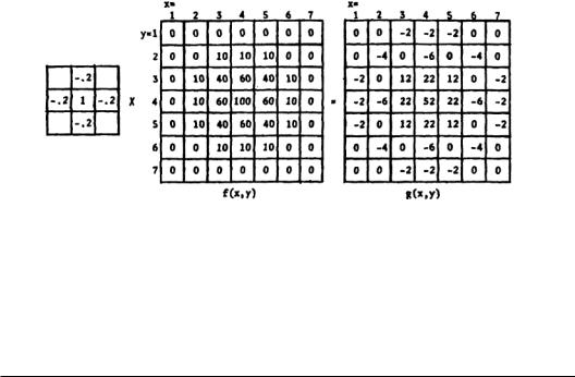
These models include both one-dimensional examination of the phenomenon and two-dimensional treatment, where a two-dimensional array is used as a stimulus. This two-dimensional treatment is justified because most of the sensory receptors of the body form two-dimensional maps (receptive fields). In principle, if a one-dimensional lateral inhibition system is linear, one can extend the analysis to two dimensions by means of superposition. The two-dimensional array can be thought of as a function f(x, y), and the lateral inhibition network itself is embodied in a separate N×N array, the central square of which has a positive value and can be thought of as a direct input from an incoming axon.The surrounding squares have negative values that are higher than the corner values, which are also negative.The method consists of multiplying the input signal values f(x, y) and their contiguous values by the lateral inhibitory network’s weighting factors to get a corresponding g(x,y). Figure 3.1 presents an example of such a process.The technique illustrated here is used in the contrast enhancement of photographs.The objective is the same as that of the nervous system: to improve image sharpness without introducing too much distortion.This technique requires storage of each picture element and lateral “inhibitory” interactions between adjacent elements. Since a picture may contain millions of elements, high-speed computers with large-scale memories are required.
At a higher level,similar algorithms can be used to evaluate decision-making mechanisms.In this case, many inputs from different sensory systems are competing for attention.The brain evaluates each one of the inputs as a function of the remaining ones.One can picture a decision-making mechanism resembling a “locator” of stimulus peaks.The final output depends on what weights are used at the inputs of a pushpull mechanism.Thus a decision can be made depending on the weights an individual’s brain
Nervous System |
3-7 |
FIGURE 3.1 An example of two-dimensional lateral inhibition. On the left, the 3 x 3 array corresponds to the values of the synaptic junctions weighting coefficients. For simplicity, the corner weights are assumed to be zero. g(x,y) represents the output matrix after lateral inhibition has been applied to the input matrix.
is applying to the incoming information about a situation under consideration.The most important information is heavily weighted, while the rest is either totally masked or weighted very lightly.
3.5 Higher Functions of the Nervous System
Pattern Recognition
One way of understanding human perception is to study the mechanism of information processing in the brain. The recognition of patterns of sensory input is one of the functions of the brain, a task accomplished by neuronal circuits, the feature extractors. Although such neuronal information is more likely to be processed globally by a large number of neurons, in animals, single-unit recording is one of the most powerful tools in the hands of the physiologist. Most often, the concept of the receptive field is used as a method of understanding sensory information processing.In the case of the visual system,one could call the receptive field a well-defined region of the visual field which, when stimulated, will change the firing rate of a neuron in the visual pathway. The response of that neuron will usually depend on the distribution of light in the receptive field.Therefore, the information collected by the brain from the outside world is transformed into spatial as well as temporal patterns of neuronal activity.
The question often asked is how do we perceive and recognize faces, objects, and scenes. Even in those cases where only noisy representations exist, we are still able to make some inference as to what the pattern represents.Unfortunately,in humans,single-unit recording,as mentioned above,is impossible. As a result, one has to use other kinds of measurements, such as evoked potentials (EPs). Although physiologic in nature, EPs are still far away from giving us information at the neuronal level.Yet EPs have been used extensively as a way of probing the human (and animal) brains because of their noninvasive character. EPs can be considered to be the result of integrations of the neuronal activity of many neurons some place in the brain.This gross potential can then be used as a measure of the response of the brain to sensory input.
The question then becomes:Can we somehow use this response to influence the brain in producing patterns of activity that we want? None of the efforts of the past closed this loop. How do we explain then the phenomenon of selective attention by which we selectively direct our attention to something of interest and discard the rest? And what happens with the evolution of certain species that change appearance according to their everyday needs? All these questions tend to lead to the fact that somewhere in the brain there is a loop where previous knowledge or experience is used as a feedback to the brain itself.This feedback then modifies the ability of the brain to respond in a different way to the same