

Biotechnology for Biomedical Engineers - Martin L. Yarmush et al
.pdf
8
Protein Engineering
Alan J. Russell
University of Pittsburgh |
8.1 |
Site-Directed Mutagenesis |
8–1 |
ChenzhaoVierheller |
8.2 |
Solvent Engineering |
8–3 |
University of Pittsburgh |
8.3 |
Conclusions |
8–1 |
|
|
|
Enzymes have numerous applications in both research and industry. The conformation of proteins must be maintained in order for them to function at optimal activity. Protein stability is dependent on maintaining a balance of forces that include hydrophobic interactions, hydrogen bonding, and electrostatic interactions. Most proteins are denatured, i.e., lose their active conformation in high-temperature environments (exceptions to this are found among the enzymes from thermophilic microorganisms). Therefore,understanding and maintaining enzyme stability are critical if enzymes are to be widely used in medicine and industry. Protein engineering [1] is used to construct and analyze modified proteins using molecular biologic, genetic engineering,biochemical, and traditional chemical methods (Table 8.1).The generation of proteins with improved activity and stability is now feasible.Recent developments in molecular biology also have enabled a rapid development of the technologies associated with protein engineering (Table 8.2).
8.1 Site-Direction Mutagenesis
Since site-directed mutagenesis was described by Hutchinson et al. [2] in 1978, it has become a powerful tool to study the molecular structure and function of proteins.The purpose of site-directed mutagenesis is to alter a recombinant protein by introducing, replacing, or deleting a specific amino acid.The technique enables a desired modification to be achieved with exquisite precision [3]. Sitedirected mutagenesis has been used to change the activity and stability of enzymes, as well as substrate specificity and affinity. Indeed, much of the biologic detergent enzyme sold in the United States (~$200 million per year) is a protein-engineered variant of the native enzyme.
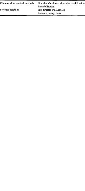
The basic idea behind site-directed mutagenesis is illustrated in Fig. 8.1. The first step of sitedirected mutagenesis involves cloning the gene for the protein to be studied into a vector.This nontrivial exercise is discussed elsewhere in this handbook.Next, an oligonucleotide is designed and synthesized. The oligonucleotide will have a centrally located desired mutation (usually a mismatch) that is flanked by sequences of DNA that are complementary to a specific region of interest.Thus the oligonucleotide is designed to bind to a single region of the target gene.The mutation can then be introduced into the gene by hydridizing the oligonucleotide to the single-stranded template. Single-stranded templates of a target gene may be obtained by either cloning the gene to a single-stranded vector,such as bacteriophage M13 [4],or by using phagemids (a chimeric plasmid containing a filamentous bacteriophage replication origin that directs synthesis of single-stranded DNA with a helper bacteriophage) [5]. Alternatively, single-stranded template may be generated by digesting double-stranded DNA with exonuclease III following nicking of the target DNA with DNase or a restriction endonuclease [6].
0-8493-1811-4/03/$0.00+$1.50 |
8-1 |
© 2003 by CRC Press LLC |
8-2 |
Biotechnology for Biomedical Engineers |
TABLE 8.1 Selected Methods for Modification of Proteins
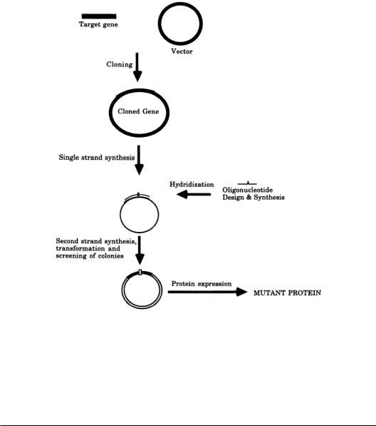
The second strand is synthesized with DNA polymerase using the oligonucleotide as a primer, and the DNA can be circularized with DNA ligase.The vector that carries the newly synthesized DNA (in which one strand is mutated) can be introduced into a bacterial host, where, because DNA duplicates in a semiconservative mode, half the newly synthesized cells containing the DNA will theoretically contain the mutation. In reality, premature DNA polymerization, DNA mismatch repair, and strand displacement synthesis result in much lower yield of mutants. Different strategies have been developed to obtain a higher level of mutation efficiency in order to minimize the screening of mutant.A second mutagenic oligonucleotide containing an active antibiotic resistance gene can be introduced at the same time as the site-directed oligonucleotide.Therefore, the wild-type plasmid will be eliminated in an antibiotic-selective medium [7] (Fig.8.2).Alternatively,introducing a second mutagenic oligonucleotide can result in the elimination of a unique restriction endonuclease in the mutant strand [8].Therefore, wild-type plasmid can be linearized with this unique restriction endonuclease, while the circular mutant plasmid can be transformed into bacteria host. Another alternative method is to perform second-strand extension in the presence of 5-methyl-dCTP, resulting in resistance of number of restriction enzymes, including HpaII, MspI, and Sau3A I, in the mutant strand only [9].The template DNA can then be nicked with these enzymes, followed by digestion with exonuclease III, to increase mutagenesis efficiency.
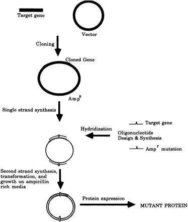
The recent development of the polymerase chain reaction provides a new approach to site-direct mutagenesis. In 1989, Ho and colleagues [10] developed a method named overlap extension (Fig. 8.3), in which four oligonucleotides are used as primers. Two of the primers containing the mutant are complementary to each other.The two other primers are complementary to the opposite strand of the ends of the cloned genes.Polymerase chain reactions are performed three times.The first two polymerase chain reactions are carried out using one end primer and one mutant primer in each reaction.The products contain one double-stranded DNA from one end to the mutation point and the other doublestranded DNA from another end to the mutation point. In other words, the two DNA products are overlapped in the mutated region.The polymerase chain reaction products are purified and used as templates for the third polymerase chain reaction with two end primers. This generates the whole length of DNA with the desired mutation.The advantage of this method is that it can be done quickly with nearly 100% efficiency.
After screening and selection, a mutation is generally confirmed by DNA sequencing.The mutant can then be subcloned into an expression vector to test the effect of mutation on the activity of the
TABLE 8.2 Potentially Useful Modifications to Proteins
Source: Modified from Primrose SB. 1991. In Molecular
Biotechnology, 2d ed. New York, Blackwell Scientific.
Protein Engineering |
8-3 |
FIGURE 8.1 Overview of site-directed mutagenesis. See text for details.
enzyme. Over the last decade, site-directed mutagenesis has become a somewhat trivial technical process, and the most challenging segment of a protein engineering research project is unquestionably the process of determining why a mutant protein has altered properties.The discussion of this particular enterprise lies beyond the scope of this brief review.
8.2 Solvent Engineering
An enzyme-catalyzed reaction can be simplified to its most basic components by considering it as the transfer of a substrate molecule from solvent to the surface of an enzyme molecule.The exchange of substrate-solvent and enzyme-solvent interactions for enzyme-substrate interactions then enables the chemistry of catalysis to take place.Protein engineering, as described above, is the process of changing the enzyme in a predictable and precise manner to effect a change on the catalytic process. Since the enzyme is only one side of the balance, however, any changes in the rest of the equation also will alter the catalytic process. Until the late 1980s, for example, substrate specificity could be altered by either protein engineering of the enzyme or by changing the substrate.Solvent engineering is now also emerging as a powerful tool in rational control of enzyme activity.Using solvents other than water has successfully led to enzymes with increased thermostability, activity against some substrates, pH dependence, and substrate and enantiospecificity.
The simplicity of the solvent-engineering approach is clear. If a protein is not inactivated by a solvent change, then its activity will be dependent on the solvent in which the enzyme and substrate
8-4 |
Biotechnology for Biomedical Engineers |
FIGURE 8.2 Selective antibiotic genes as strategy for improvement of site-directed mutagenesis. See text for details.
are placed.This strategy has been discussed in detail in recent reviews [11], and we will only summarize the most pertinent information here.
Enzymes that have been freeze-dried and then suspended in anhydrous organic solvents, in which proteins are not soluble,retain their activity and specificity [12].The enzyme powders retain approximately a monolayer of water per molecule of enzyme during the freeze-drying process. As long as this monolayer of water remains associated with the enzyme in an organic environment, the structure of the enzyme is not disrupted [13], and hence enzyme activity in essentially anhydrous organic solvents can be observed.
The physical properties of a solvent in which an enzyme is placed can influence the level of activity and specificity in a given reaction.For instance,alcohol dehydrogenase can catalyze oxidation-reduction reactions equally well in buffer and heptane, but the level of activity in heptane is sharply dependent on the thermodynamic activity of water in the system.In general,activity can be increased by increasing the hydrophobicity of the solvent used.Interestingly,however,the specificity (both stereoand substrate) of an enzyme or antibody dispersed in an organic solvent is generally greater in more hydrophilic solvents [14].Water is involved as a substrate in many of the chemical modification processes that lead to irreversible inaction of proteins. Not surprisingly, therefore, proteins in anhydrous environments are stable at temperatures exceeding 100°C [15].
Considerable attention is now being given to developing the predictability of the solvent-engineering approach. Elucidating the structure-function-environment relationships that govern the activity and
Protein Engineering |
8-5 |
FIGURE 8.3 Site-directed mutagenesis by overlap extension. See text for details.
specificity of an enzyme is a crucial step for learning how to apply the power of solvent engineering. In recent years, supercritical fluids (materials at temperatures and pressures above their critical point) have been used as a dispersant for enzyme-catalyzed reactions. In addition to being excellent process solvents, the physical properties of supercritical fluids are pressure-dependent.Thus it may be possible to detect which physical properties of a nonaqueous medium have a role in determining a given enzyme function [16].
8.3 Conclusions
The goals of protein and solvent engineering are similar. Ideally, one should be able to predictably alter a given property of an enzyme by either changing the enzyme or its environment. In reality, both approaches are still somewhat unpredictable. Both protein engineering and solvent engineering have been used successfully to alter the protein properties that appear in Table 8.1. Further research is needed,however,before the results of any given attempt at such biologic engineering can be predicted.
References
1.Alvaro G,Russell AJ.1991.Methods Enzymol 202:620.
2.Hutchinson C,Phillips S,Edgell M.1978.J Biol Chem 253:6551.
3.Moody P,Wilkinson A.1990.Protein Engineering, pp 1–3.IRL Press.
4.Zoller M, Smith M. 1983. Methods Enzymol 100:468.
5.Dente L,Cesareni G,Cortese R. 1983.Nucl Acid Res 11:1645.
6.Rossi J, Zoller M. 1987.In D Oxender, C Fox (eds), Protein Engineering, pp 51–63. NewYork,Alan R Liss.
7.Promega Protocols and Application Guide, 2d ed,pp 98–105.1991.
8.DengW, Nickoloff J. 1992. Anal Biochem 200:81.
9.Vandeyar M,Weiner M,Hutton C, Batt C. 1988.Gene 65:129.
10.Ho N,Hunt H, Horton R, et al.1989. Gene 77:51.
8-6 |
Biotechnology for Biomedical Engineers |
11.Russell AJ, Chatterjee S, Rapanovich I, Goodwin J. 1992. In A Gomez-Puyon (ed), Biomolecules in Organic Solvents, pp 92–109.Boca Raton,Fla,CRC Press.
12.Zaks A,Klibanov AM. 1985. Proc Natl Acad Sci USA 82:3192.
13.Affleck R, Xu Z-F, SuzawaV, et al. 1992. Proc Natl Acad Sci USA 89:1100.
14.Kamat SV,Beckman EJ,Russell AJ.1993. J Am Chem Soc 115:8845.
15.Zaks A,Klibanov AM.1984.Science 224:1249.
16.Kamat S, Iwaskewycz B, Beckman EJ, Russell AJ. 1993. Proc Natl Acad Sci USA 90:2940.

9
Monoclonal Antibodies and Their Engineered Fragments
|
9.1 |
Structure and Function of Antibodies |
9–2 |
|
9.2 |
Monoclonal Antibody Cloning Techniques |
9–4 |
|
|
HybridomaTechnology • Repertoire CloningTechnology |
|
|
9.3 |
Monoclonal Antibody Expression Systems |
9–9 |
|
|
Bacterial Expression • Expression in Lymphoid and |
|
|
|
Nonlymphoid Systems (Transfectoma Technology) |
• |
Srikanth Sundaram |
|
Expression inYeast • Expression in Baculovirus • |
Expression |
|
in Plants |
|
|
Rutgers University |
9.4 |
Genetically Engineered And Their Fragements |
9–12 |
David M.Yarmush |
9.5 |
Applications of Monoclonal Fragments |
9–15 |
Rutgers University |
9.6 |
Summary |
9–17 |
Antibodies are a class of topographically homologous multidomain glycoproteins produced by the immune system that display a remarkably diverse range of binding specificities.The most important aspects of the immune system are that it is diverse and driven to produce antibodies of the highest possible antigen affinity.The primary repertoire of antibodies consists of about 109 different specificities, each of which can be produced by an encounter with the appropriate antigen.This diversity is known to be produced by a series of genetic events each of which can play a role in determining the final function of the antibody molecule.After the initial exposure to the antigen, additional diversity occurs by a process of somatic mutation so that, for any selected antigen, about 104 new binding specificities are generated. Thus the immunologic repertoire is the most diverse system of binding proteins in biology. Antibodies also display remarkable binding specificity. For example, it has been shown that antibodies are able to distinguish between ortho-, meta-, and para-forms of the same haptenic group [Landsteiner,1945].This exquisite specificity and diversity make antibodies ideal candidates for diagnostic and therapeutic agents.
Originally, the source of antibodies was antisera, which by their nature are limited in quantity and heterogeneous in quality.Antibodies derived from such sera are termed conventional antibodies (polyclonal antibodies). Polyclonal antibody production requires methods for the introduction of immunogen into animals, withdrawal of blood for testing the antibody levels, and finally exsanguination for collection of immune sera. These apparently simple technical requirements are complicated by the necessity of choosing a suitable species and immunization protocol that will produce a highly immune animal in a short time. Choice of animal is determined by animal house facilities available, amount of antiserum required (a mouse will afford only 1.0 to 1.5 ml of blood; a goat can provide several liters), and amount of immunogen available (mice will usually respond very well to 50 µg or less of antigen; goats may require several milligrams). Another consideration is the phylogenic relationship between the animal
0-8493-1811-4/03/$0.00+$1.50 |
9-1 |
© 2003 by CRC Press LLC |
9-2 |
Biotechnology for Biomedical Engineers |
from which the immunogen is derived and that used for antibody production. In most cases, it is advisable to immunize a species phylogenetically unrelated to the immunogen donor, and for highly conserved mammalian proteins, nonmammals (e.g., chickens) should be used for antibody production. The polyclonal antibody elicited by an antigen facilitates the localization, phagocytosis, and complementmediated lysis of that antigen; thus the usual polyclonal immune response has clear advantages in vivo. Unfortunately, the antibody heterogeneity that increases immune protection in vivo often reduces the efficacy of an antiserum for various in vitro uses.Conventional heterogeneous antisera vary from animal to animal and contain undesirable nonspecific or cross-reacting antibodies. Removal of unwanted specificities from a polyclonal antibody preparation is a time-consuming task,involving repeated adsorption techniques, which often results in the loss of much of the desired antibody and seldom is very effective in reducing the heterogeneity of an antiserum.
After the development of hybridoma technology [Köhler and Milstein,1975],a potentially unlimited quantity of homogeneous antibodies with precisely defined specificities and affinities (monoclonal antibodies) became available, and this resulted in a step change in the utility of antibodies. Monoclonal antibodies (mAbs) have gained increasing importance as reagents in diagnostic and therapeutic medicine, in the identification and determination of antigen molecules, in biocatalysis (catalytic antibodies), and in affinity purification and labeling of antigens and cells.
9.1 Structure and Function of Antibodies
Antibody molecules are essentially required to carry out two principal roles in immune defense. First, they recognize and bind nonself or foreign material (antigen binding).In molecular terms,this generally means binding to structures on the surface of the foreign material (antigenic determinants) that differ from those on the host. Second, they trigger the elimination of foreign material (biologic effector functions).In molecular terms, this involves binding of effector molecules (such as complement) to the antibody-antigen complex to trigger elimination mechanisms such as the complement system and phagocytosis by macrophages and neutrophils, etc.
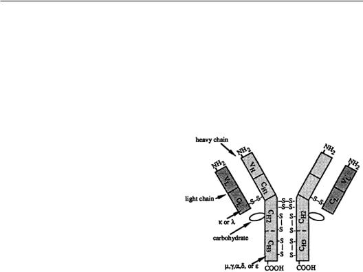
In humans and other animals, five major immunoglobulin classes have been identified. The five classes include immunoglobulins G (IgG),A (IgA), M (IgM), D (IgD), and E (IgE).With the exception of IgA (dimer) and IgM (pentamer),all other antibody classes are monomeric.The monomeric antibody molecule consists of a basic four-chain structure, as shown in Fig. 9.1.There are two distinct types of chains: the light (L) and the heavy chains (H).
The chains are held together by disulfide bonds. The light and heavy chains are held together by interchain disulfides, and the two heavy chains are held together by numerous disulfides in the hinge region of the heavy chain.The light chains have a molecular weight of about 25,000 Da,while the heavy chains have a molecular weight of 50,000 to 77,000 Da depending on the isotope. The L chains can be divided into two subclasses, kappa (k) and lambda (l), on the basis of their structures and amino acid sequences. In humans, about 65% of the antibody molecules have k chains, whereas in rodents,they constitute over 95% of all antibody molecules. The light chain consists of two structural domains:The carboxy-terminal half of
the chain is constant except for certain allotypic and isotype variations and is called the CL (constant: light chain) region, whereas the amino-terminal half of the chain shows much sequence variability and is known as the VL (variable: light chain) region.The H chains are unique for each immunoglobulin
Monoclonal Antibodies and Their Engineered Fragments |
9-3 |
class and are designated by the Greek letter corresponding to the capital letter designation of the immunoglobulin class (a for the H chains of IgA, g for the H chains of IgG). IgM and IgA have a third chain component, the J chain, which joins the monomeric units.The heavy chain usually consists of four domains:The aminoterminal region (approximately 110 amino acid residues) shows high sequence variability and is called the VH (variable: heavy chain) region, whereas there are three domains called CH1, CH2, and CH3 in the constant part of the chain.The CH2 domain is glycosylated, and it has been shown that glycosylation is important in some of the effector functions of antibody molecules.The extent of glycosylation varies with the antibody class and,to some extent,the method of its production. In some antibodies (IgE and IgM), there is an additional domain in the constant part of the heavy chain called the CH4 region.The hinge region of the heavy chain (found between the CH1 and CH2 domains) contains a large number of hydrophilic and proline residues and is responsible for the segmental flexibility of the antibody molecule.
All binding interactions to the antigen occur within the variable domains (VH andVL).Each variable domain consists of three regions of relatively greater variability, which have been termed hypervariable (HV) or complementarity-determining regions (CDR) because they are the regions that determine complementarity to the particular antigen. Each CDR is relatively short, consisting of from 6 to 15 residues. In general, the topography of the six combined CDRs (three from each variable domain) produces a structure that is designed to accommodate the particular antigen.The CDRs on the light chain include residues 24 to 34 (Ll), 50 to 56 (L2), and 89 to 73 (L3), whereas those on the heavy chain include residues 31 to 35 (Hl), 50 to 65 (H2), and 95 to 102 (H3) in the Kabat numbering system.The regions between the hypervariable regions are called the framework regions (FR) because they are more conserved and play an important structural role.The effector functions are, on the other hand,mediated via the two C-terminal constant domains, namely, the CH2 and CH3.
Each variable region is composed of several different genetic elements that are separate on the germ line chromosome but rearrange in the B cell to produce a variable-region exon. The light chain is composed of two genetic elements, a variable (V) gene and a joining (J) gene.TheVK gene generally encodes residues 1 to 95 in a kappa light chain, and the JK gene encodes residues 96 to 107, which is the carboxyl end of the variable region. Thus the kappa V gene encodes all the first and second hypervariable regions (Ll and L2) and a major portion of the third hypervariable region (L3). In the human system, there are estimated to be about 100 differentVK genes and about 5 functional JK genes, and the potential diversity is increased by the imprecision of theVJ joining.The heavy-chain variable region is similarly produced by the splicing together of genetic elements that are distant from each other in the germ line.The mature VH region is produced by the splicing of a variable gene segment
(V) to a diversity segment (D) and to a joining (J) segment.There are about 300VH genes, about 5 DH genes, and 9 functional JH genes [Tizard, 1992].With respect to the CDRs of the heavy chain, Hl and H2 are encoded by theVH gene, while the H3 region is formed by the joining of theV, D, and J genes. Additional diversity is generated by recombinational inaccuracies,lightand heavy-chain recombination, and N-region additions.
The three-dimensional structure of several monoclonal antibodies has been determined via x-ray crystallography and has been reviewed extensively [Davies et al., 1990]. Each domain consists of a sandwich of (b sheets with a layer of four and three strands making up the constant domains and a layer of four and five strands making up the variable domains.The CDRs are highly solvent exposed and are the loops at the ends of the variable region ß strands that interact directly with the antigen. The superimposition of severalVL andVH structures from available x-ray crystallographic coordinates using the conserved framework residues shows that the framework residues of the antibody variable domains are spatially closely superimposable. The CDRs varied in shape and length, as well as in sequence. Similarities in the structures of the CDRs from one antibody to another suggest that they are held in a fixed relationship in space, at least within a domain.Also, the N and C terminal of the CDRs were rigidly conserved by the framework residues.Among the CDRs, L1, L2, H2, and H3 show the most conformational variability.