

Biotechnology for Biomedical Engineers - Martin L. Yarmush et al
.pdf5-6 |
Biotechnology for Biomedical Engineers |
to neurotransmitters [Dobie and Rubel, 1989]. Second, observation of the Henson’s cells, passive cells that are intimately connected to OHCs,shows that spontaneous vibrations are produced by the Henson’s cells in mammals and likely in the OHCs as well. These vibrations exhibit spectral peaks that are appropriate in frequency to their locations on the BM [Khanna et al,1993].Third, action of the OHCs as amplifiers leads to spontaneous otoacoustic emissions [Kim, 1984; Lyon and Mead, 1989] and to changes in the response of the auditory system [Lyon and Mead, 1989; Geisler, 1992]. Fourth, AC excitation of the OHCs of mammals produces changes in length [Cooke, 1993].
The OHCs appear to affect the response of the auditory system in several ways.They enhance the tuning characteristics of the system to sinusoidal stimuli, decreasing thresholds and narrowing the filter’s bandwidth [Dobie and Rubel, 1989].They likely influence the damping of the BM dynamically by actively changing its stiffness.
Spiral Ganglion Cells and the Auditory Nerve
Anatomy
The auditory nerve of the human contains about 30,000 fibers consisting of myelinated proximal processes of spiral ganglion cells (SGCs).The somas of spiral ganglion cells (SGCs) lie in Rosenthal’s canal, which spirals medial to the three scalae of the cochlea. Most (93%) are large, heavily myelinated, type I SGCs whose distal processes synapse on IHCs.The rest are smaller type II SGCs, which are more lightly myelinated. Each IHC has, on average, a number of fibers that synapse with it, 8 in the human and 18 in the cat, although some fibers contact more than one IHC. In contrast, each type II SGC contacts OHCs at a rate of about 10 to 60 cells per fiber.
The auditory nerve collects in the center of the cochlea, its modiolus, as SGC fibers join it. Lowfrequency fibers from the apex join first, and successively higher frequency fibers come to lie concentrically on the outer layers of the nerve in a spiraling fashion before it exits the modiolus to enter the internal auditory meatus of the temporal bone.A precise tonotopic organization is retrained in the concentrically wrapped fibers.
Physiology
Discharge spike patterns from neurons can be recorded extracellularly while repeating tone bursts are presented.A threshold level can be identified from the resulting rate-level function (RLF).In the absence of sound and at lower, subthreshold levels, a spontaneous rate of discharge is measured. In the nerve this ranges from 50 spikes per second to less than 10.As intensity is raised, the threshold level is encountered, where the evoked discharge rate significantly exceeds the spontaneous discharge rate. The plot of threshold levels as a function of frequency is the neuron’s threshold tuning curve. The tuning curves for axons in the auditory nerve show a minimal threshold (maximum sensitivity) at a characteristic frequency (CF) with a narrow frequency range of responding for slightly more intense sounds.At high intensities, a large range of frequencies elicits spike discharges. RLFs for nerve fibers are monotonic (i.e., spike rate increases with stimulus intensity),and although a saturation rate is usually approached at high levels,the spike rate does not decline. Mechanical tuning curves for the BM and neural threshold tuning curves are highly similar (Fig. 5.3). Mechanical frequency analysis in the cochlea and the orderly projection of fibers through the nerve lead to correspondingly orderly maps for CFs in the nerve and the nuclei of the central pathways.
Sachs andYoung [1979] found that the frequency content of lower intensity vowel sounds is represented as corresponding tonotopic rate peaks in nerve activity, but for higher intensities this rate code is lost as fibers tend toward equal discharge rates. At high intensities spike synchrony to frequencies near CF continue to signal the relative spectral content of vowels, a temporal code.These results hold for high- spontaneous-rate fibers (over 15 spikes per second), which are numerous. Less common, low-spontaneous- rate fibers (less than 15 spikes per second) appear to maintain the rate code at higher intensities,suggesting different coding roles for these two fiber populations.
Auditory System |
5-7 |
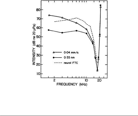
FIGURE 5.3 Mechanical and neural turning curves from the BM and auditory nerve, respectively. The two mechanical curves show the intensity and frequency combinations for tones required to obtain a criterion displacement or velocity, while the neural curve shows the combinations needed to increase neural discharge rates a small amount over spontaneous rate.
5.3 The Central Auditory System
Overview
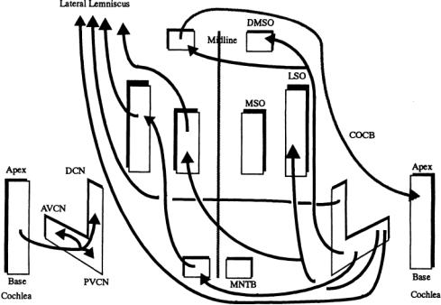
In ascending paths, obligatory synapses occur at the CN, IC, MGB, and AC, but a large number of alternative paths exist with ascending and descending internuclear paths and the shorter intranuclear connections between neighboring neurons and subdivisions within a major nuclear group. Each of the centers listed contains subpopulations of neurons that differ in aspects of their morphologies, discharge patterns to sounds,segregation in the nucleus,biochemistry,and synaptic connectivities.The arrangement of the major ascending auditory pathways is schematically illustrated in Fig. 5.4. For references, see Altschuler et al. [1991].
Neural Bases of Processing
The Cochlear Nuclei
Anatomy of the Cochlear Nuclei. The CN can be subdivided into at least three distinct regions, the anteroventral CN (AVCN), the posteroventral CN (PVCN), and the dorsal CN (DCN). Each subdivision has one or more distinctive neuron types and unique intraand internuclear connections.The axon from each type I SGC in the nerve branches to reach each of the three divisions in an orderly manner so that tonotopic organization is maintained. Neurons with common morphologic classifications are found in all three divisions, especially granule cells, which tend to receive connections from type II spiral ganglion cells.
Morphologic classification of neurons based on the shapes of their dendritic trees and somas show that the anterior part of the AVCN contains many spherical bushy cells, while in its posterior part both
5-8 |
Biotechnology for Biomedical Engineers |
FIGURE 5.4 A schematic of major connections in the auditory brainstem discussed in the text.All structures and connections are bilaterally symmetrical, but connections have been shown on one side only for clarity. No cell types are indicated, but the subdivisions of origin are suggested in the CN. Note that the LSO and MSO receive inputs from both CN.
globular bushy cells and spherical bushy cells are found. Spherical bushy cells receive input from one type I ganglion cell through a large synapse formation containing end bulbs of Held, while the globular cells may receive inputs from a few afferent fibers.These endings cover a large part of the soma surface and parts of the proximal dendrite, especially in spherical bushy cells, and they have rounded vesicles presynaptically, indicating excitatory input to the bushy cells, while other synaptic endings of noncochlear origins tend to have flattened vesicles associated with inhibitory inputs. Stellate cells are found throughout the AVCN, as well as in the lower layers of the DCN.The AVCN is tonotopically organized, and neurons having similar CFs have been observed to lie in layers or laminae [Bourk et al., 1981]. Isofrequency laminae also have been indicated in other auditory nuclei.
The predominant neuron in the PVCN is the octopus cell, a label arising from its distinctive shape with asymmetrically placed dendrites. Octopus cells receive cochlear input from type I SGCs on their somas and proximal dendrites.Their dendrites cross the incoming array of cochlear fibers, and these often branch to follow the dendrite toward the soma.
The DCN is structurally the most intricate of the CN. In many species, four or five layers are noticeable, giving it a “cortical” structure, and its local circuitry has been compared with that of the cerebellum. Fusiform cells are the most common morphologic type.Their somas lie in the deeper layers of the DCN, and their dendrites extend toward the surface of the nucleus and receive primarily noncochlear inputs. Cochlear fibers pass deeply in the DCN and turn toward the surface to innervate fusiform and giant cells that lie in the deepest layer of the DCN.The axons of fusiform and giant cells project out of the DCN to the contralateral IC.
Intracellular recording in slice preparation is beginning to identify the membrane characteristics of neuronal types in the CN.The diversity of neuronal morphologic types, their participation in local circuits, and the emerging knowledge of their membrane biophysics are motivating detailed compartmental modeling [Arle and Kim, 1991].
Auditory System |
5-9 |
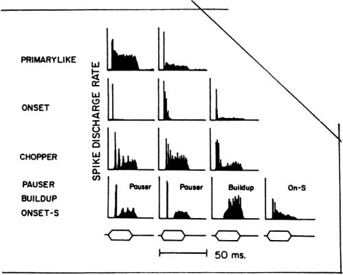
FIGURE 5.5 Peristimulus time histogram patterns obtained in the CN and nerve.Repeated presentations of a tone burst at CF are used to obtain these estimates of discharge rate during the stimulus. (Adapted fromYoung, 1984.)
Spike Discharge Patterns. Auditory nerve fibers and neurons in central nuclei may discharge only a few times during a brief tone burst, but if a histogram of spike events is synchronized to the onset of the tone burst, a peristimulus time histogram (PSTH) is obtained that is more representative of the neuron’s response than any single one.The PSTH may be expressed in terms of spike counts, spike probability, or spike rate as a function of time, but all these retain the underlying temporal pattern of the response. PSTHs taken at the CF for a tone burst intensity roughly 40 dB above threshold have shapes that are distinctive to different nuclear subdivisions and even different morphologic types.They have been used for functionally classifying auditory neurons.
Figure 5.5 illustrates some of the major pattern types obtained from the auditory nerve and CN. Auditory nerve fibers and spherical bushy cells in AVCN have primary-like patterns in their PSTHs, an elevated spike rate after tone onset, falling to a slowly adapting level until the tone burst ends. Globular bushy cells may have primary-like, pri-notch (primary-like with a brief notch after onset), or chopper patterns. Stellate cells have nonprimary-like patterns. Onset response patterns, one or a few brief peaks of discharge at onset with little or no discharges afterward,are observed in the PVCN from octopus cells. Chopper,pauser, and buildup patterns are observed in many cells of the DCN.For most neurons of the CN, these patterns are not necessarily stable over different stimulus intensities; a primary-like pattern may change to a pauser pattern and then to a chopper pattern as intensity is raised [Young, 1984].
Functional classification also has been based on the response map, a plot of a neuron’s spike discharge rate as a function of tonal frequency and intensity.Fibers and neurons with primary-like PSTHs generally have response maps with only an excitatory region of elevated rate.The lower edges of this region approximate the threshold tuning curve. Octopus cells often have very broad tuning curves and extended response
5-10 |
Biotechnology for Biomedical Engineers |
maps,as suggested by their frequency-spanning dendritic trees.More complex response maps are observed for some neurons, such as those in the DCN. Inhibitory regions alone, a frequency-intensity area of suppressed spontaneous discharge rates,or combinations of excitatory regions and inhibitory regions have been observed. Some neurons are excited only within islands of frequency-intensity combinations, demonstrating a CF but having no response to high-intensity sounds.In these cases,an RLF at CF would be nonmonotonic; i.e., spike rate decreases as the level is raised. Response maps in the DCN containing both excitatory and inhibitory regions have been shown to arise from a convergence of inputs from neurons with only excitatory or inhibitory regions in their maps [Young andVoigt, 1981].
Superior Olivary Complex (SOC)
The SOC contains 10 or more subdivisions in some species. It is the first site at which connections from the two ears converge and is therefore a center for binaural processing that underlies sound localization.There are large differences in the subdivisions between mammalian groups such as bats, primates, cetaceans, and burrowing rodents that utilize vastly different binaural cues. Binaural cues to the locus of sounds include interaural level differences (ILDs), interaural time differences (ITDs), and detailed spectral differences for multispectral sounds due to head and pinna filtering characteristics.
Neurons in the medial superior olive (MSO) and lateral superior olive (LSO) tend to process ITDs and ILDs, respectively.A neuron in the MSO receives projections from spherical bushy cells of the CN from both sides and thereby the precise timing and tuning cues of nerve fibers passed through the large synapses mentioned.The time accuracy of the pathways and the comparison precision of MSO neurons permit the discrimination of changes in ITD of a few tens of microseconds.MSO neurons project to the ipsilateral IC through the lateral lemniscus.Globular bushy cells of the CN project to the medial nucleus of the trapezoid body (MNTB) on the contralateral side, where they synapse on one and only one neuron in a large, excitatory synapse,the calyx of Held.MNTB neurons send inhibitory projections to neurons of the LSO on the same side, which also receives excitatory input from spherical bushy cells from the AVCN on the same side. Sounds reaching the ipsilateral side will excite discharges from an LSO neuron, while those reaching the contralateral side will inhibit its discharge.The relative balance of excitation and inhibition is a function of ILD over part of its physiological range, leading to this cue being encoded in discharge rate.
One of the subdivisions of the SOC, the dorsomedial periolivary nucleus (DMPO), is a source of efferent fibers that reach the contralateral cochlea in the crossed olivocochlear bundle (COCB). Neurons of the DMPO receive inputs from collaterals of globular bushy cell axons of the contralateral ACVN that project to the MNTB and from octopus cells on both sides.The functional role of the feedback from the DMPO to the cochlea is not well understood.
Nuclei of the Lateral Lemniscus (NLL)
The lateral lemniscus consists of ascending axons from the CN and LSO.The NLL lie within this tract, and some, such as the dorsal nucleus (DNLL), are known to process binaural information, but less is known about these nuclei as a group than others, partially due to their relative inaccessibility.
Inferior Colliculi (IC)
The IC are paired structures lying on the dorsal surface of the rostral brainstem. Each colliculus has a large central nucleus (ICC), a surface cortex, and paracentral nuclei. Each colliculus receives afferents from a number of lower brainstem nuclei,projects to the MGB through the brachium, and communicates with the other colliculus through a commissure. The ICC is the major division and has distinctive laminae in much of its volume.The laminae are formed from disk-shaped cells and afferent fibers.The disk-shaped cells,which make up about 80% of the ICC’s neuronal population,have flattened dendritic fields that lie in the laminar plane. The terminal endings of afferents form fibrous layers between laminae.The remaining neurons in the ICC are stellate cells that have dendritic trees spanning laminae. Axons from these two cell types make up much of the ascending ICC output.
Tonotropic organization is preserved in the ICCs laminae, each corresponding to an isofrequency lamina. Both monaural and binaural information converges at the IC through direct projections from

Auditory System |
5-11 |
the CN and from the SOC and NLL. Crossed CN inputs and those from the ipsilateral MSO are excitatory. Inhibitory synapses in the ICC arise from the DNLL, mediated by gamma-amino-butyric acid (GABA), and from the ipsilateral LSO, mediated by glycine.
These connections provide an extensive base for identifying sound direction at this midbrain level,but due to their convergence,it is difficult to determine what binaural processing occurs at the IC as opposed to being passed from the SOC and NLL. Many neurons in the IC respond differently depending on binaural parameters.Varying ILDs for clicks or high-frequency tones often indicates that contralateral sound is excitatory.Ipsilateral sound may have no effect on responses to contralateral sound,classifying the cell as E0, or it may inhibit responses, in which case the neuron is classified as EI, or maximal excitation may occur for sound at both ears,classifying the neuron as EE. Neurons responding to lower frequencies are influenced by ITDs, specifically the phase difference between sinusoids at the ears. Spatial receptive fields for sounds are not well documented in the mammalian IC, but barn owls, who use the sounds of prey for hunting at night, have sound-based spatial maps in the homologous structure. The superior colliculus, situated just rostral to the IC, has spatial auditory receptive field maps for mammals and owl.
Auditory Thalamocortical System
Medial Geniculate Body (MGB).The MGB and AC form the auditory thalamocortical system.As with other sensory systems, extensive projections to and from the cortical region exist in this system,The MGB has three divisions, the ventral, dorsal, and medial The ventral division is the largest and has the most precise tonotopic organization.Almost all its input is from the ipsilateral ICC through the brachium of the IC. Its large bushy cells have dendrites oriented so as to lie in isofrequency layers,and the axons of these neurons project to the AC, terminating in layers III and IV.
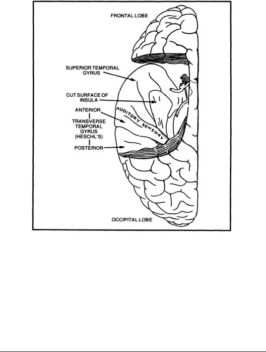
Auditory Cortex. The auditory cortex (AC) consists of areas of the cerebral cortex that respond to sounds. In mammals, the AC is bilateral and has a primary area with surrounding secondary areas. In nonprimates,the AC is on the lateral surface of the cortex,but in most primates,it lies within the lateral fissure on the superior surface of the temporal lobe. Figure 5.6 reveals the area of the temporal lobe involved with auditory function in humans. Tonotopic mapping is usually evident in these areas as isofrequency surface lines.The primaryAC responds most strongly and quickly to sounds.In echolocating bats, the cortex has a large tonotopic area devoted to the frequency region of its emitted cries and cues related to its frequency modulation and returned Doppler shift [Aitkin, 1990].
The cytoarchitecture of the primary AC shows layers I (surface) throughVI (next to white matter), with the largest neurons in layers V and VI. Columns with widths of 50 to 75 µm are evident from dendritic organization in layers III and IV, with fibers lying between the columns.A description of cell types is beyond this treatment.
Discharge patterns in the AC for sound stimuli are mainly of the onset type. Continuous stimuli often evoke later spikes, after the onset discharge, in unanesthetized animals.About half the neurons in the primary AC have monotonic RLFs, but the proportion of nonmonotonic RLFs in secondary areas is much higher. A number of studies have used complex sounds to study cortical responses. Neural responses to species-specific cries, speech, and other important sounds have proven to be labile and to a great extent dependent on the arousal level and behavioral set of the animal.
Cortical lesions in humans rarely produce deafhess, although deficits in speech comprehension and generation may exist.Audiometric tests will generally indicate that sensitivity to tonal stimuli is retained. It has been known for some time that left-hemisphere lesions in the temporal region can disrupt comprehension (Wernicke’s area) and in the region anterior to the precentral gyrus (Broca’s area) can interfere with speech production. It is difficult to separate the effects of these areas because speech comprehension provides vital feedback for producing correct speech.
5.4 Pathologies
Hearing loss results from conductive and neural deficits. Conductive hearing loss due to attenuation in the outer or middle ear often can be alleviated by amplification provided by hearing aids and may be
5-12 |
Biotechnology for Biomedical Engineers |
FIGURE 5.6 A view of the human cerebral cortex showing the auditory cortex on the superior surface of the left temporal lobe after removal of the overlying parietal cortex.
subject to surgical correction. Sensorineural loss due to the absence of IHCs results from genetic deficits,biochemical insult,exposure to intense sound,or aging (presbycusis). For some cases of sensorineural loss, partial hearing function can be restored with the cochlear prosthesis, electrical stimulation of remaining SGCs using small arrays of electrodes inserted into the scala tympani [Miller and Spelman, 1990]. In a few patients having no auditory nerve,direct electrical stimulation of the CN has been used experimentally to provide auditory sensation. Lesions of the nerve and central structures occur due to trauma, tumor growth, and vascular accidents.These may be subject to surgical intervention to prevent further damage and promote functional recovery.
5.5 Models of Auditory Function
Hearing mechanisms have been modeled for many years at a phenomenologic level using psychophysical data. As physiologic and anatomic observations have provided detailed parameters for peripheral and central processing, models of auditory encoding and processing have become more quantitative and physically based. Compartmental models of single neurons, especially SGCs and neurons in the CN, having accurate morphometric geometries,electrical properties,membrane biophysics,and local circuitry are seeing increasing use.
Auditory System |
5-13 |
References
Aitkin L.1990.TheAuditory Cortex:Structural and Functional Bases ofAuditory Perception. London,Chapman and Hall.
Altschuler RA,Bobbin RP,Clopton BM,Hoffman DW (Eds).1991.Neurobiology of Hearing:The Central Auditory System. NewYork, Raven Press.
Arle JE,Kim DO.1991.Neural modeling of intrinsic and spike-discharge properties of cochlear nucleus neurons. Biol Cybern 64:273.
Bekesy G. von. 1960. Experiments in Hearing. NewYork, McGraw-Hill.
BourkTR, Mielcarz JP, Norris BE. 1981.Tonotopic organization of the anteroventral cochlear nucleus of the cat. Hear Res 4:215.
Cooke M. 1993. Modelling Auditory Processing and Organisation. Cambridge, England, Cambridge University Press.
Cooper NP, Rhode WS. 1992. Basilar membrane mechanics in the hook region of cat and guinea-pig cochleae:Sharp tuning and nonlinearity in the absence of baseline position shifts.Hear Res 63:163.
Crawford AC, Fettiplace R. 1985.The mechanical properties of ciliary bundles of turtle cochlear hair cells. J Physiol 364:359.
Dobie RA, Rubel EW. 1989.The auditory system: Acoustics, psychoacoustics, and the periphery. In HD Patton et al (Eds), Textbook of Physiology, Vol 1: Excitable Cells and Neurophysiology, 21st ed.
Philadelphia, Saunders.
Fay RR. 1988. Hearing in Vertebrates:A Psychophysics Databook. Winnetka, Hill-Fay Associates.
Geisler CD. 1992.Two-tone suppression by a saturating feedback model of the cochlear partition. Hear Res 63:203.
Hudspeth AJ.1987.Mechanoelectrical transduction by hair cells in the acousticolateralis sensory system.
Annu Rev Neurosci 6:187.
Hudspeth AJ,Corey DP.1977.Sensitivity,polarity,and conductance change in the response of vertebrate hair cells to controlled mechanical stimuli. Proc Natl Acad Sci USA 74:2407.
Khanna SM,Keilson SE,Ulfendahl M,Teich MC.1993.Spontaneous cellular vibrations in the guineapig temporal-bone preparation. Br J Audiol 27:79.
Kim DO.1984.Functional roles of the innerand outer-hair-cell subsystems in the cochlea and brainstem. In CI Berlin (Ed), Hearing Science: Recent Advances. San Diego, Calif, College-Hill Press.
Kinsler LE, Frey AR. 1962. Fundamentals of Acoustics. NewYork,Wiley.
LePage EL. 1991. Helmholtz revisited: Direct mechanical data suggest a physical model for dynamic control of mapping frequency to place along the cochlear partition.In Lecture Notes in Biomechanics. NewYork, Springer-Verlag.
Lyon RF,Mead C.1989.Electronic cochlea.In C Mead (Ed),AnalogVLSI and Neural Systems. Reading, Mass.Addison-Wesley.
Miller JM, Spelman FA (Eds). 1990. Cochlear Implants: Models of the Electrically Stimulated Ear. NewYork, Springer-Verlag.
Nadol JB Jr. 1988. Comparative anatomy of the cochlea and auditory nerve in mammals. Hear Res 34:253.
Sachs MB,Young ED. 1979. Encoding of steady-state vowels in the auditory nerve: representation in terms of discharge rate. J Acoust Soc Am 66:470.
Young ED,Voigt HF. 1981.The internal organization of the dorsal cochlear nucleus. In J Syka and L Aitkin (Eds), Neuronal Mechanisms in Hearing, pp 127–133. NewYork, Plenum Press.
Young ED. 1984.Response characteristics of neurons of the cochlear nuclei. In CI Berlin (Ed),Hearing Science: Recent Advances. San Diego, Calif. College-Hill Press.

6
The Gastrointestinal System
|
6.1 |
Introduction |
6-1 |
|
6.2 |
Gastrointestinal Electrical Oscillations |
6-1 |
|
6.3 |
A Historical Perspective |
6-3 |
|
|
Minute Rhythms • Hour Rhythms • Terminology |
|
|
6.4 |
The Stomach |
6-6 |
|
|
Anatomical Features • Gastric ECA •The Electrogastrogram |
|
|
6.5 |
The Small Intestine |
6-7 |
|
|
Anatomical Features • Small Intestinal ECA • Small Intestinal MMC |
|
Berj L. Bardakjian |
6.6 |
The Colon |
6-8 |
|
Anatomical Features • Colonic ECA |
|
|
University of Toronto |
6.7 |
Epilogue |
6-9 |
6.1 Introduction
The primary function of the gastrointestinal system (Fig. 6.1) is to supply the body with nutrients and water. The ingested food is moved along the alimentary canal at an appropriate rate for digestion, absorption, storage, and expulsion. To fulfill the various requirements of the system, each organ has adapted one or more functions. The esophagus acts as a conduit for the passage of food into the stomach for trituration and mixing.The ingested food is then emptied into the small intestine, which plays a major role in the digestion and absorption processes. The chyme is mixed thoroughly with secretions and it is propelled distally (1) to allow further gastric emptying, (2) to allow for uniform exposure to the absorptive mucosal surface of the small intestine, and (3) to empty into the colon.The vigor of mixing and the rate of propulsion depend on the required contact time of chyme with enzymes and the mucosal surface for efficient performance of digestion and absorption. The colon absorbs water and electrolytes from the chyme, concentrating and collecting waste products that are expelled from the system at appropriate times.All of these motor functions are performed by contractions of the muscle layers in the gastrointestinal wall.
6.2 Gastrointestinal Electrical Oscillations
Gastrointestinal motility is governed by myogenic, neural, and chemical control systems (Fig. 6.2).The myogenic control system is manifest by periodic depolarizations of the smooth muscle cells,which constitute autonomous electrical oscillations called the electrical control activity (ECA) or slow waves [Daniel and Chapman, 1963].The properties of this myogenic system and its electrical oscillations dictate to a large extent the contraction patterns in the stomach, small intestine, and colon [Szurszewski, 1987].The ECA controls the contractile excitability of smooth muscle cells since the cells may contract only when depolarization
0-8493-1811-4/03/$0.00+$1.50 |
|
© 2003 by CRC Press LLC |
6-1 |